Keywords
Post-operative outcome, meningioma, predictive factor
This article is included in the Oncology gateway.
Post-operative outcome, meningioma, predictive factor
Brain tumors are lesions that cause space-occupying effects, which grow from the brain parenchyma, meninges, or in the skull cavity. Tumors or neoplasms in brain tissue are called primary brain tumors if they originate from the brain parenchyma, namely glial cell neurons (astrocytes, oligodendrocytes, etc.), while tumors in the intracranial cavity originating from other organs (metastases) such as lung and breast cancer are called secondary brain tumors.1
Meningioma is the second most common intracranial neoplasm, meaning it accounts for up to 30 percent of all intracranial tumors and occurs in about 4 out of every 100,000 people. Meningiomas are tumors of the meninges, protective membranes that protect the brain and spinal cord, but are more common intracranially than intraspinal. Most meningiomas are benign, whereas malignant meningiomas are rare. Meningiomas are more common in women than men, especially in the age group between 60-70 years, and tend to be found in several members of the same family. This tumor most often affects women, with a female to male ratio of 2:1.2,3
Surgery remains the main treatment for meningioma at Dr. Soetomo General Academic Hospital. Gender, age, preoperative circumstances, hormonal family planning risk factors, tumor anatomic location, operating time, Simpson grading, and anatomical pathology findings are among the variables that affect the outcome of surgery. The objective of this study was to predict factors associated with meningioma surgery outcomes.
Ethical approval with approval number 0430/LOE/301.4.2/IV/2021 was obtained from the ethical research committee from Universitas Airlangga.
All patients signed informed consent for their medical records to be used for medical research purposes. The ethical committee gives permission as long as no personal information was mentioned. The consent procedure was carried out in several stages based on the rules of the ethics committee of the Faculty of Medicine, Airlangga University, Dr. Soetomo Academic General Hospital:
1. A cover letter was requested from the Department of Neurosurgery, Faculty of Medicine, Airlangga University, Dr. Soetomo Academic General Hospital, to research after the supervising doctor approved the research proposal.
2. Ethical Clearance: this research was submitted to the ethics committee of the Faculty of Medicine, Airlangga University, Dr. Soetomo Academic General Hospital, to obtain ethical approval for data collection.
3. Observations were made.
4. Data obtained from observations were processed and analyzed.
This study describes the results of surgery performed on meningioma patients at Dr. Soetomo General Academic Hospital in a retrospective analytic descriptive manner. The research locations are the Emergency Room (ER), Inpatient Installation, and Outpatient Installation of Dr. Soetomo General Academic Hospital. The study was conducted from January 2014 to December 2020.
The study included patients with clinical, radiological and anatomic pathologies examination confirming meningioma who underwent surgery at Dr. Soetomo General Academic Hospital from 2014 to 2020, each patient has an administrative identification number that is automatically recorded in the patient registration database. The patient registration database is an internal database that registers all data from Dr. Soetomo General Academic Hospital. It can only be accessed by data managers and on request for research purposes. Patients with anatomic pathology results other than meningiomas and incomplete medical records were excluded from this study.
SPSS IBM 25 was used to analyze the data. Using binomial logistic regression, predictors of postoperative complications are then analysed. If a significance level or p-value is found to be lower than 0.05, an independent variable is thought to have an impact on the dependent variable. The odds ratio (OR) is used to evaluate the impact of the independent variable on the dependent variable.
In this study, the number of samples collected from all medical record data of patients who underwent meningioma surgery from January 2014 to December 2020 was 440 samples, with the highest number in 2015 (18.9% of all cases), and the lowest in 2020, (4.1% of all cases) as shown in Figure 1.
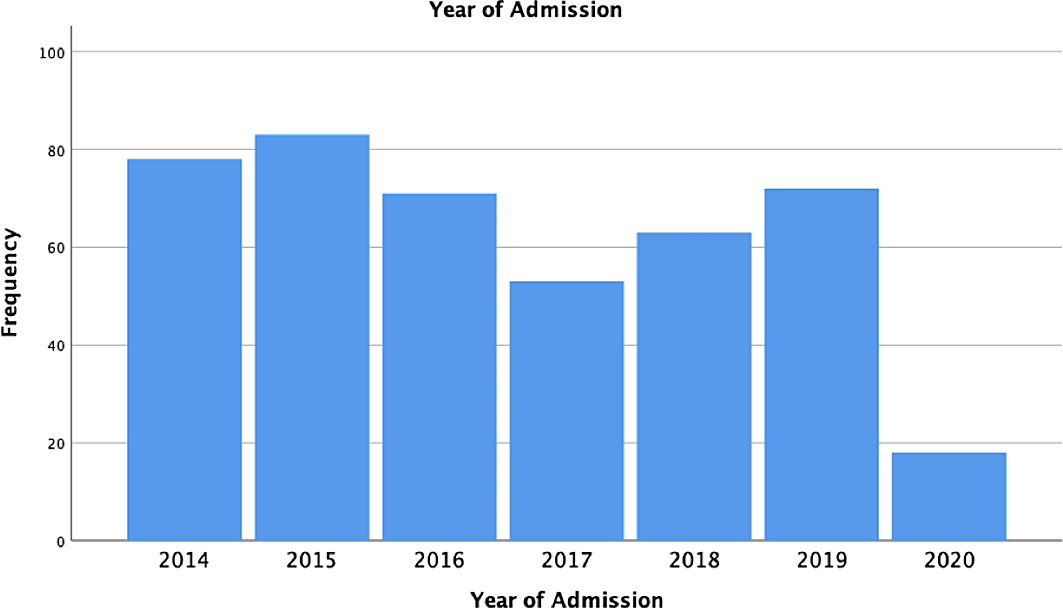
Table 1 summarises the patient’s characteristics. It was found that the highest age group was 36-45 years, seen in 173 patients (39.4%). The patients were predominantly female with 397 patients (90.4%). The most common symptoms at admission were headache (93.3%) followed by neurological deficits (76.5%), decreased consciousness (23%), seizures (19.6%), intratumoral haemorrhage (5.7%), and asymptomatic (4.3%). The systemic disease was found in 240 patients (54.7%).
The use of hormonal contraception was found in 241 patients or 54.9% of the entire population. Injections every three months were the most frequently used contraceptive method in this population, with a total of 114 patients (47.3%).
The most common type of surgery was elective surgery with 359 patients (81.8%). Most meningiomas are discovered at the skull's base, as seen in 259 patients (59.0%), followed by convexity (31.2%), and parasagittal (10.3%). Simpson grade 2 is the highest tumor excision rate with a total of 241 patients (54.9%).
The results of anatomical pathology in this population were grouped according to WHO grading and subtypes. In this study, it was found that the transitional subtype (WHO Grade I) was the most common finding, totalling 230 patients (52.4%) (Table 2). The grading group in this study was WHO Grade I (87.7%), followed by WHO Grade II as many as 45 patients (10.3%) and WHO Grade III as many as 8 patients (1.8%) (Table 3).
| Frequency | Percentage (%) | |
|---|---|---|
| Transitional | 230 | 52.4 |
| Fibroblastic | 52 | 11.8 |
| Atypical | 35 | 8.0 |
| Microcystic | 47 | 10.7 |
| Angiomatous | 15 | 3.4 |
| Chordoid | 2 | 0.5 |
| Meningothelial | 25 | 5.7 |
| Anaplastic | 12 | 2.7 |
| Rhabdoid | 4 | 0.9 |
| Psammomatous | 12 | 2.7 |
| Secretory | 4 | 0.9 |
| Frequency | Percentage (%) | |
|---|---|---|
| WHO Grade I | 385 | 87.7 |
| WHO Grade II | 45 | 10.3 |
| WHO Grade III | 8 | 1.8 |
Post-operative complications reported were post-operative hematoma, post-operative infection, neurologic deterioration, and mortality within 30 days (Table 4). Post-operative hematomas were found in 11 patients (2.5%). Post-operative infection was found in 13 patients (3%). Neurological deterioration was found in 17 patients (3.9%) and 30-day mortality occurred in 10 patients (2.3%).
Post-operative hematoma
Emergency surgery and Simpson Grade IV were significant predictive factors for post-operative hematomas (Table 5). Patients who underwent non-elective or emergency surgery had a 4.5 times greater risk of developing a post-operative hematoma than patients who underwent elective surgery (P = 0.025). Compared with Simpson Grade I, patients with Simpson Grade IV had a 33.5 times higher risk of developing a post-operative hematoma (P = 0.016).
Post-operative infection
Emergency surgery was a significant predictive factor for postoperative infection (Table 6). Patients undergoing emergency surgery had a 4.7 times higher risk for postoperative infection than those undergoing elective surgery (P = 0.015).
Post-operative neurological deterioration
Post-operative hematoma and emergency surgery were significant predictive factors for neurologic deterioration (Table 7). The risk of neurologic deterioration was increased 5-fold in patients undergoing emergency surgery compared with those undergoing elective surgery (P = 0.022). In patients with postoperative hematoma, the risk of neurologic deterioration was increased 235-fold (P = 0.000).
Mortality within 30-day post-operatively
Post-operative hematoma and emergency surgery were significant predictive factors for mortality within 30-day post-operatively (Table 8). The risk of 30-day mortality increased 18-fold in patients undergoing emergency surgery (P = 0.002) and 40-fold in patients with postoperative hematoma (P = 0.001).
Meningiomas become more common with increasing age, with the median age at diagnosis being 66 years. The incidence rate in patients aged over 40 years was 18.69 per 100,000 population, while 0.16 per 100,000 in those aged 0 to 19 years. Women were more likely to have benign and malignant meningiomas, with incidence rates of 2.33 and 1.12, respectively. Meningiomas are more common in boys than girls between 0 and 19 years. Children are more likely to have high-grade meningiomas, which have a higher chance of recurrence and lower overall mortality.4
In this study, there were 440 patients whose epidemiological characteristics could be analysed. Of meningioma patients who underwent surgery at Dr. Soetomo General Academy Hospital in a total of seven years study, the highest number was in 2015 (18.9% of all cases), and the lowest was in 2020 (4.1% of all cases). There was a decrease in surgeries due to the COVID-19 pandemic. The decrease in the number of operations was caused by the spread of COVID-19 in Surabaya. To prevent the spread of COVID-19, the Dr Soetomo Academic General Hospital reduced operations during the pandemic. In addition, the decline in collective operations was due to people's hesitation to undergo hospital treatment during the pandemic. The oldest age group was in the 36-45 year age group. It is following the findings in Ogasawara's research, which states that the incidence is higher in the 40-year age group.4
There were more women than men in this study, with 397 women making up 90.4% of the sample and 41 men making up 9.3%. This is also in line with data from the Central Brain Tumor Registry of the United States (CBTRUS), which showed that women had a more than two-fold higher incidence of brain tumors, with age-adjusted incidence rates of 8.36 and 3.61 for women and men, respectively, per 100,000 person-years.5
Initial meningioma symptoms may occur based on the location of the tumor mass. The symptoms of meningiomas are typically dangerous because they frequently do not infiltrate and grow slowly. Common presentations include seizures brought on by mass effect and/or direct tumor involvement, focal neurologic deficits (cranial nerves), and headaches resulting from elevated intracranial pressure. In this study, headache (93.3%) was the most prevalent symptom, followed by a neurological deficit (76.5%), loss of consciousness (23%), seizures (19.6%), intratumoral haemorrhage (5.7%), and asymptomatic conditions (4.3%). This result is consistent with the study by Lemee et al., in which only 5.4% of patients reported no symptoms while 60.2% of patients had neurologic deficits and 29.6% had seizures.6
Additionally, 240 (54.7%) patients in this study had a systemic disease. According to research by Muskens et al., the incidence of systemic diseases like hypertension and diabetes is positively correlated with the development of meningiomas. Individuals with a history of hypertension and diabetes show an even stronger correlation.7,8
As many as 259 of the 440 patients in this study had meningiomas at the base of their skulls (59.0%), followed by convexity meningiomas, which occurred in 137 patients (31.2%) and parasagittal meningiomas, which occurred in 45 patients (10.3%). Lemee et al. reported similar findings, stating that 690 patients, or 47% of the total patient population, had meningioma located at the base of the skull.6 According to Ogasawara et al., the location in the base of the skull is where meningiomas most frequently occur, accounting for 15-43% of all cases. Parasagittal (13–22%) and convexity (20–37%) are two additional locations that are quite frequent.4,9
Meningiomas are graded according to the WHO tumor classification system. The most recent WHO recommendation (2016) divides meningiomas into 15 subtypes into three categories based on histological criteria. This scoring system has important implications for the treatment strategy because it correlates with the risk of recurrence and overall survival. About eighty percent of meningioma cases are WHO grade I, with benign histology and unresponsive behaviour. Atypical malignant histology in meningiomas of WHO categories II and III, which made up 17.7% and 1.7% of all meningiomas, respectively, suggested a more aggressive clinical course.5 WHO Grade I was discovered to be the most prevalent histopathological finding in this study (385 of 440 patients, or 87.7%), followed by WHO Grade 2 (20.3%) and WHO Grade 3 (1.8%). The most prevalent histopathological subtype finding, transitional, included in WHO Grade 1, was observed in 230 out of 440 patients (52.4%).
Post-operative hematoma
Compared to surgery for other types of brain tumors, removal of an intracranial meningioma carries a higher risk of postoperative hematoma.10,11 A total of 11 patients (2.5%) had post-operative hematomas. Lemee et al., Sicking et al., Nittby, Lee et al., and Gerlach et al. reported a postoperative hematoma rate of 2.7%, 5.0%, 1.3%, 9.8%, and 7.1%, respectively.6,11–13 According to the literature, one of the reasons meningioma surgery has a higher postoperative hematoma incidence rate than surgery for other benign brain tumors is meningioma-derived hyperfibrinolysis.11
It was discovered that Simpson Grade III and emergency surgery performed were predictors of postoperative hematoma. Lemee et al. reported different findings, with age serving as the only significant predictor of postoperative hematoma.6 It is possible that changes in the blood vessels themselves and platelet function, particularly their spread, are linked to postoperative bleeding in the elderly.12 Age, gender, postoperative anticoagulation for comorbidities, tumor location, histology, infiltration into dura mater and arachnoid sinuses, tumor evacuation rate, and peri-operative coagulation factors were the risk factors examined in the study by Lee et al. In the study by Lee et al., it was found that the age was over 70 years, and the platelet count was less than 150,000 after surgery.11 Because most patients with postoperative bleeding do not have a clear cause of hematoma, factors affecting postoperative hematomas vary depending on the literature, both in surgical technique and hemostatic parameters.12,14
Post-operative infection
Out of 440 patients in our study, 13 experienced postoperative infections (3%). Non-elective or emergency surgery time was found to be a factor that raised the risk of postoperative infection by 4.7 times compared to patients who underwent elective surgery (P = 0.015). Each study reported a different incidence of postoperative infection. The infection rate in a study by Conolly et al. was 2.19 percent.15 A postoperative infection rate of 2.6 percent was reported by Lemee et al.6
In contrast, Lemee et al. study's found that the meningioma's location at the base of the skull significantly raises the risk of postoperative infection. Skull base meningiomas frequently require a higher level of expertise and technical proficiency than their counterparts, so they are typically operated by senior neurosurgeons with more experience. Convexity meningiomas, for example, may be considered easier to treat surgically and given to a senior resident or junior nurse in contrast to non-cranial base tumors. This difference in surgical experience may primarily impact the management of the dural defect and watertight dural closure. The deeper location of skull base meningiomas in comparison to more superficial locations, such as convexity meningiomas, may also contribute to their lower infection rate.9
Post-operative neurological deterioration
Seventeen patients in this study experienced neurological deterioration (3.9%). Emergency surgery performed and the presence of a postoperative hematoma both had a significant impact on neurological decline. The postoperative neurologic deterioration rate in the study by Lemee et al. was 3.9 percent, which is consistent with the results of this study. The presence of postoperative hematoma increased the risk of neurologic deterioration after surgery by 4 times in the study by Lemee et al.6
Mortality within 30-days post-operatively
The incidence of death within 30 days was 2.3% in this study. Emergency surgery and postoperative hematoma were factors that affected mortality within 30 days. Patients undergoing emergency surgery had an 18-fold higher risk of death at 30 days (P = 0.002), and patients with postoperative hematomas had a 40-fold higher risk (P = 0.001).
The 30-day mortality endpoint was used in this study as the means to describe postoperative mortality. Standard measures like 30-day mortality should be given precedence over other criteria like in-hospital death or surgical death, whose definition and the result can change depending on the type of tumor and therapeutic management. It is necessary to consider all potential causes of death that occur within 30 days of surgery, including accidents.
Meningioma surgery has been linked to several reports of 30-day mortality. In contrast to Bartek et al., who found 0.6 % mortality within 30 days, Lemee et al. study's found 5.4% mortality within 30 days. Two percent mortality within 30 days was found in research by Mariam Slot et al.6,16,17 This suggests that our study's 30-day mortality rate is comparable to reports in the literature.
According to Lemee, age was a significant factor in determining 30-day mortality, with each year of age increase leading to a 7% increase in mortality. According to Gerlach et al., post-operative hematoma increases mortality in meningioma patients. Additionally, according to several studies, patients with meningiomas had the highest rates of postoperative hematomas.6,12
In conclusion, the analysis of operative outcomes in meningioma patients at Dr. Soetomo General Academic Hospital found that patients undergoing emergency surgery had a 4.5 times greater risk of developing a postoperative hematoma than patients undergoing elective surgery. In addition, patients who underwent Simpson grade IV surgery had a 33.5 times higher risk for post-operative hematoma than patients with Simpson Grade I Patients undergoing emergency surgery had a 4.7 times higher risk for postoperative infection than patients undergoing elective surgery. The risk of neurologic deterioration was increased 5-fold in patients undergoing emergency surgery compared with those undergoing elective surgery. In patients with postoperative hematoma, the risk of neurologic deterioration was increased 235-fold. The risk of 30-day mortality increased 18-fold in patients undergoing emergency surgery and 40-fold in patients with postoperative hematoma.
This study uses secondary information from medical records (retrospective). Due to the need for complete data, this retrospective method using medical records requires extensive data collection. It is advised that prospective methods be used in research to lessen bias. Investigating the long-term side effects of meningioma surgery is also necessary.
Figshare: Data Base Tumor 2014-2020, https://doi.org/10.6084/m9.figshare.20517372.v2.18
This project contains the following underlying data:
Data are available under the terms of the Creative Commons Zero “No rights reserved” data waiver (CC0 1.0 Public domain dedication).
| Views | Downloads | |
|---|---|---|
| F1000Research | - | - |
|
PubMed Central
Data from PMC are received and updated monthly.
|
- | - |
Provide sufficient details of any financial or non-financial competing interests to enable users to assess whether your comments might lead a reasonable person to question your impartiality. Consider the following examples, but note that this is not an exhaustive list:
Sign up for content alerts and receive a weekly or monthly email with all newly published articles
Already registered? Sign in
The email address should be the one you originally registered with F1000.
You registered with F1000 via Google, so we cannot reset your password.
To sign in, please click here.
If you still need help with your Google account password, please click here.
You registered with F1000 via Facebook, so we cannot reset your password.
To sign in, please click here.
If you still need help with your Facebook account password, please click here.
If your email address is registered with us, we will email you instructions to reset your password.
If you think you should have received this email but it has not arrived, please check your spam filters and/or contact for further assistance.
Comments on this article Comments (0)