Keywords
Invasive micropapillary breast carcinoma, clinical breast examination, ultrasound, mammography, diagnosis, radiology, histology, case report
This article is included in the Oncology gateway.
Invasive micropapillary breast carcinoma, clinical breast examination, ultrasound, mammography, diagnosis, radiology, histology, case report
The histopathological type of breast cancer provides important prognostic information about the natural history and overall outcome of the disease. Of all breast cancer types, invasive micropapillary carcinoma (IMPC) is a rare subtype of invasive ductal carcinoma (IDC) characterized histologically by the formation of micropapillaries within the cystic spaces.1–3 This is due to its distinct presentation, particularly lymphovascular invasion. IMPC was first identified in 1993 and classified separately by the World Health Organization (WHO) in 2003, before being subjected to the general rule for the clinical and pathological registration of breast cancer in 2008.2,3 It accounts for about 2–8% of all breast cancers and is considered to be more aggressive than the other IDC types.1,3 It is also listed as a special histological type in the 2003 and 2012 editions of the WHO classification of breast tumors.4,5 This case report followed the CAse REport (CARE) guidelines as a guiding principle.6,7
A 41-year-old woman black African housewife, presented with a left breast lump for two months. She had no family history of breast cancer and had never had breast surgery.
The patient was informed and consented to the use of her medical records and clinical images. She understood that she had a rare subtype of cancer and believed that using it as a case study would advance science and improve clinical diagnosis and outcomes for patients with IMPC in the future. Additionally, hospital management gave permission and ethics clearance for this case report was obtained from the Walter Sisulu University Ethic committee (WSU Ethics approval No: 056/2022) on the 14th of June 2022.
During clinical breast examination, there was no nipple discharge or associated pain. The left breast mass was clinically palpable with no skin changes or nipple retraction. The mass was firm and fixed to the overlying skin, but it was not attached to the underlying structures. There were palpable mobile lymph nodes in the left axilla.
Mammography revealed an oval, spiculated, high-density mass in the upper outer quadrant of the left breast (Figure 1). This was associated with ancillary findings of focal trabecular and Cooper's ligament thickening. There were no associated microcalcifications. Axillary lymphadenopathy was present on the left side.
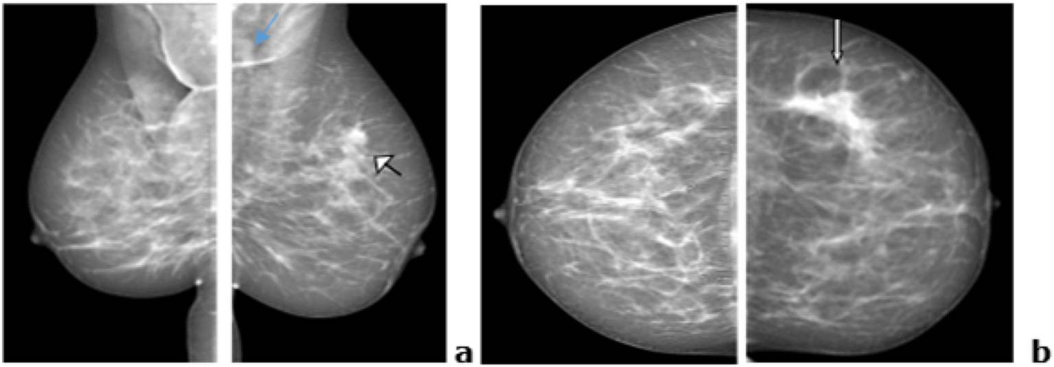
Spiculated, irregular mass-highly suspicious (white arrows). ACR-BIRADS score 5 (American College of Radiology - Breast Imaging and Data System).
High resolution ultrasound (HRUS) showed an oval, not-well circumscribed spiculated, parallel mass which was heterogeneous but predominantly hypoechoic with no posterior features at 2 o’clock (Figure 2). The mass measured 20 × 15 × 21 mm and was in the superficial glandular layer, 4cm from the nipple with moderate vascularity at color Doppler. A second smaller mass was seen next to the larger one which was also hypoechoic and oval shaped with lobulated margins, measuring 8 × 5 × 6 mm. This was thought to be a fibroadenoma or an infiltrated lymph node. Round hypoechoic nodes with eccentric mediastinum and diffusely thickened cortex were seen in the left axillar (Figure 3). The radiological diagnosis was ACR BIRADS category 5 (American College of Radiology- Breast Imaging Reporting and Data System).8,9 As a result, biopsy of the lesion was recommended. Differential diagnoses included invasive ductal carcinomas and lobular carcinomas.


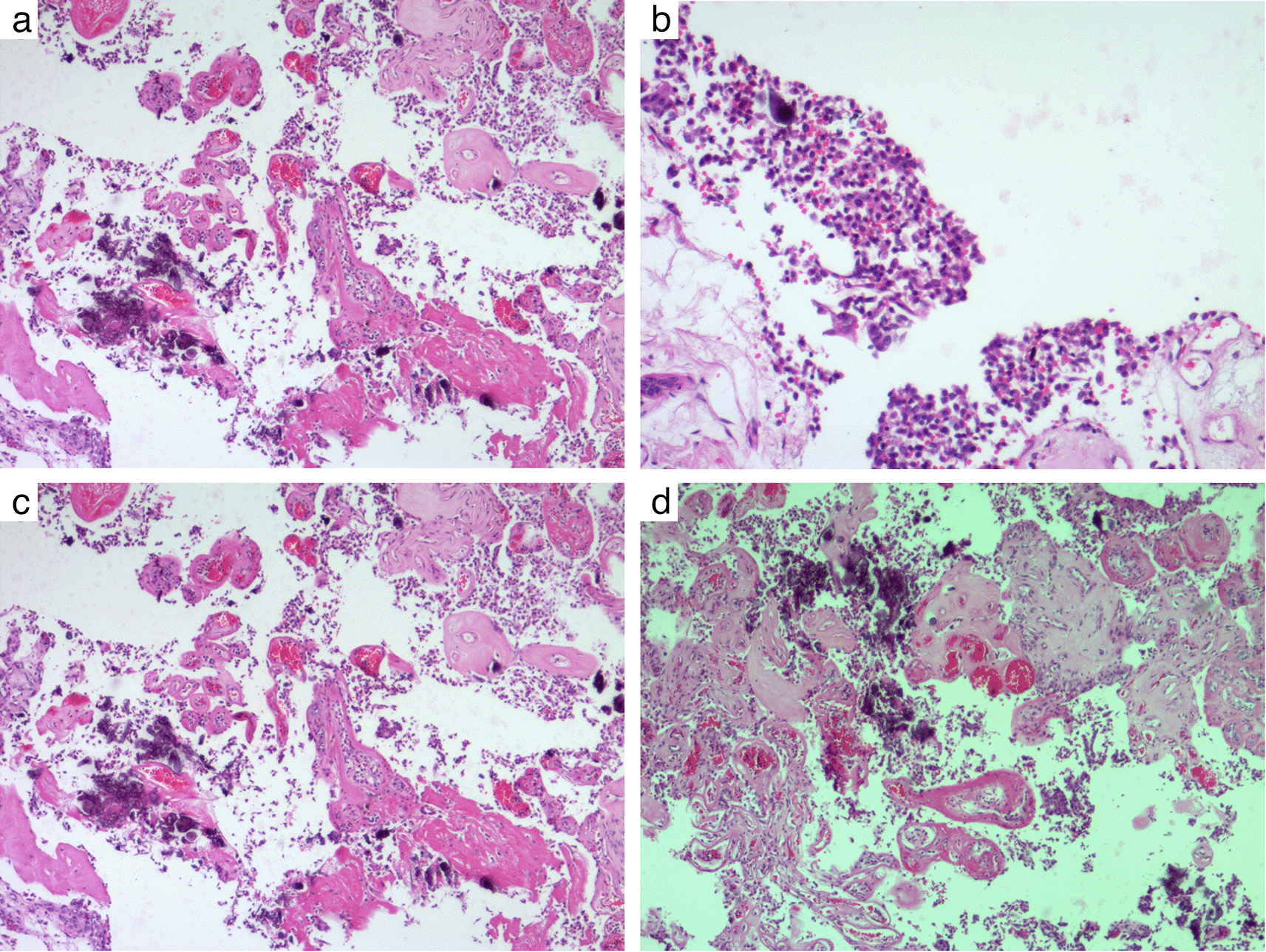
The patient underwent an incision biopsy at which a 5 × 4 × 2.5 cm irregular, yellow piece of tissue with a 2.5 × 2 × 1.5 cm firm mass was sent to the laboratory for histopathological analysis. On microscopy, sections showed an invasive tumor with a distinctly micropapillary growth pattern –fibrovascular cores lined by epithelial cells located in cystic spaces (Figure 4a and b). The tumor cells showed significant nuclear pleomorphism with lymphovascular invasion and prominent fibrovascular cores with no surrounding fibrous capsule (Figure 4c and d). In addition, there were areas of high-grade ductal carcinoma in situ with a combination of fibro adenomatous change. On immune histochemistry, the tumor stained strongly diffusely positive for estrogen and progesterone receptors and negative for human epidermal growth factor receptor 2 (HER2). The surgical margins were reported to be positive, and the modified Bloom-Richardson grade was reported as III.10 Though the positron emission tomography (PET) scan could have been more informative, unfortunately, this was not available in our center. Hence, distant metastatic work up was done using computed tomography (CT)-abdomen and pelvis at which no suspicious lesions were found. The patient subsequently underwent total right breast mastectomy and axillary lymph node dissection followed by chemotherapy. The patient received cyclophosphamide 600 mg/m2 doxorubicin 60 m2 and 5-Fluoracil 600 mg/m2. All the drugs were given intra venously every 21 days for six cycles. Since the patient’s receptor status was positive for estrogen and progesterone, she is now on tamoxifen 20 mg once a day at night.
HRUS of the mastectomy bed, axillary and supraclavicular lymph nodes, as well as mammography of the right breast, were performed as part of the 12 and 24-month follow-up investigations. The ultrasound and mammography revealed no signs of local recurrence or contra lateral disease.
Breast cancer is the most common female malignancy worldwide and is a heterogeneous disease with diverse biological patterns and multiple pathological subtypes.11,12 IMPC is a rare, distinct pathologic intraductal carcinoma subtype known to be aggressive at initial presentation, with a significant propensity for lymph node metastasis and lymphovascular invasion.3 There are two subtypes, mixed (most common) and pure, which is rare.3,11 There is scanty radiological and clinical information on the entity.11,13–15 The unique properties of IMPC make it a breast cancer subtype that breast physicians and imaging professionals should recognize, treat cautiously, and follow up because of its potential for recurrence and poor prognosis.16 Patients with IMPC often present with a painless palpable breast mass,4,11,12,17 which we also observed in the patient in this report. Studies have shown that axillary lymph node involvement is common at presentation.12,16 Some patients may present with axillary lumps before the breast mass is clinically palpable (occult breast cancer).12 Morphologically, the tumor resembles micropapillary tumors of the urinary bladder, colon, lungs, and major salivary glands, where again it shows aggressive behavior with a poor prognosis.2,16
Histopathologically, IMPC is characterized by tubuloalveolar or pseudopapillary structures that lack a fibrovascular core and are surrounded by clear void spaces, as previously mentioned.1–3,18 The micropapillary component in each tumor is variable. However, regardless of the extent of the micropapillary component, tumors with any amount of this particular histological pattern exhibit a more aggressive clinical behavior with a high degree of lymphovascular involvement compared to other types of invasive ductal carcinoma.2,4,17 Immunohistochemical findings in individuals diagnosed with IMPC are more likely to be positive for the expression of estrogen and progesterone hormone receptors than in patients with other types of breast cancer.1–3,17–19 This is consistent with our findings for the patient discussed here.
So far, there are no typical IMPC imaging identifiers.11 The sonography of the primary lesion shows no typical features. The most common finding in the literature is a poorly circumscribed, homogeneous, hypoechoic mass.4,10,13 One case report was identified reporting a hyperechoic lesion at ultrasound, a feature commonly seen in benign lesions.18 In a study by Jones et al.13 they found that 67% of the patients had sonographically suspicious axillary lymph nodes. Axillary ultrasound is useful in assessing lymph node involvement in patients with IMPC due to the high incidence of lymphovascular invasion by the tumor.2,4,11,13 Adrada et al.4 documented pathologic supra- and infraclavicular nodules in some patients in their study, suggesting that investigation of metastases in IMPC patients should routinely include these sites. Ultrasound can show more lesions in IMPC patients than mammography. Color Doppler may show centripetal increased vascularity, a finding suggestive of malignancy.13
On mammography, IMPC often presents as an irregular, high-density mass with spiculed or lobulated borders.4,11,13 Mammographic findings in most studies, although with small sample sizes due to the rarity of the tumor, generally suggest a malignant lesion.11,13,17 Microcalcifications were a common feature in most patients with IMPC. The characteristics of the microcalcifications were indeterminate in most studies or rather suggestive of malignancy.11,17 In some patients, the tumor was occult on mammography, as in the study by Adrada et al.4 and Jones et al.13 only architectural distortion was seen on mammography.
Just like mammography and ultrasound, an irregular mass is the most common feature of IMPC on magnetic resonance imaging (MRI). However, MRI is more accurate in determining the anatomical extent of the tumor and more sensitive to the presence of multiple lesions, if present. IMPC demonstrates rapid initial washout kinetics for patients with invasive ductal carcinoma with micropapillary features in the available literature.4,12,16,17 Some cases showed a non-mass-like enhancement characteristic, while lymph node assessment for nodal involvement is more accurate on MRI. IMPC typically shows malignant MRI characteristics and contrasted MRI is able to show the enlargement of the microcapillary bed in breast tumors with a higher sensitivity than mammography or ultrasound.12,17 However, despite the high MRI sensitivity, IMPC cannot be distinguished from other non-micropapillary breast lesions.12,16,17
Follow-up of patients with IMPC requires screening of the remaining breast, but more important is evaluation for recurrences in the mastectomy bed and involvement of the nodes at the different three levels of breast lymphatic drainage. In this regard, HRUS plays a major role in the follow-up of IMPC patients and should be specifically requested by the treating physician. Detection of IMPC by breast imagers and physicians is important as it is believed to have a poorer prognosis than other types of IDC.3,17,18 The major challenges with IMPC is the presentation at an advanced stage usually stage 3 or 4, due to early nodal involvement and lymphovascular invasion.3,16
The treatment of choice for IMPC is mastectomy plus axillary lymph node removal with adjuvant chemotherapy in cases with tumor size greater than 1 cm and node positive.20 A high proportion of patients with IMPC express hormone receptor positivity estrogen, progesterone and HER2, this makes them beneficiaries of targeted hormone therapy, which improves overall outcome despite the aggressiveness of the disease.16
As is typical of most case reports, the main limitation is the inability to establish a cause-and-effect relationship. A major strength, however, is the generalizability of these clinical implications, given that the reported case is a rare type of breast cancer with similar clinical features to the few documented breast IMPCs worldwide
Invasive micropapillary carcinoma is a distinct subtype and a poorly recognized variant of invasive ductal carcinoma. It has significant lymphovascular invasion, extensive lymph node involvement, and a high rate of local recurrence. Malignant imaging features include a high-density irregular mass on mammography, a hypoechoic, non-circumscribed irregular or speculated mass at ultrasound, and an irregular enhancing mass on MRI. Ultrasound is helpful in assessing local and regional recurrences, and MRI is critical in determining the extent and presence of multifocal lesions prior to surgery. If the breast cancer management team recognizes IMPC, it is possible to identify a group of patients with a poor prognosis who may require adjuvant treatment with nodal and post-mastectomy bedside assessment for recurrence at follow-up.
| Views | Downloads | |
|---|---|---|
| F1000Research | - | - |
|
PubMed Central
Data from PMC are received and updated monthly.
|
- | - |
Provide sufficient details of any financial or non-financial competing interests to enable users to assess whether your comments might lead a reasonable person to question your impartiality. Consider the following examples, but note that this is not an exhaustive list:
Sign up for content alerts and receive a weekly or monthly email with all newly published articles
Already registered? Sign in
The email address should be the one you originally registered with F1000.
You registered with F1000 via Google, so we cannot reset your password.
To sign in, please click here.
If you still need help with your Google account password, please click here.
You registered with F1000 via Facebook, so we cannot reset your password.
To sign in, please click here.
If you still need help with your Facebook account password, please click here.
If your email address is registered with us, we will email you instructions to reset your password.
If you think you should have received this email but it has not arrived, please check your spam filters and/or contact for further assistance.
Comments on this article Comments (0)