Keywords
lipoma, spindle cells, spindle cell lipoma, popliteal fossa, case report
This article is included in the Cell & Molecular Biology gateway.
lipoma, spindle cells, spindle cell lipoma, popliteal fossa, case report
Spindle cell lipoma (SCL) is an infrequent benign tumor of the subcutaneous fat tissue. It usually touches the shawl region, but rarely the scalp, the face, or the extremities.1 SCL of the knee represents less than 1% of all spindle cell lipomas; to date only a few cases are reported in the literature.1–3 No cases of SCL of the popliteal fossa have been reported. The classical reported tumors have mostly been between 1 and 5cm in size which are significantly smaller than the one presented in this case. We present herein the first and largest spindle cell lipoma of the popliteal fossa to our knowledge. This case report has been reported in line with the criteria.
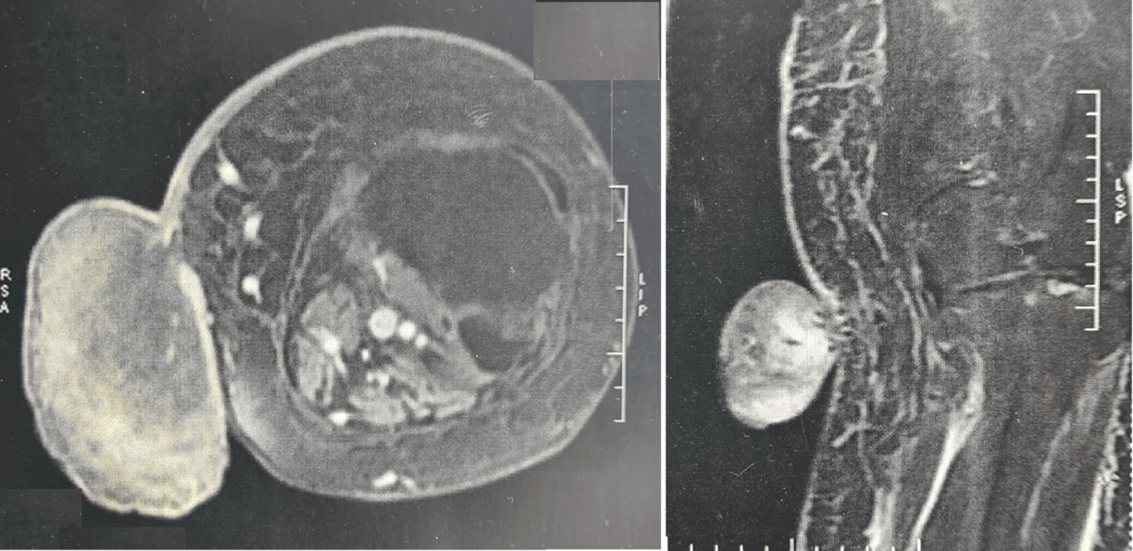
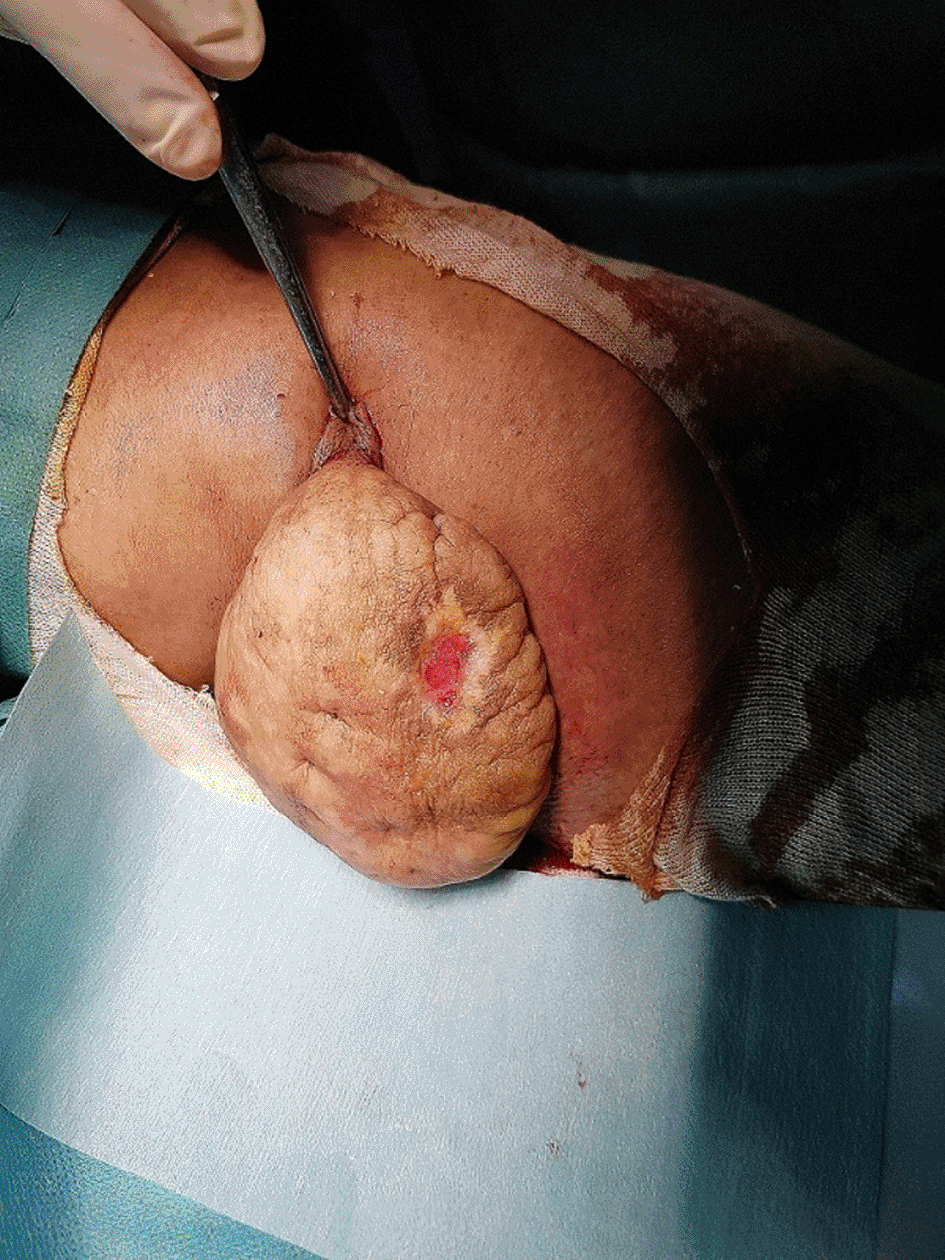
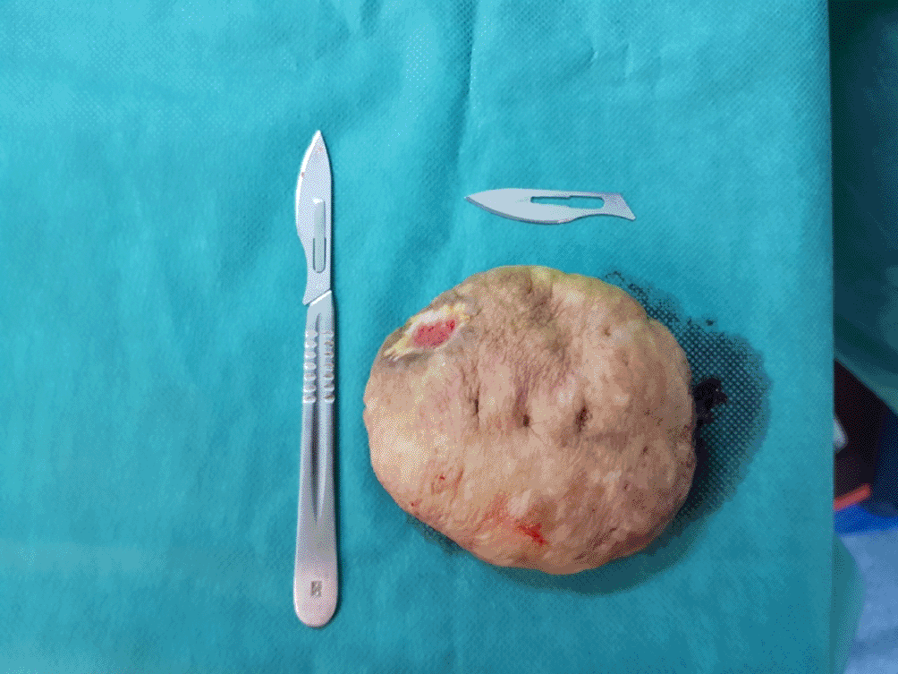
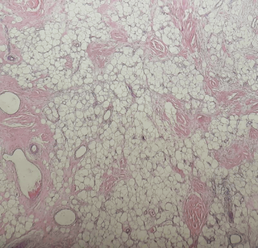
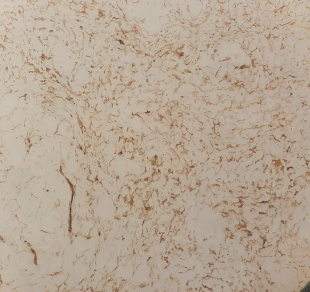
A 75-year-old woman, with a medical history of hypertension, presented with a swelling of the left knee evolving for four years since November 2017. The mass was initially sub-centimetric and gradually increased in size leading the patient to seek medical attention. Physical examination showed a 12 cm firm, well-limited, and painless mass of the left popliteal fossa (Figure 1). This mass was mobile, pediculated with a central ulceration leaking pus. An ultrasound scan of the left knee was performed showing a well-limited, non-vascularized skin mass of 10 cm, with no popliteal cysts or vascular lesions. MRI scan of the left leg revealed a tumor mass in the popliteal fossa measuring 98 x 55 x 8 mm with some non-dilated vessels draining to the long saphenous vein. There was no infiltration of the underlying tissues (Figure 2). The patient underwent a surgical total resection of the mass. Postoperative care was uncomplicated. The intervention was performed by a senior orthopedic surgeon in the Orthopedic Surgery Department of the Internal Security Forces Hospital, La Marsa, Tunisia (Figure 3). Gross examination showed a mass of 11 x 9 x 6 cm with a cutaneous ulceration of 3 x 4 cm. There was no evidence of macroscopic extension to the underlying subcutaneous tissue. Histological examination showed a benign mesenchymal proliferation associating foci of well-differentiated and mature adipocytes and myxoid and collagenous patches of variable thickness. These patches were composed of spindle-shaped cells with elongated nuclei without any atypical character or mitosis figures, mixed with collagenous clusters. Numerous mast cells were also seen. No vascular thrombi, lipoblasts, or necrosis were seen. There were no signs of malignancy. On immunohistochemical study, this tumor proliferation diffusely expressed CD34. Desmin expression was negative and the Ki67 proliferation index was <1% (Figures 4–6) This morphological and immunophenotypic appearance was compatible with a spindle cell lipoma. The resection was total and there was no tumor residue. In July 2022, after one year of follow-up, the patient had no signs of local recurrence. She was able to do all her daily activities without any sensory-motor disorder.
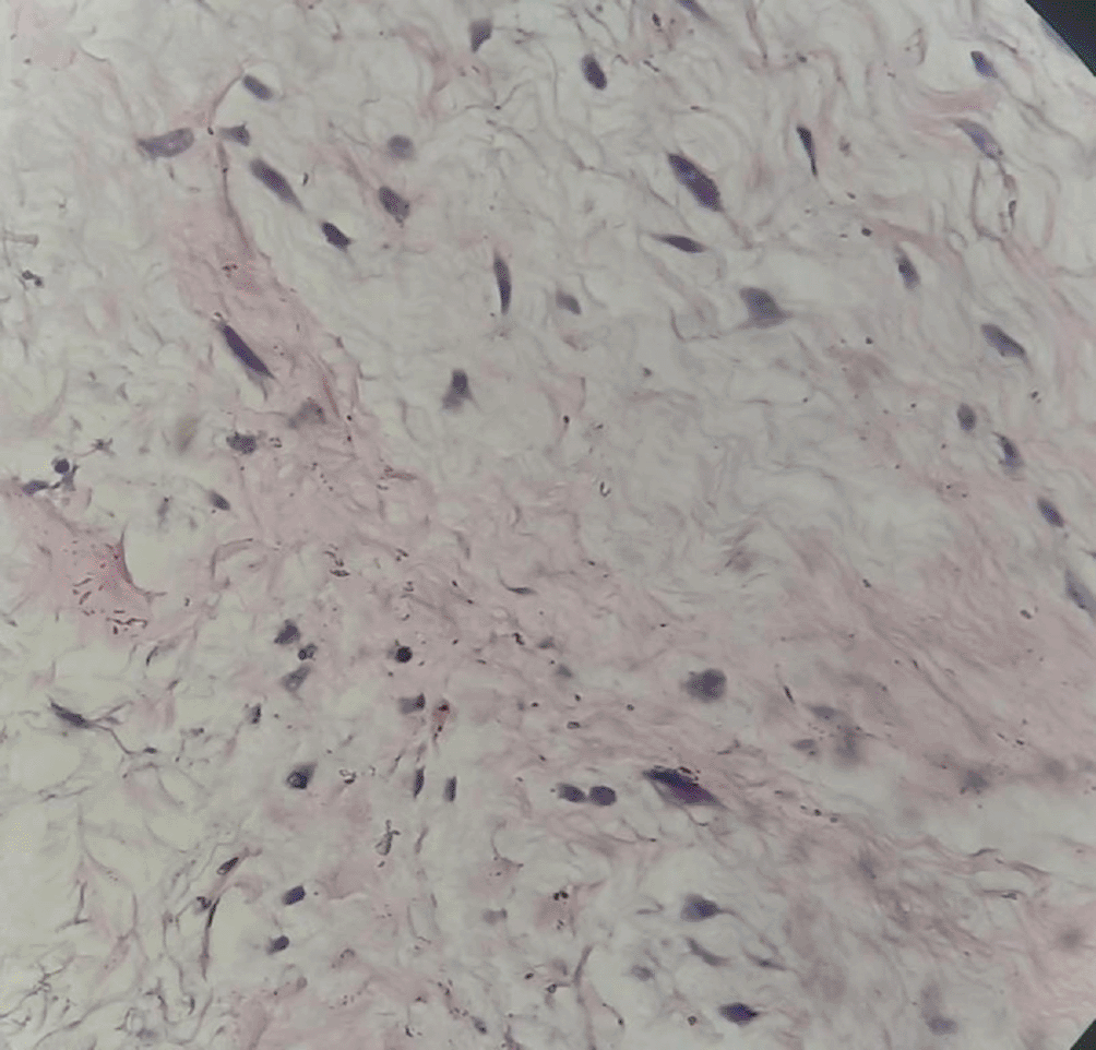
SCL is an uncommon histological variant of lipoma (representing 1.5% of all lipomas) first described by Enzinger and Harvey in 1975.4,5 It is a benign tumor of the subcutaneous fat tissue that occurs mostly in the posterior side of the upper trunk such as the neck, the back, and the shoulders. Atypical locations are possible, it may touch the face, the scalp, the upper and lower extremities, the retroperitoneum, but rarely the knee or the popliteal fossa.6–10 Only four cases of SCL of the knee have been reported, these cases are summarized in Table 1. SCL has a noteworthy predilection for men with a percentage of 90% occurring usually in the second and third decades.4 Clinically, the spindle cell lipoma presents as a gradually increasing painless mass with a long history (more than one year in 75% of cases). The tumor may reach a considerable size leading the patient to seek medical attention. Spindle cell lipomas usually have clear boundaries, are firm, mobile and without associated cutaneous lesions, ulcerations are rarely noted.7,11 SCL usually measures between 1 and 5 cm in greatest dimension, rarely exceeding 10 cm. The rare cases with extraordinary size or atypical anatomical location require further investigations to rule out more worrisome malignancies.9,12–15
| Authors | Age (years) | Sex | Site | Size (cm) |
|---|---|---|---|---|
| Goto et al. 20043 | 58 | F | Right knee | 8 x 4 x 1 |
| Chen et al. 20191 | 48 | M | Right knee | 9 x 6 x 3 |
| Jelinek et al. 20202 | 48 | M | Knee | 4 |
| Jelinek et al. 20202 | 63 | M | Knee | 7 |
| Studied case | 75 | F | Left popiliteal fossa | 11 x 9 x 6 |
Radiologically, the images of the SCL are variable and no pathognomonic signs are identified. In most cases, tumor images are composed of a relatively equal ratio of spindle cells and fatty tissue. This ratio is variable, and the aspect may overlap that of liposarcoma or hibernoma. Intense enhancement of the non-adipose component supports the diagnosis of spindle cell lipoma.2,16 Histologically, SCL have different textures, from soft to tough, usually without necrosis or bleeding. Most of the tumors are made of spindle-shaped cells, rope-like collagen fibers and mature adipocytes surrounded by a fibrous capsule. Mast cells are numerous in a significant number of cases, small aggregates of lymphocytes are seen only occasionally. Different amounts of adipocytic component are present and may be accompanied by a myxoid or collagenous matrix. Therefore, multiple subtypes of spindle cell lipoma may exist such as fibrous, myxoid, fat-rich, low-fat, and pseudo angiomatous type.1,17 Most cells do not have atypia or high mitotic activity. Immunohistologically, spindle cell lipomas usually show diffuse and strong expression of CD34, and to a lesser degree vimentin. Proliferation index Ki-67 is <3% in 99% of cases. Due to the diversity of histological morphotypes of SCL, it is sometimes diagnosed as other benign or malignant tumors such as solitary fibrous tumor and shuttle cell liposarcoma, therefore immunohistochemical staining combined with molecular pathology examination can confirm the diagnosis. Benign spindle cell lesions are usually underrecognized and can represent potential pitfalls of malignancy, particularly in a small biopsy. The main differential diagnoses of spindle cell lipoma are elastrofibroma, solitary fibrous tumors, angiomyolipoma, myxoid liposarcoma (for the myxoid type of SCL) and mammary myofibroblastoma.16,18 Spindle cell lipoma and pleomorphic lipoma share many known non-pathognomonic genetic chromosomal abnormalities specifically losses at 16q and 13q chromosomes, but these abnormalities are not constant and do not interfere with the diagnosis.12,19 The standard treatment of SCL is the surgical marginal resection, post excision recurrence rate is stated to be <1% with adequate marginal resection.3,9
| Views | Downloads | |
|---|---|---|
| F1000Research | - | - |
|
PubMed Central
Data from PMC are received and updated monthly.
|
- | - |
Provide sufficient details of any financial or non-financial competing interests to enable users to assess whether your comments might lead a reasonable person to question your impartiality. Consider the following examples, but note that this is not an exhaustive list:
Sign up for content alerts and receive a weekly or monthly email with all newly published articles
Already registered? Sign in
The email address should be the one you originally registered with F1000.
You registered with F1000 via Google, so we cannot reset your password.
To sign in, please click here.
If you still need help with your Google account password, please click here.
You registered with F1000 via Facebook, so we cannot reset your password.
To sign in, please click here.
If you still need help with your Facebook account password, please click here.
If your email address is registered with us, we will email you instructions to reset your password.
If you think you should have received this email but it has not arrived, please check your spam filters and/or contact for further assistance.
Comments on this article Comments (0)