Keywords
Percutaneous Catheter Drainage, PCD, Intermittent needle aspiration, INA, Fluid collections, Abscess, Image-guidance, Pigtail catheter.
This article is included in the Manipal Academy of Higher Education gateway.
Percutaneous Catheter Drainage, PCD, Intermittent needle aspiration, INA, Fluid collections, Abscess, Image-guidance, Pigtail catheter.
Thoracic, abdominal, and pelvic fluid collections are commonly encountered clinical entities. Image-guided percutaneous continuous catheter drainage is an interventional radiology procedure that is now considered to be the standard curative therapy for thoracic, abdominal, and pelvic fluid collection or abscesses.1 Percutaneous catheter drainage (PCD) has over time replaced open surgical drainage (OSD) in all but the most complicated or inaccessible cases.2 Most of the fluid collections permit easy accessibility. However, deep-seated collections or collections in close proximity to adjacent structures could prove to be inaccessible and call for procedural modifications and/or alternate approaches. The feasibility of the drainage is dependent on the consistency of the collection.3 Thus, PCD is a multifactorial procedure that is based on the size, location, character, and configuration of the fluid collection.4
PCD bridges the gap between non-invasive and surgical intervention with minimally invasive, image-guided drainage by utilizing ultrasound (US) or computerized tomography (CT).5,6 PCD using US guidance is the preferred procedure when the fluid collection is large and is localized superficially. In contrast, deeper collections are better evaluated under CT guidance. However, the major advantages of using US over CT are attributed to the lack of radiation, lower cost, flexibility, portability, and shorter period. Pigtail catheters with a single lumen and multiple side holes are generally the preferred catheters in the continuous drainage method of these collections.7
This study aims to evaluate the efficacy, feasibility, safety, and relevant clinical outcomes of image-guided percutaneous (pigtail) catheter drainage of thoracic, abdominal, and pelvic fluid collections. It also aims to assess the causes of complications and the failure of PCD.
This study is a prospective analysis conducted over a span of two years (Aug 2019 to Sep 2021) by the Department of Radiodiagnosis, Kasturba Medical College, Mangalore, India. The study was conducted only after obtaining the required permission from the Institutional Ethics Committee (IEC) (Ethics approval number: IEC KMC MLR 10-19/471). This study included 83 patients diagnosed with thoracic, abdominal, and pelvic fluid collections referred by clinicians from the emergency department, general wards, intensive care unit (ICU), or outpatient departments (OPD) for percutaneous pigtail catheter drainage in the hospital. The diagnosis of abscess or fluid collection in the corresponding region was done by either a US scan or CT scan. The approval and consent for performing the PCD procedure were taken by the referring clinician from the patient. The sample size was estimated using the following formula:
Where, = sample size; z = 1.96 at 95% confidence interval (CI); P = prevalence from the reference article (88% or 0.88); Q = 1-P (0.12); and D = effect size (7% or 0.07). With 95% CI, 5% level of significance, and 88% prevalence, the sample size required for this study was found to be 83 (. Therefore, a minimum of 83 cases were taken into consideration for this study. Data sampling was done by non-probability convenience sampling and the outcomes measured included efficacy, feasibility, safety, the collapse of the cavity after PCD without requiring further drainage, and clinical improvement of the patient.
Data were entered into a Microsoft Excel datasheet and was analyzed using SPSS 22 version software (IBM SPSS Statistics, Somers NY, USA). Categorical data was represented in the form of frequencies and proportions. The Chi-square test was used as a test of significance for qualitative data. P-value of <0.05 was considered statistically significant after assuming all the rules of statistical tests. MS Excel and MS Word were used to obtain various types of graphs such as bar diagrams and pie diagrams.
The US equipment included: Ultrasound – GE healthcare LOGIQS7 EXPERT, Ultrasound-GE VOLUSON 730 EXPERT 4D, Ultrasound-GE HEALTHCARE LOGIQS8 EXPERT, Ultrasound – SAMSUNG HS40, Ultrasound Philips HD15 machine, and Ultrasound Philips HD7 machine. The CT equipment included BRIGHT SPEED Elite 16 slice MDCT GE medical systems, CT BRIVO 385-ESCITE-16 slice – GE, and Somatom GO now 32 slice.
Pigtail catheters were used for PCD. Figure 1 shows the parts of a pigtail catheter.
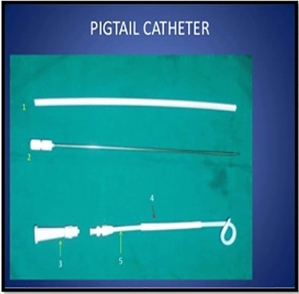
(1, Needle protection sheath; 2, Puncture needle; 3, Connector; 4, Outer sheath; 5, Catheter).
Indications for Intermittent Needle Aspiration (INA) included patients who had a safe and feasible access pathway under imaging guidance with simple fluid collections.
Indications for Pigtail catheter drainage included necrotic fluid collections with the presence of internal septations, loculations, debris, or air pockets within.
Indications for surgical drainage included inadequate drainage of fluid/necrotic collection after PCD, persistent fever/deteriorating symptoms, and bowel complications. (e.g., fistula formation, obstruction, etc.)
All the procedures were performed by certified radiologists after taking appropriate consent from the patient(s). Written informed consent was obtained and all required pre-procedural preparation and investigation was done before the procedure. Prothrombin time (PT), activated partial prothrombin time (aPTT), bleeding and clotting time, and international normalized ratio (INR) was assessed before the procedure. All the procedures were accessed percutaneously and performed under local anesthesia. The following steps were incorporated in performing the PCD procedure:
• The patient(s) were placed in a convenient suitable position for drainage.
• The site of entry on the skin was painted and draped in a sterile surgical field.
• After having precisely located the abscess/fluid collection on either the US or CT scan, the clinical radiologist initially assessed the diameter/volume of the collection and determined the safest access pathway to avoid injury to adjacent structures (e.g., vascular structures, solid organs, bowel, etc.) (Figure 2).
• Subsequently, the skin and subcutaneous tissue were anaesthetized with local anaesthesia accordingly.
• An 18/20/22-gauge (G) lumbar puncture (LP) needle was inserted under image guidance into the site of fluid collection and the tip of the LP needle was well-positioned in the fluid cavity.
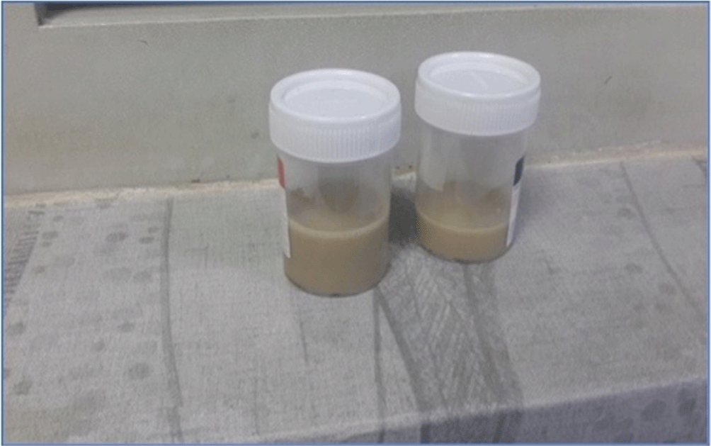
• The aspirated fluid was collected in sterile bottles and sent to the laboratory for further diagnostic analysis (Figure 3). Intermittent needle aspiration (INA) was attempted till no further fluid was aspirated.
• In the case of deep-seated collections and/or fluid collections with thick echogenic contents on the US or high attenuation values on CT, pigtail catheterization was attempted in the same sitting and fluid drainage was allowed – This group of patients was categorized into Group A.
o Procedure for pigtail catheterization (Figure 4)
▪ After choosing appropriate pigtail catheter brands and sizes, a minor skin incision was made on the site of the fluid collection or on the site of a previously inserted needle to fit in the catheter.
▪ A multipurpose drainage catheter (polyurethane pigtail catheter) was advanced into the fluid-filled cavity (Figure 4A and 4B)
▪ Post pigtail catheter placement for continuous drainage, check imaging was done to confirm appropriate positioning of the catheter (Figure 4C)
▪ Once a well-seated catheter was confirmed, it was secured with a surgical suture (Figure 4D)
▪ Subsequently, the catheter tube was connected to a drainage bag and the fluid was allowed to drain continuously (Figure 4E and 4F)
▪ Daily care of the catheter was provided to the patient by both the clinician and the radiologist in regard to the output and monitoring of the vitals of the patient
▪ Follow-up imaging was done to confirm a significant reduction in the fluid cavity. The catheter was then withdrawn when the clinical status of the patient had improved with the output of less than 10 mL/day (provided the catheter had not clogged)
• In cases where needle aspiration alone was attempted, US was done after 48 hours of INA (on day 3) to look for any residual fluid collection. On follow-up US imaging, if there was reaccumulation or inadequate drainage of the fluid cavity (i.e., reduction of the fluid cavity by less than 75% from the original volume), pigtail catheterization was attempted. This group of patients was categorized into Group B. (Cases with significant resolution of the fluid collection were excluded from the study).
• If the residual fluid collection was found to be >75% of its original volume, it was considered to be a failure of the procedure and was scheduled for OSD. This group of patients were categorized into Group C.


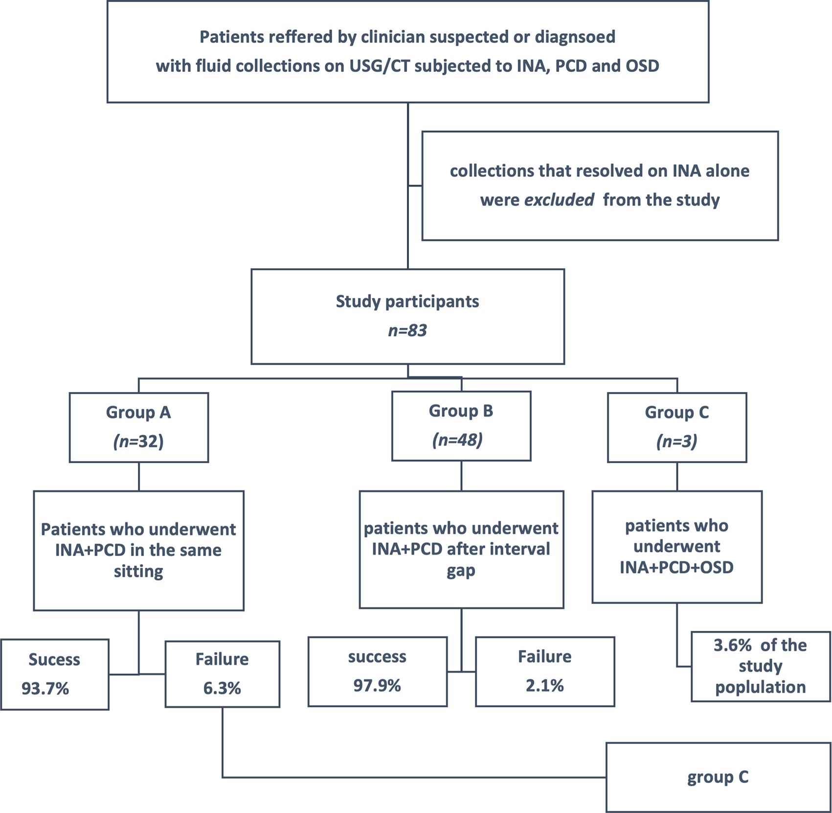
‘Table 1’ is a summary of the categorization of patients into different intervention groups.
INA, Intermittent Needle Aspiration; PCD, Pigtail Catheter Drainage; OSD, Open Surgical Drainage.
| Group A | Group B | Group C |
|---|---|---|
| Patients subjected to INA + PCD in the same setting | Patients subjected to INA followed by PCD after an interval gap of 48 hours | Patients subjected to INA + PCD + OSD |
The majority of the patients were aged between thirty to fifty with a mean age of 46.73 years and a standard deviation of 15.92 (46.73±15.92 years). The youngest patient encountered was three years old and the oldest patient encountered was 76 years old. The majority of the patients in this study were males (68.67%) (compared to females at 31.33%). The male-to-female ratio was 2.2:1. Table 2 provides details regarding the demography of the patients in this study.
The abdomen was the most commonly encountered location of fluid collection in our study, with 61.45% of the study subjects. CT was the most commonly utilized imaging modality to guide the pigtail catheter, followed by US, and both CT and US. 10F was the most frequently used catheter size (53.01%), followed by 12F and 8.5F. Regarding the nature of the fluid drained, purulent fluid was the most commonly drained fluid (57.83%), followed by serious fluid (18.07%). Table 3 shows further details regarding the distribution of disease and intervention characteristics.
Patients were categorized into three groups, namely, groups A, B, and C based on the intervention received by them. Group A included patients subjected to INA and PCD in the same setting and comprised a total of 32 patients (38.53%, n=83). Group B included patients subjected to INA followed by PCD after an interval gap of 48 hours (on day 3), and comprised of 48 patients (57.83%, n=83). Group C included patients who were subjected to INA, PCD, and OSD, and comprised of 3 patients (3.61%, n=83).
The majority of study subjects (68.67%) showed a more than 75% reduction in the residual collection from their original volume of the collection. The complications were classified as major and minor complications based on the severity. Major complications (3.6%) included the reaccumulation of fluid and pneumothorax. 72.29% of the subjects reported pain after the procedure, making it the most widely reported minor complication in our study. Catheter-related complications (1.2%) included blockage of the catheter. However, 26.51% of the patients reported no complications. Figure 5 shows the distribution of various complications among study subjects. Figure 6 shows details regarding the distribution of residual collection among study subjects.


Group A, INA + PCD in the same setting; Group B, INA followed by PCD after 48 hours; Group C, INA and PCD followed by OSD.
| Characteristic | Distribution | Site of fluid collection/abscess | p-value | ||
|---|---|---|---|---|---|
| Abdomen (n=51) % | Thorax (n=26) % | Pelvis (n=6) % | |||
| Age (years) | <20 | 1.96% | 7.69% | 0.00% | 0.126 |
| 21 – 30 | 5.88% | 11.54% | 33.33% | ||
| 31 – 40 | 23.53% | 19.23% | 33.33% | ||
| 41 – 50 | 33.33% | 11.54% | 16.67% | ||
| 51 – 60 | 17.65% | 11.54% | 16.67% | ||
| >60 | 17.65% | 38.46% | 0.00% | ||
| Gender | Female | 29.41% | 23.08% | 83.33% | 0.015* |
| Male | 70.59% | 76.92% | 16.67% | ||
| Categorized group | A | 54.90% | 11.54% | 16.67% | 0.001* |
| B | 43.14% | 84.62% | 66.67% | ||
| C | 1.96% | 3.85% | 16.67% | ||
| Catheter size | 10F | 31.37% | 88.46% | 83.33% | <0.001* |
| 12F | 60.78% | 11.54% | 16.67% | ||
| 8.5F | 7.84% | 0.00% | 0.00% | ||
| Complications | Fluid reaccumulation | 0.00% | 3.85% | 0.00% | 0.330 |
| Blockage of catheter | 0.00% | 3.85% | 0.00% | 0.330 | |
| Presacral hematoma | 1.96% | 0.00% | 0.00% | 0.728 | |
| Perinephric hematoma | 1.96% | 0.00% | 0.00% | 0.728 | |
| Pneumothorax | 0.00% | 7.69% | 0.00% | 0.106 | |
| No complications | 25.49% | 34.62% | 0.00% | 0.216 | |
| Pain | 74.51% | 61.54% | 100.0% | 0.140 | |
| Residual collection | No residual collection | 21.57% | 38.46% | 16.67% | 0.106 |
| >75% reduction in collection from original volume | 76.47% | 50.00% | 83.33% | ||
| <75% reduction in collection from original volume | 1.96% | 11.54% | 0.00% | ||
Age distribution among sites of the collection - On comparing the sites of fluid collection among both male and female populations of different age groups, the p-value was found to be not statistically significant (p > 0.05). This suggested that there was no significant difference in age distribution among sites of collections (x2 = 15.163, df = 10, p = 0.126).
Sex distribution among sites of collections – Among the female population, pelvis was the most frequent site of fluid collection/abscess (83.33%). Whereas, among the male population, the most frequent site of fluid collection/abscess was the thorax (76.92%), followed by the abdomen (70.59%). Upon conducting statistical analysis, it was found that there existed a statistically significant difference (p = <0.05) among the sex of the subjects and sit of fluid collection (x2 = 8.453, df = 2, p = 0.015*).
Categorized group distribution among site of the collection – Among subjects categorized into group A (INA + PCD in the same setting), the abdomen was the most frequent site of fluid collection (54.90%). In subjects categorized into group B (INA followed by PCD after 48 hours), the most common site of the fluid collection was found to be in the thorax (84.62%). The pelvis was the most frequent site of collection (16.67%) in subjects categorized into group C (INA and PCD followed by OSD). Upon conducting statistical analysis, it was found that there exists a statistically significant difference (p < 0.05) in the categorized group of subjects and site of fluid collection (x2 = 17.631, df = 4, p = 0.001*).
Distribution of size of the catheter inserted among site of the collection – The size of the catheters used in our study was based on the operator and institutional preferences. 12F catheter was most frequently utilized (60.78%) in cases of abdominal fluid collection, 10F catheter was most frequently utilized for cases of thoracic (88.46%), and pelvic (83.33%) collections. We found there to be a statistically significant difference (p < 0.05) in the distribution of the size of the catheter inserted in the site of fluid collection (x2 = 25.12, df = 4, p <0.001*).
Distribution of complications and residual collection in the site of the fluid collection – There was no statistically significant difference found between the distribution of complications and residual collection among sites of fluid collection (p > 0.05).
The outcome of our study was determined in terms of the success and failures of intervention provided to subjects. The two groups – A and B were compared with respect to the residual collection. Group A showed a 93.7% success rate and a 6.3% failure rate. Similarly, group B showed a 97.9% success rate and a 2.1% failure rate. However, upon conducting statistical analysis, there was no significant difference (p > 0.05) in the categorized group distribution among outcomes. (x2 = 0.924, df = 1, p = 0.337).
Several studies have evaluated and proved the efficacy and feasibility of image-guided drainage of thoracic, abdominal, and pelvic fluid collections of varying aetiologies.1–5,7–9 These studies differ in their recommendations for the technique needed to effectively manage fluid accumulations/abscesses, namely INA versus PCD (either in terms of short-term or long-term efficacy with respect to the site of the fluid collections). This study reports, to our knowledge, on the variety of fluid collections with respect to different locations in the body (abdominal, thoracic, and pelvic) and the effectiveness, feasibility, and safety of image-guided PCD in the management of fluid collections in the abdomen, thorax, and pelvis.
Demography - In our study, the majority of the study subjects (n=83) were aged between 30 to 50 (25.3%), were males (68.9%), and had intra-abdominal collections (61.5%), followed by thoracic (31.3%), and pelvic (7.2%) collections. These characteristics of our study were similar to the ones reported by Abusedera et al,1 wherein, they observed that among 84 study subjects (45 males and 39 females), males were 45.1±16.9 years old, while females were 45.5±13.9 years old. Similarly, Raj et al,10 in their study of subjects with intra-abdominal collections, reported that the majority of their subjects were aged less than 60 (74.2%), 90.3% were males and 9.7% were females. Therefore, from previous literature and our current study, it is evident that the most common demography presenting with abdominal, thoracic, or pelvic collections is that of middle-aged subjects with a male preponderance.
INA vs PCD – Numerous authors have investigated the effectiveness of either aspiration or PCD in managing fluid collections.1,4,5,11,12 Such studies have found that INA alone can be utilized in cases where the abscesses are smaller than 4 cm.5 PCD and antibiotics are the therapy of choice for most abdominal and pelvic abscesses.1,13,14 Abusedera et al.1 and Aziz F et al.4 observed that catheter drainage is the most effective technique for the management of intra-abdominal and pelvic fluid collections. Whereas, according to Wroblicka and Kuligowska,15 INA and lavage should be the first line of treatment for multiloculated abscesses in the abdomen and/or pelvis. In this study, the clinical radiologist performing the procedure decided in consultation with the referring clinician whether INA and PCD should be performed in the same sitting or after an interval gap of 48 hours. This study found that when done as an initial step in the management of fluid collections, INA allows for easy and free drainage of the fluid, while guiding an access pathway for catheter placement and collection of uncontaminated samples for further diagnostic evaluation in the laboratory. However, in cases of fluid collections with echogenic contents or high-fluid density, thick-walled collections, deep-seated collections, or abscesses that required immediate evacuation, a pigtail catheter was introduced in the same sitting, and PCD was done. In cases where the residual collection was present after unsuccessful attempts at repeated INA, a catheter was introduced after an interval gap of 48-78 hours (on day 3), after sonographic evaluation of the residual collection.
Size of catheters – Raj et al.10 in their study observed that CT was used in 51.6% of cases and USG was used in 49.4% of the cases as modes of image-guidance, and the most commonly used size of the catheter was 10F (54.8%), followed by 12F (35.5%), 14F (6.5%), finally 8F (3.2%). This is similar to our current study, wherein, 61.4% of the patients were subjected to CT-guided catheterization, 36.1% patients were subjected to US-guided catheterization, and 2.4% patients were subjected to both US and CT imaging. Additionally, our study also included 10F sized catheters as the most frequently used (53.01%), followed by 12F (42.17%), and 8.5F (4.82%). However, it is worth noting that although the 10F catheter was widely used in drainage of thoracic (88.46%) and pelvic (83.33%) collections, 12F was more frequently used in cases of abdominal collections (60.78%), followed by 10F (31.37%), and 8.5F (7.84%). In our study, the models and sizes of the catheter(s) used were selected according to the operator’s convenience and preference for the characteristics of the fluid collection. According to Aziz et al,4 the thoracostomy tube is a key therapeutic technique in draining pleural fluid collections, however, because of the force required to breach the chest wall (insertion of chest tube by blunt dissection or by use of a trocar), there is a risk of morbidity. Gammie et al.16 succeeded in developing a flexible polyurethane pigtail catheter that can be used at the bedside to drain small pleural effusions. They found that such pigtail catheterization was a less traumatic alternative to traditional catheterization techniques. Furthermore, this is accomplished with catheters with a relatively small-bore.17–19 The procedure is less traumatic for the patient than inserting a large tube, and the catheters are pliable enough to be comfortable once in place as long as they are irrigated regularly. In a retrospective analysis of 88 patients conducted by Tsai et al.,20 27 patients developed pneumothorax following pigtail catheter insertion and resolution was seen in 22 of them. In our study, post-catheterization pneumothorax was reported in two patients with 12F sized pigtail catheters. However, the observed pneumothoraxes were small, apical, and spontaneously resolving. Those subjects with 10F pigtail catheter had no reported pneumothorax. Thus, 10F is considered suitable for drainage of thoracic collections.
Group categorization and outcome of patients – In our study, 38.55% of the patients were categorized into group A (INA + PCD in the same setting), 57.83% of the patients were categorized into group B (INA followed by PCD after an interval gap of 48-72 hours), and 3.61% patients were categorized into group C (INA + PCD + OSD). Groups A and B were compared for the amount of residual collection after PCD. There was no significant difference in the categorized group distribution among outcomes. The PCD of fluid collection in the thorax, abdomen, or pelvis with immediate or delayed INA showed similar results in terms of outcome, suggesting that INA could postpone the PCD in patients with fluid collections. Only three patients (3.6% of 83 subjects) in our study were categorized into group C (patients subjected to OSD because the INA and PCD failed to evacuate the fluid cavity).
In one of the cases presenting with an enterocutaneous fistula of ileum secondary to Crohn’s disease, pigtail catheterization complemented the surgery by aiding the evacuation of the abscess, followed by resection and anastomosis of the involved bowel loops. Abscesses needed immediate intervention, which was carried out by INA followed by PCD in the same sitting.
In a case of acute necrotizing pancreatitis, two pigtail catheters were introduced to drain out the accumulated collections. The procedure involved the use of one pigtail to drain the fluid collected and another catheter to irrigate the cavity with normal saline. The first catheter was introduced through the peritoneal cavity, while the second was introduced through the retroperitoneal cavity. The residual collection was significantly reduced on the follow-up CT imaging. Thus, dual pigtail catheterization for turbid fluid collection and air-containing collections was successful. Figure 7 shows the progression of this patient with acute necrotizing pancreatitis managed with dual pigtail catheterization. It is an alternative to surgical drainage in the treatment of acute necrotizing pancreatitis.21
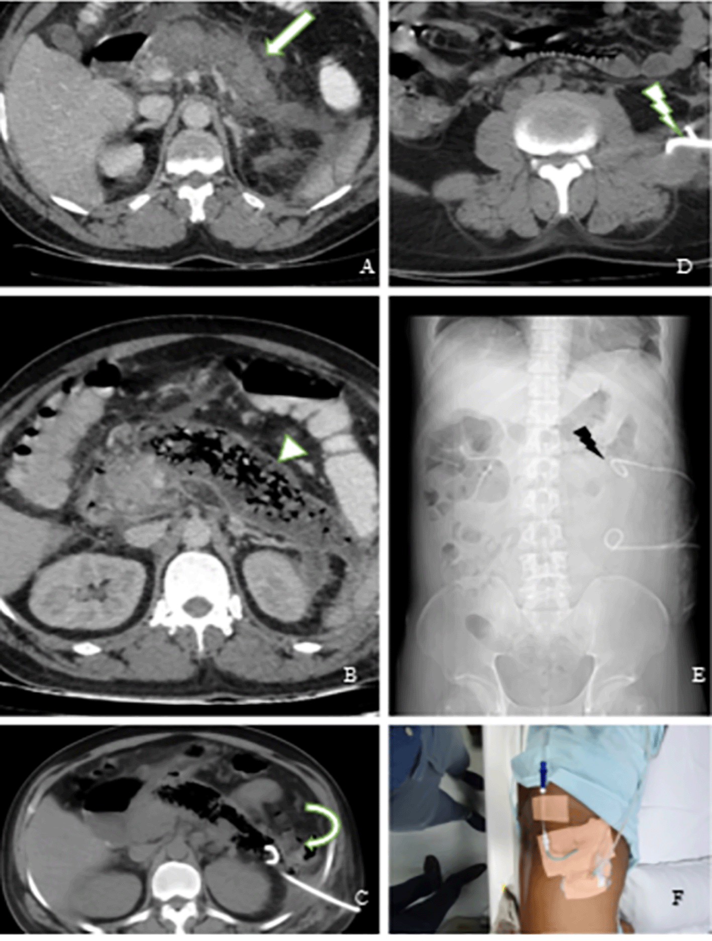
Fig. 7A – axial CECT image with an arrow showing peripancreatic fluid collection extending to the paraspinal region on the left side; Fig. 7B – CECT image done after 3 weeks shows necrotic collection with extensive air pockets within suggesting superadded infection; Fig. 7C – check CT image with a curved arrow showing the well-positioned tip of the pigtail catheter in the pancreatic tail region; Fig. 7D – axial CT images showing the second pigtail catheter tip in the left para-psoas collection; Fig. 7E – scout image showing two well-positioned pigtail catheters; Fig. 7F – post-catheterization photograph of the patient.
The two groups – A and B were compared based on the amount of residual collection with a success rate of 93.7% in group A, and 97.9% in group B. The failure rate in both groups was low (6.3% failure rate in group A and 2.1% failure rate in group B). The outcomes of categorized group distribution did not differ significantly. The INA of fluid collection in the thorax, abdomen, and pelvis with immediate or delayed PCD yielded similar findings in terms of outcome, suggesting that INA could defer PCD in patients with fluid collections. Gerzof et al.22 reported that the use of image-guided PCD to treat liver abscesses has a success rate of 70 to 100%. Similarly, Raj R et al,10 reported that PCD had a success rate of 77.4% and a 22.6% failure rate. These higher rates of procedure failure were observed mainly in cases where the patient was either elderly, female, or when a CT scan was used as a guiding technique. Lower success rate with CT-guided drainage could be attributed to the selection bias which favoured CT for deeper-seated and less accessible collections, especially of the pancreas, which is another confounding factor.
To our knowledge, there are no studies that categorized the patients into intervention groups based on the type of procedure and timing of catheterization. Hence, our study provides unique findings in this aspect. Figure 8 depicts a summary of our design and categorization of patients into different intervention groups in the form of a flow chart.
Complications – Major complications such as pneumothorax, liver perforation, or hemothorax are more likely to occur with chest tubes. Unlike chest tubes, pigtail catheters have a low risk of such complications.3 According to a study by Roberts et al.,18 5-6% of patients with pigtail catheter implantations developed significant complications such as hemothorax, pneumothorax, and liver perforation, and 20% had catheter-related complications such as dislodgement, failure to drain, kinking, blockage, and disconnection of the catheter. In the present study, we classified complications as major and minor complications. Major complications (3.6%) included reaccumulation of fluid in the cavity and pneumothorax. Minor complications (68.6%) included pain at the catheter insertion site, hematoma at the insertion site, and catheter-related complications. Pain was the most commonly reported minor complication in our study. Hematomas at the site of insertion were observed in two of the subjects– one patient (1.2%) developed a presacral hematoma, and another patient (1.2%) developed a perinephric hematoma. One patient with empyema in the left hemithorax also developed blockage of the 12F catheter, which was then removed, replaced, and upsized to a 14F catheter at the same site. 22.6% of the patients in our study, however, reported no major or minor complications.
In the present study, both INA and PCD were safe, effective, and minimally invasive techniques to drain abdominal, thoracic, and pelvic fluid collections in the absence of an indication for surgical intervention. INA should be considered an initial step of management for fluid drainage, which acts as a guide for the access pathway for catheter placement for PCD. We conclude that PCD is very effective in draining various types of fluid collections, most commonly serous and purulent fluids. In the majority of cases from our study, image-guided PCD proved to be a time-saving and efficient intervention, making it an intervention of choice in a variety of fluid collections/abscesses.
Open Science Framework: Efficacy, Feasibility, and Safety of Percutaneous Image-Guided Catheter Drainage of Thoracic, Abdominal, and Pelvic Fluid Collections https://doi.org/10.17605/OSF.IO/HPC5G.23
This project contains the following underlying data:
Data are available under the terms of the Creative Commons Zero “No rights reserved” data waiver (CC0 1.0 Public domain dedication).
| Views | Downloads | |
|---|---|---|
| F1000Research | - | - |
|
PubMed Central
Data from PMC are received and updated monthly.
|
- | - |
Provide sufficient details of any financial or non-financial competing interests to enable users to assess whether your comments might lead a reasonable person to question your impartiality. Consider the following examples, but note that this is not an exhaustive list:
Sign up for content alerts and receive a weekly or monthly email with all newly published articles
Already registered? Sign in
The email address should be the one you originally registered with F1000.
You registered with F1000 via Google, so we cannot reset your password.
To sign in, please click here.
If you still need help with your Google account password, please click here.
You registered with F1000 via Facebook, so we cannot reset your password.
To sign in, please click here.
If you still need help with your Facebook account password, please click here.
If your email address is registered with us, we will email you instructions to reset your password.
If you think you should have received this email but it has not arrived, please check your spam filters and/or contact for further assistance.
Comments on this article Comments (0)