Keywords
p53, predictor, endocrine therapy resistance, luminal breast cancer
This article is included in the Oncology gateway.
p53, predictor, endocrine therapy resistance, luminal breast cancer
Endocrine Therapy (ET) resistance in Luminal Breast Cancer (BC) is a concerning issue. Approximately 30-40% of Luminal BC are ET resistant, which leads to a higher recurrence rate and worsened prognosis. Although it has been extensively studied, till now there is no single predictive biomarker has been established to predict which patient will develop ET resistance during the 5-years-course of endocrine therapy.1–3
Such predictive biomarker will be advantageous both for clinician and patients, as patients which probably have bigger chance for endocrine therapy resistance could be monitored closely. Perhaps later in the future it could help to effectively change the course of the therapy before recurrence is established (and it becomes too late), as well as to help clinicians to identify which patients will not have ET benefit in the first place.1,2 And as we know, current trend in clinical trials of BC treatment are moving into personalized and tailored therapy for each cases, therefore finding predictive biomarker to predict the ET resistance will be important issue for such theurapeutic program development as well.4
Endocrine therapy resistance is complex molecular process which involved many process to develop. Several hypotheses has been developed regarding addressing such process, and finding such predictive biomarker. The resistance could develop at the start of the endocrine therapy (de novo or intrinsic resistance) or develop later in the course of the endocrine therapy. The hypotheses are ranging from the loss of hormonal receptor (HR) caused by ESR1 gene mutation and epigenetic mechanism,5–8 altered expression of co-factors (such as NF-kB, AIB1, SRC-1),9–11 crosstalk between ER and growth factors signaling (such as Her2neu, Insulin-like growth factor-1 receptor (IGF-1R))4,6,8,10,12,13 absent or reduced expression of negative regulator such as p21 and p27,14–16 metabolic resistance caused by polymorphism or loss of CYP2D6 (main enzymes responsible for converting tamoxifen into its active metabolites),2,4,8,10,17,18 NF1 mutation lead to MAPK pathway activation,19–24 APOBEC mutation associated with PI3KCA mutation,25 as well as tumor microenvironment roles such as NF-kB and TNF-α.9,11,26–29
The molecular mechanism of Estrogen Receptor (ER) and Progesterone Receptor (PR) actions are studied extensively for their association with ET resistance in Luminal BC. These molecular mechanisms additionally become an essential basis in rationalizing treatments such as Cyclin-CDK (Cyclin-Dependent Kinase) inhibitor and PI3K/Akt/mTOR inhibitor, which have been internationally accepted as current adjuvant treatments for Luminal BC with recurrence after ET resistance. Their actions therefore are basic and fundamental knowledge to find a logical explanation of endocrine therapy resistance, and most of the hypotheses above could be explained by the disruption of the ER and PR mechanism of actions, resulting increased cellular proliferation and decreased apoptosis.4,6,8–16,27,30
p53 mutation is one of the most frequent genetic alterations in BC, found in approximately 28.3%-35% of overall BC patients, with higher incidence in Luminal B BC (30-55%), Her-2neu overexpressive (70%) and TNBC group (80%).31–33 p53 mutation in hormonal positive BC will result in distinct poor prognosis, and especially seen in Luminal B BC with higher frequency and stronger association to poor prognosis compared to Luminal A BC.32,34 The mutation of this profound tumor suppressor gene may occur at the early onset of Luminal BC or progressively at the later course of the disease due to cancer cells’ ability to form more mutations in the advanced stage.20,31,35–37
p53 mutation has been known for more than four decades and its extensive roles span from cell cycle regulation, DNA repair, apoptosis process, cell metabolism, as well as immune response in tumor microenvironment.21,37–41 This versatile tumor suppressor gene has been studied in many cancers including breast cancer, and its protein accumulation as well as its mutation is conclusively found in numerous endocrine resistance breast cancer studies.20,35,36,42,43
This review will explore the current knowledge of ER and PR molecular mechanisms and their impact in initiating ET resistance in Luminal BC. Furthermore, we will discuss the apparent effect of p53 mutation in their molecular mechanisms, consequently aggravating ET resistance.
Estrogen is a steroid hormone that acts in several tissues such as skin, liver, bone, and breast. In breast tissue, Estrogen’s potent mitogenic effect will generate breast epithelial proliferation, alveolar growth, fat deposition, and fibrous tissue development during puberty, pregnancy, and lactation phases. These distinctive changes in the breast are affected by Estrogen, which works alongside Progesterone and other growth factors.44
The active form of Estrogen in breast tissue, Estradiol, and its metabolites have been acknowledged as essential factors of early malignant transformation, such as DNA single strand breaks and chromosomal impairment. Furthermore, it may lead to uncontrolled cell proliferation, accompanied by the development of cellular signalling collaborating in the cancerous cells’ progression. All events mentioned above will benefit the growth of cancer cells, and all of them are depended on the molecular mechanism of ER in BC cells.45,46
Estrogen Receptor has a paramount role in BC cells, as described above. Hence it becomes the main target of the endocrine therapy such as ovarian blockade, SERM (Selective Estrogen Receptor Modulator, i.e., Tamoxifen), and SERD (Selective Estrogen Receptor Degrader, i.e., Fulvestran).10,22
Being a nuclear receptor family member, ER-α and ER-β are the two different types of Estrogen Receptors. In breast tissue, the ER-α has a dominant role. Meanwhile, ER-β is still considered controversial and has an unclear role.47 Another estrogen receptor type is the G-coupled Estrogen Receptor (GPER), which is paramount for estrogen molecular action via the membranous mechanism.48
ER-α coded by ESR-1 gene in chromosome 14, with an identical structure as other nuclear receptors, consists of 4 structural and functional domains. These domains are the amino-terminal domain (A/B domain), DNA binding domain/DBD, hinge region (D domain), and Ligand-Binding Domain/LBD.49
After entering the cytoplasm of the cells, Estrogen will form a dimer form then bind to the C-domain of the ER at its 12th helix. Afterward, this complex (Estrogen dimer and ER) will be transported to the nucleus by D-domain.50
Subsequently after entering the nucleus, DBD with the aid of co-activators will bind to Estrogen’s target genes that contain Estrogen Response Elements (EREs). The known co-activators are steroid receptor co-activator-1/SRC-1, SRC-2, SRC-3 (also known as AIB1/Amplified in Breast-Cancer 1). The binding of ER and EREs will activate the transcription of Estrogen’s target genes.4,51,52 This process is the so-called classic mechanism of ER molecular action, depicted in the animation below along with other mechanisms. This mechanism is the first known ER molecular action and has become the theoretical basis for applying the traditional endocrine therapy in luminal BC such as ovarian blockade, SERM, and SERD.10,24
The estrogen receptor molecular actions are complicated and involve the intersecting apoptotic along with the survival pathways such as PI3K/Akt/mTOR, MAPK/ERK, resulting in the same similar target genes such as Cyclin-CDK, growth factor, and its activators.10,53 Currently, there are four known molecular mechanisms of ER actions: classical (genomic), non-classical, non-genomic (membranous), and ligand-independent (estrogen-independent). These four mechanisms are pictured in Figure 1.
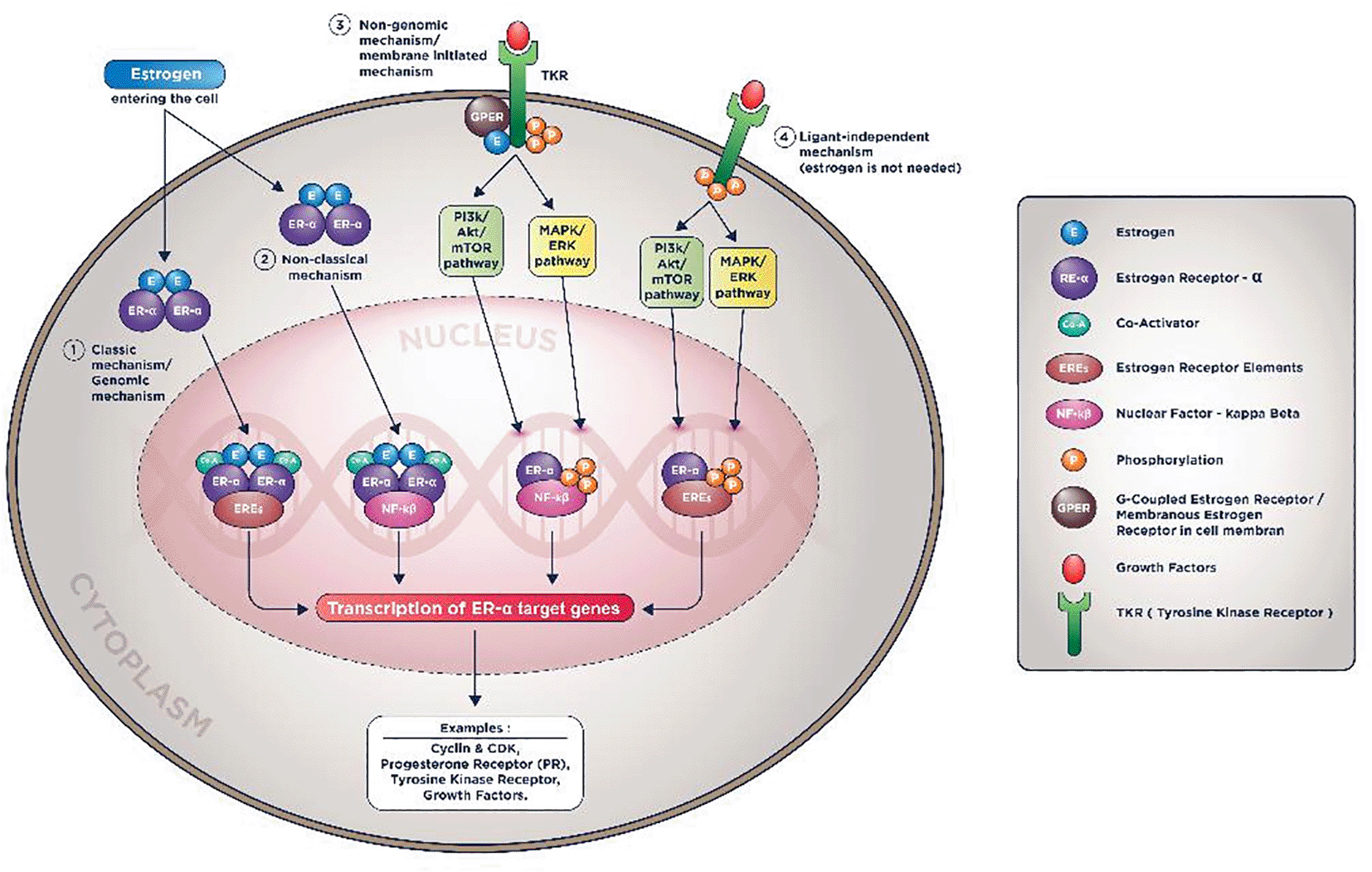
In the non-classical mechanism (2nd mechanism in Figure 1), Estrogen could activate genes transcription that don’t contain EREs with help from tethering co-factors such as NF-kB (Nuclear Factor-Kappa Beta), activator protein 1 (AP-1), or specificity protein 1 (SP-1).4,10
In the membranous mechanism (3rd mechanism in animation), Estrogen will not be required to enter the nucleus to do the genomic action since ER receptors conduct all the inciting processes in the cell membrane. In the last mechanism, even Estrogen is not required to induce its target genes transcription (hence the name ligand-independent mechanism).4,10
These mechanisms will induce the exact effect on breast cancers cells: accentuating proliferative pathways and diminishing pro-apoptotic pathways. These non-classic, membranous, and particularly ligand-independent mechanisms result in cancer cells being more resistant to endocrine therapy. It is as if these estrogen receptors’ mechanism of action is being “hijacked” by the cells; the cancer cells are manipulating it to their benefit that is to replicate more and being less sensitive to apoptotic signals.5,10,22,48
The final result of these processes are constant activation of the estrogen receptor target genes although there were no more estrogen molecules available (i.e., due to ovarian blockade or inhibition by aromatase inhibitor), and although its receptor being blocked or degraded (i.e., due to inhibition by SERM/SERD).5,10,22,48
Progesterone is a steroid hormone produced by the corpus luteum in the human ovarium, which its primary duty is to prepare the female body for gestation. In breast epithelial cells, its role is indispensable in affecting ducto-alveolar changes in phases such as puberty, luteal phase (pre-menstrual period), pregnancy, and lactation.54
Compared to Estrogen, the role of Progesterone in breast cancer cells, especially endocrine therapy, and its resistance is less distinctive and less studied. The cyclic level of Progesterone and its hundreds of active metabolites available in the female body are the main difficulties in testing this hormone. Furthermore, the target genes of ER and PR are overlapping, thus adding the complexity of this issue.55 But still, one cannot ignore the fact that the combination of Progesterone and Estrogen will add mitogenic effect to BC cells in the animal model, as epidemiological observations had shown.56–58
PR was transcribed by three means. First, its transcription is induced by Estrogen as PGR (gene for encoding the PR) is one of the Estrogen target genes. Estrogen has been proven to be required in maintaining PR levels in breast and endometrium epithelial cells.59 Second, cancer cells could induce PR transcription mediated by Insulin Growth Factor-1 (IGF-1) and MAPK/ERK activity. Even more, at high Progestin concentration, these growth factors will be re-induced and thus will re-activate the ER-α phosphorylation in the ligand-independent mechanism of ER (review above animation), resulting in more PR transcription.60
Some of the Progesterone target genes are also known to overlap with Estrogen target genes such as Cyclin-CDK, RANKL (Receptor activator of nuclear factor-kappa-Β ligand), and other growth factors. Hence these crosstalk mechanisms between ER and PR are crucial for breast cancer cells carcinogenesis and endocrine therapy resistance.5,60
Like Estrogen, the action of Progesterone in cells is entirely dependent on its receptors, and it consists of both nuclear and membranous receptor types. There are two types of nuclear PR: PR-A and PR-B. Both nuclear receptors exist in breast epithelial cells in variable amounts and activity. Furthermore, it is unclear which nuclear receptor is more dominant in breast epithelial cells.55
Identical to ER, PR has N-terminal domain, Ligand Binding Domain /LB, Progesterone will bind the PR, DNA Binding Domain/DBD in which target genes contain Progesterone Receptor Elements (PREs) will bind.61
After entering the cell’s cytoplasm, Progesterone will form a dimer, bind to PR in LBD, subsequently enter the nucleus, and bind to PREs with the aid of a co-factor. This process will activate the transcription of PR target genes. This mechanism is known as the PR action’s classical/direct genomic mechanism.55 Other mechanisms known are the non-classical/direct non-genomic and membranous mechanisms, depicted in Figure 2.16,55
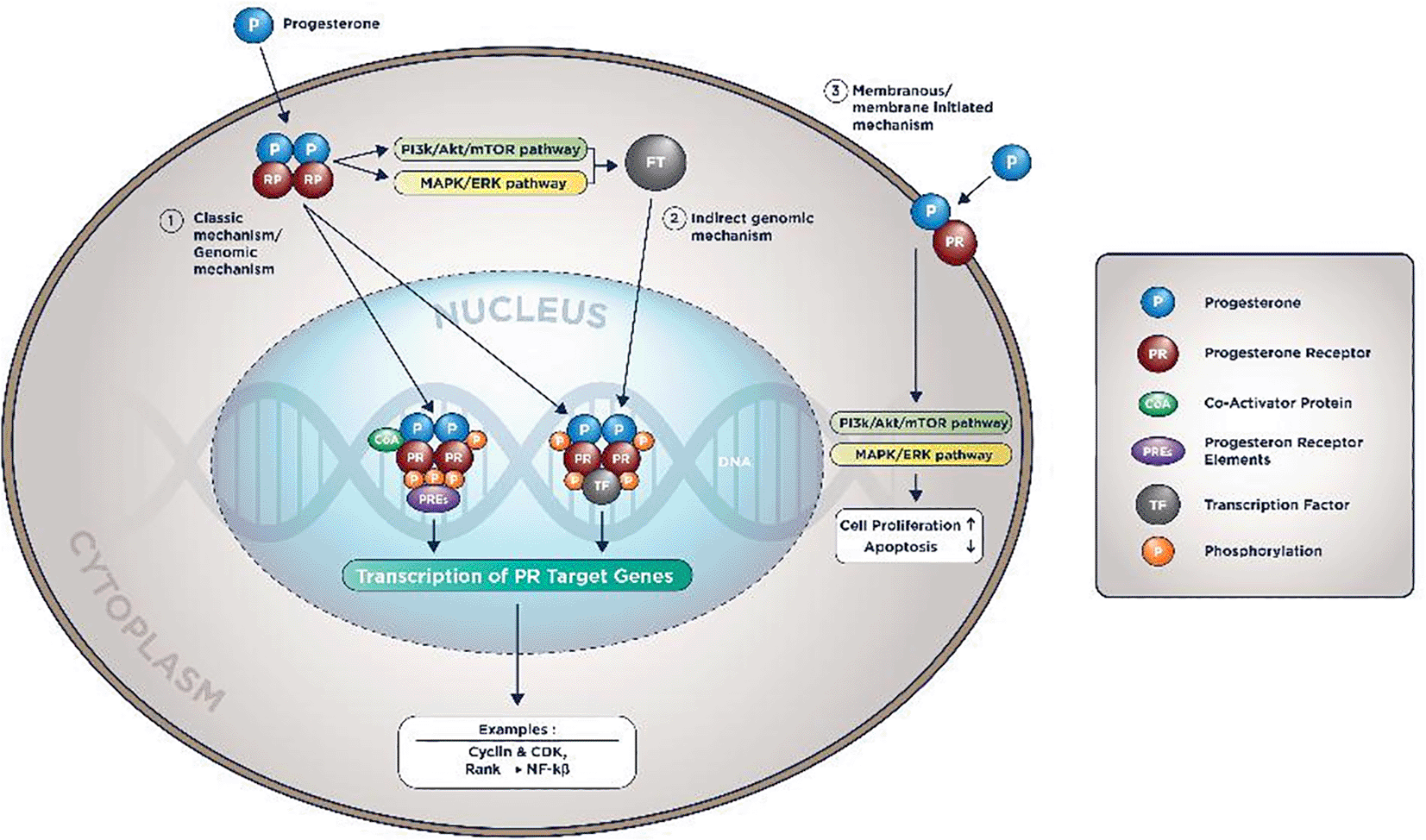
In the indirect genomic mechanism, progesterone could activate genes that do not contain PREs as long there are tethering co-factors. In the membranous mechanism, PI3K/Akt/mTOR pathway and MAPK/ERK are also activated by progesterone. Therefore, it will accentuate the proliferative pathways and diminish the pro-apoptotic signals.16,55
The complicated cellular signaling tightly regulates the cell cycle to maintain the regular cell proliferation rate and minimize error in DNA synthesis. In this cell cycle, abnormal cells with DNA error will be ceased in G1-S transition critical point.62
Essentially, this critical G1-S transition point is determined by the interaction of Cyclin-D1& CDK4/6. In conditions without inhibition, this interaction will release E2F protein from its bond with Retinoblastoma Protein (RB Protein). The E2F protein will further trigger the cell to enter the S phase. Then consequently, abnormal cells with DNA will be duplicated.63
This mechanistic complex is one of the most often disrupted cellular signaling. It is found in endocrine therapy-resistant breast cancer cells, as Cyclin D1 (CCND1) and CDK4 become the target genes of Estrogen and Progesterone. Previously, Cyclin-CDK is still transcribed by the cancer cells, although the Estrogen production has been diminished and their receptors have been blocked.22,51,52 Relevantly, CDK 4/6 inhibitor has been approved in clinical guidelines as an adjunctive for endocrine therapy in Luminal BC, both pre- and post-menopausal patients.13,64
In normal cellular regulation, cells with abnormal DNA will be forced to enter G0 phase by the p21 protein, a protein transcribed and regulated by p53. This p21 protein will inhibit the CyclinD1-CDK4/6 complex, resulting in the cell entering the G0 phase and starting the DNA repairing process. This well-regulated system earned p53 the old nickname: “guardian of the genome”.65,66
PI3K/Akt/mTOR pathway is a series of consecutive intracellular signaling that will activate proliferation and prevent apoptotic events. Genetic accumulation in this pathway and mutation of its inhibitor (PTEN/Phosphatase and TENsin homolog deleted on chromosome 10) are found about 70% from the whole BC population.12,67
PI3K is an intracellular lipid kinase enzyme that will phosphorylate phosphatidylinositol molecule in the cell membrane, subsequently turning phosphatidylinositol-4,5-bisphosphate (PIP2) into phosphatidylinositol-3,4,5-trisphosphate (PIP3).67 Afterward, PIP3 will facilitate interaction between phosphoinositide-dependent kinase 1 (PDK-1) and Akt in the cell cytoplasm, resulting in a phosphorylated Akt. This phosphorylated Akt will activate Forkhead box O transcription factor (FoxO) that inhibits pro-apoptosis genes and also activates mechanistic targets of rapamycin (mTOR) complexes.12
The mTOR complexes are consist of 2 active forms: activated mTORC1 and mTORC2. mTORC1 will activate genes involved in carcinogeneses like protein synthesis, pro-survival genes, and cell growth. mTORC2 will specifically enhance phosphorylation, further causing Akt hyperactivation.12,67
In ER-α molecular actions, PI3K/Akt/mTOR will be activated in the non-classical, membranous, and ligand-independent mechanism.10,48,53,68 A likely, PI3K/Akt/mTOR will also be activated in PR molecular actions.16,55 Additionally, mTOR1 will activate S6K that will help to phosphorylate RE-α, further activating the functional domain of RE-α. Likewise, the Akt activates the NF-kB that functions as a co-factor in the non-classical and membranous mechanism of ER-α molecular actions.21
It is well known that PTEN, a classical tumor suppressor gene, will reverse PIP3 to PIP2; hence the subsequent Akt/mTOR activation will not occur. In the absence of PTEN, PI3K/Akt/mTOR pathway will be hyperactivated.69
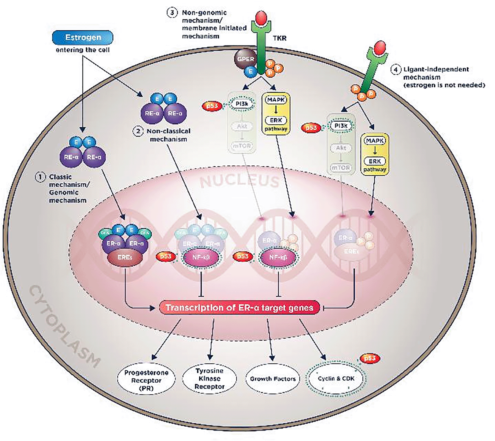
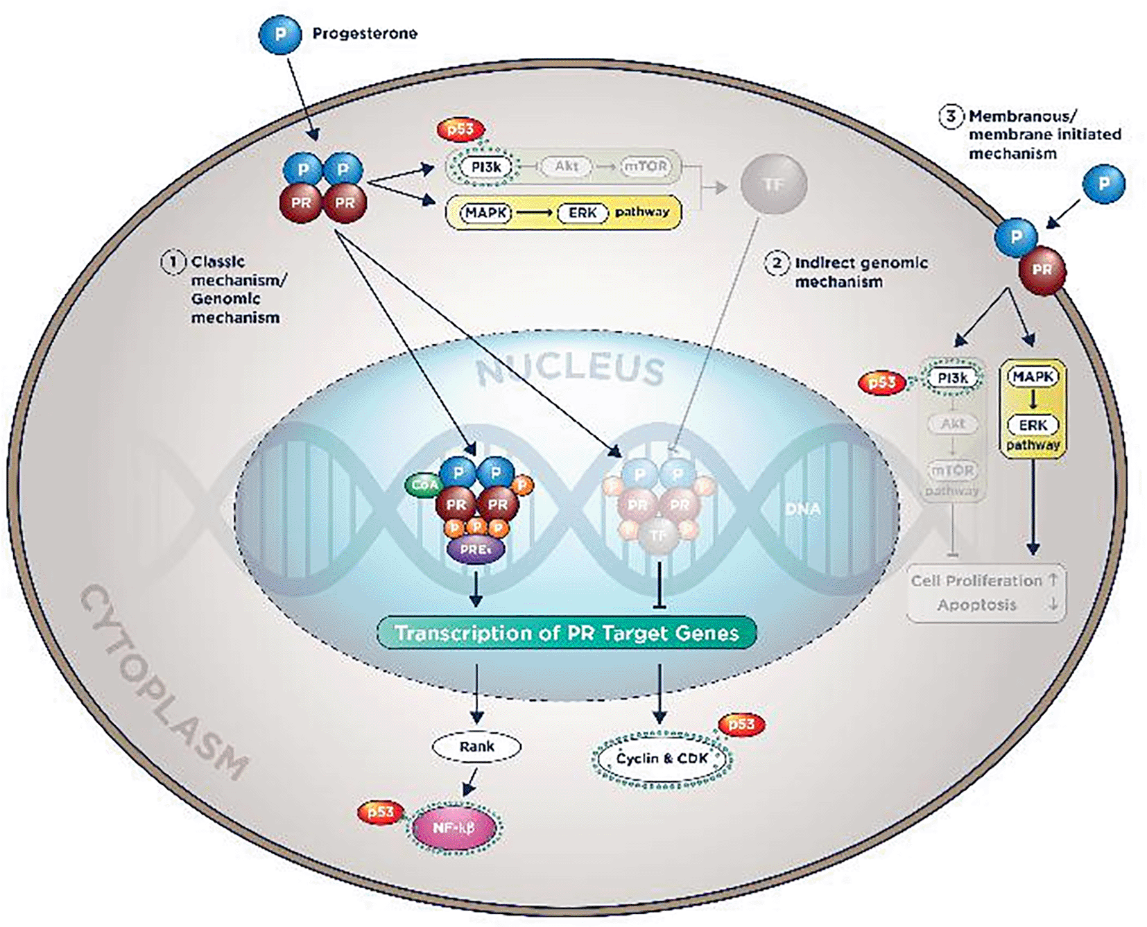
The wild-type p53 protein will activate PTEN gene transcription. In cells with mutant p53, PTEN gene mRNA expression will be drastically reduced compared to cells with wild-type p53 status.70Another seminal finding by Jung, et al. 2018 in cell culture studies show cells with PTEN loss will cause PI3K/Akt/mTOR hyperactivation, causing mTORC1 and mTORC2 enhancement, and both will phosphorylate and activate wild-type p53 protein, which causes p21 transcription. p21 protein will further induce cells to premature senescence condition.71
Nuclear Factor-kappa Beta (NFKB) is a transcription factor family consisting of 5 subtypes: p50, p52, p65 (RelA), RelB, and c-Rel. With a vast target gene involved crucially in chronic inflammation and cellular proliferation, NF-kB is studied extensively in many cancers, including endocrine-resistant Luminal BC. In cell culture studies, treatment with NF-kB inhibitor will evoke the endocrine therapy sensitivity and toxicity; therefore, it has not been tested on humans.9
NF-kB target genes considered instrumental in endocrine therapy resistance evolution are Cyclin D1, D2, D3 dan E, anti-apoptotic protein Bcl-2, MDM-2, and PDL-1 (Programmed Death Ligand-1).9,27,72 In ER molecular actions explained above; the NF-KB works as a co-factor in non-classical and membranous mechanisms so that Estrogen’s target genes are still transcribed although Estrogen has been blocked.9,72–76
The p53 and NF-kB have a negative association, for one cannot exist if the others are activated. The function is also contradictive; NF-kB will cause cell proliferation, anti-apoptotic, and enhance chronic inflammation, whereas p53 will regulate the cell cycle and trigger pro-apoptotic events when needed.72,77
The antagonistic mechanisms are various. Some studies noted wild-type p53 protein act as the direct promoter inhibitor of NF-kB target genes, therefore inhibiting the transcription of the NF-kB target genes. Others stated they compete with each other to get transcription co-factor 300. Wild-type p53 protein will also be known to inhibit the IKK enzyme (Inhibitor of Kappa Kinase, an enzyme to activate the active form of NF-kB). Without IKK, NF-kB couldn’t enter the nucleus and induce its target genes transcription.72,77,39
TNF-α, the multi-functional mediator in inflammation and cells apoptosis, is also noted in Luminal BC for its role in enhancing cellular proliferation via NF-kB activation.78,79 The wild-type p53 was also recognized in turning off TNF-α induced NF-kB activation. This action is achieved by binding and blocking the work of Disabled homolog 2-interacting protein (DAB2IP), a protein that will activate TNF-α to trigger NF-kB activation.78
Summary of all p53 works in ER-α and PR molecular actions could be seen in Figures 3 and 4.
Although p53 is prominently correlated with poorer clinical features such as high proliferation index and higher grade and stadium, its usage in Luminal BC is still arguably limited.80 One possible cause is that the presence of ER seems to supress the p53 mutation itself.81 From epidemiological point of view, p53 mutation indeed is more frequently found in HER-2 enriched group and the Triple Negative BC group rather than Luminal BC. However, p53 mutation when found in Luminal BC is not without importance. In fact study by Lee, et al. 2013 from 7739 patients shown us that p53 mutation is correlated with higher proliferative index such as Ki-67 in Luminal A BC, and when combined together they effect long term survival of the patients.82
Since it has been found 40 years ago, p53 protein has been published in thousands studies in numerous cancer either using protein detection or genetic testing. These factors along with versatility of p53 function in cells makes the test for p53 are widely known and readily available in most laboratories, therefore p53 is an ideal predictor to be chosen.38
p53 protein accumulation is easily detected by immunohistochemistry (IHC) as a surrogate marker of its mutation. However p53 immunohistochemistry testing in BC could present and correlates either with our without positive gene mutations tested, further affecting its capability as biomarker in BC.81 It is an obstacle that also frequently found in other cancers such as ovarian and gastric cancer.83,84
No clear cut off of p53 positivity in IHC assay also have been noted as the cause and affecting p53 usage as predictive biomarker in BC.85 Study by Kikuchi, et al. 2013 had tried to address this cut-off issue, found that when we set the cut-off of p53 immunoreactivity into ≥50% then it could be useful to predict clinical behaviour in Luminal Breast Cancer, especially Luminal B type (p<0.0001).86 This finding is also confirmed by another epidemiological study of 7226 patients by Abubakar, et al. in 2019.34
Furthermore, p53 protein accumulation has been a limitation as a predictor due to many p53 protein isoforms formed within the tissue. These isoforms are many, with each was said to have its roles in molecular effect in cancer cells.31,81,87,88 This limitation has been countered with the suggestion of genetic testing such as PAM50 or Mammaprint that would replace the p53 protein accumulation testing. Although it has been deemed more accurate than IHC assay, the genetic testing is expensive and not readily available in most laboratories, becoming the deterrent factors for choosing this testing in clinical setting.89
Endocrine therapy is a very beneficial therapy for Luminal BC patient. Its usage will decrease 15-years mortality rate up to 30-40%, consequently its resistance will pose the patients to dismal prognosis. As mentioned above, an established predictive biomarker will help clinicians to identify which patients will not have ET benefit in the first place, therefore their adjuvant therapy should be changed to other modalities to reduce recurrence and increase overall survival rate.2 In future perspective, predictive biomarker that could anticipate ET is certainly needed towards developing personalized and tailored therapy for Luminal BC patients.2,30
Developing and planning studies to identify such biomarker is not easy since the complexities of the endocrine therapy resistance theories mentioned before. Estrogen metabolism in premenopausal and postmenopausal women are also different, hence the endocrine therapy given are different, therefore these group cannot be investigated together.5,28,48 With previous reasons mentioned, meticulously planned study embedded in RCT with carefully chosen patients and prospective analysis probably is best to identify such biomarker, explained very well in seminal study by Beelen, et al.4
Additionally, p53 mutation could occurred as early as pre-carcinogenesis period, in early stage of BC and in late/metastatic disease.20 Consequently it will be compulsory to test p53 mutation along the course of the disease and observe whether it correlate with ET resistance later.
p53 is also known to have particular effects in each type of breast cancer (luminal A/B, with or without HER2 positive status) due to BC heterogeneity.90 Several studies have been made to address this issue, and concluded that Luminal B breast cancer is the most probable BC group in which p53 mutation could be useful as predictive biomarker to predict ET resistance occurence.82,90 This fact is also supported by epidemiological data that showed p53 mutation was found in higher in Luminal B BC compared than Luminal A BC.33,34,91
Another interesting development is Neoadjuvant Endocrine Therapy (NET), which currently being studied in clinical trial, reportedly has advantages in downstaging and increasing Breast Conserving Therapy (BCT) success.92 In the future NET could reduce hospital stay for Luminal BC patients and outreach the undertreated patients group. When patients could not go to the hospital for various reasons, then endocrine therapy could provide more accessible and comfortable neo-adjuvant treatment than chemotherapy or radiotherapy.92 Therefore the needs to find such predictive biomarker become more pressing and indispensable, and we have to explore p53 mutation as plausible biomarker.
p53 is an important biomarker to be considered as an ideal candidate to anticipate ET resistance in the future since its role within pathways involved in the ER and PR molecular mechanisms is paramount and cannot be ignored. Its limitation as a predictor could be countered using proper genetic testing rather than protein marker. Well planned studies will be a prerequisite to conclude whether p53 truly useful as predictive biomarker for ET resistance in Luminal BC patients especially Luminal B group, with adequate period of observation.
FH worked the Conceptualization, Data Curation, Formal Analysis, Funding Acquisition, Investigation, Methodology, Resources, Software, Visualization, Writing – Original Draft Preparation. YA involved in Conceptualization, Data Curation, Formal Analysis, Supervision, Validation, Writing – Review & Editing. SS involved in Project Administration, Resources, Software, Supervision, Writing – Review & Editing. FH wrote the draft of the article, YA and SS helped with final manuscript preparation. BH involved in Conceptualization, Project Administration, Software, Supervision, Validation, Writing – Review & Editing. All figures and animations are original to this manuscript, composed by FH and approved by YA, SS and BH. All authors read and approved the final manuscript.
| Views | Downloads | |
|---|---|---|
| F1000Research | - | - |
|
PubMed Central
Data from PMC are received and updated monthly.
|
- | - |
Provide sufficient details of any financial or non-financial competing interests to enable users to assess whether your comments might lead a reasonable person to question your impartiality. Consider the following examples, but note that this is not an exhaustive list:
Sign up for content alerts and receive a weekly or monthly email with all newly published articles
Already registered? Sign in
The email address should be the one you originally registered with F1000.
You registered with F1000 via Google, so we cannot reset your password.
To sign in, please click here.
If you still need help with your Google account password, please click here.
You registered with F1000 via Facebook, so we cannot reset your password.
To sign in, please click here.
If you still need help with your Facebook account password, please click here.
If your email address is registered with us, we will email you instructions to reset your password.
If you think you should have received this email but it has not arrived, please check your spam filters and/or contact for further assistance.
Comments on this article Comments (0)