Keywords
COVID-19, Isolation chamber, SARS-CoV-2, Temperature, Transmission
This article is included in the Emerging Diseases and Outbreaks gateway.
COVID-19, Isolation chamber, SARS-CoV-2, Temperature, Transmission
In this new version, we have added the information that this research is useful for people living in the tropics in the Introduction section. We have added more important information in the Methods section such as: (a) the description for virus propagation; and (b) information related to the SARS-CoV-2 used in this study such as the origin and the lineage. In Discussion section we have removed the hypothesis that lifting the ambient temperature around an infected patient for one hour to 40oC might reduce the risk of infection to health care workers. We have added more limitations of our study including: (a) we did not assess the role of other factors that might influence the SARS-CoV-2 transmission such as humidity; and (b) the sensitivity of the virus to temperature was tested in a liquid state only and we did not assess the aerosol state.
To read any peer review reports and author responses for this article, follow the "read" links in the Open Peer Review table.
Coronavirus disease 2019 (COVID-19), caused by severe acute respiratory syndrome coronavirus 2 (SARS-CoV-2), has inflicted disruptions in many aspects of health systems globally. SARS-CoV-2 is an enveloped, non-segmented, positive sense, single-stranded RNA virus with a genome of approximately 30 kilobases.1,2 The virus is mainly transmitted by nasopharyngeal droplets of an infected person; however, it could also be transmitted through aerosol.3 The viability of the virus in the environment is influenced by several factors, including climatic parameters such as humidity and temperature.4 Temperature has been proposed as a factor that affects SARS-CoV-2 transmission.5 Previous studies found that the temperature effected the transmission of the SARS-CoV-2 in which high environmental temperature reduced the number of COVID-19 cases.6–8 A study in State of Rio de Janeiro, Brazil found that the maximum and average of temperature were correlated negatively with COVID-19 infection.6 Data from 117 countries also found a negative association between temperature and COVID-19 transmissibility in which an increase of 1°C could decrease COVID-19 prevalence by approximately 5.4%.7 Several other investigations, however, have shown no evidence of a substantial influence of temperature on SARS-CoV-2 transmission.9,10
Nevertheless data reveals that SARS-CoV-2 is highly susceptible to heat.11 A recent study has shown that the virus could survive for at least 14 days at 4°C while only two days at 37°C and five minutes at 70°C.11 Another study suggested that, at 40°C SARS-CoV-2-infected epithelial cells have reduced viral transcription and replication.12 Although studies on the effect of elevated temperature on SARS-CoV-2 have been carried out, the temperature ranges used are limited.13 In addition, the effect of temperature on viruses originating from Indonesia has not yet been published. As part of our project to optimize the isolation chamber for COVID-19 patients, we determined the effect of temperature on the resistance of SARS-CoV-2 originating from Indonesia by evaluating the viral viability with a range of temperatures from room temperature (21-23°C) to 65°C. Understanding viral survivability is critical for developing a temperature optimized isolation chamber that could minimize the risk of infection to healthcare workers and optimize energy consumption while ensuring comfort for patients. In addition, the information of this study might important for those who are living in the tropics.
SARS-CoV-2 strain 20201012747 isolated from Jakarta, Indonesia was used in this study. The virus originated from a patient with severe COVID-19 manifestation. The virus was kindly supplied by the Eijkman Institute for Molecular Biology. Before used in this study, the virus has been passaged twice on Vero E6 cells (ECACC, Vero C1008) (RRID: CVCL_0574). The virus was classified as an ancestral SARS-CoV-2 strain and isolated on 12 October 2020.
The Vero E6 cells were maintained in Modified Eagle Media (MEM) (Cat. no. 11090081) supplemented with 10% heat-inactivated fetal bovine serum (FBS) (Cat. no. 26140095), 3.5 mM Na2CO3 (Cat. no. 25080094), 1% penicillin-streptomycin-amphotericin B (Cat. no. 15240062), 25 mM 4-(2-hydroxyethyl)-1-piperazineethanesulfonic acid (HEPES) (Cat. no. 15630080), 1% non-essential amino acid (Cat. no. 11140050), and 2 mM L-glutamine (Cat. no. 25030081). All were from Gibco (Thermo Fisher Scientific, MA, USA). The cells were seeded into 12-well clear bottom plates (Cat. no. 3513, Corning, USA) for plaque assay.
To expose to different temperatures, 0.6 mL of SARS-CoV-2 stock in 1.5 mL sterile tubes were incubated at room temperature RT (21-23°C), 25, 30, 35, 40, 45, 50, 55, 60, or 65°C for 1 hour in a digital block heater (Cat. no. 5382000031, Eppendorf, Germany). A separated experiment was conducted for each temperature; three replicates were used for each temperature and each replicate was repeated three times.
After the incubation at different temperatures, the viruses were diluted with a 10-fold serial dilution with 2% MEM (10-1 to 10-12) and 0.1 mL was inoculated onto 95-100% confluent monolayers of Vero E6 cells for 1 hour at 37°C with 5% CO2 with manual gentle shaking every 20 mins. After 1 hour of incubation, each well was then covered with 1 mL of 2% carboxymethyl cellulose (Cat. no. 17854-1KG, Merck, Germany) containing MEM, 2% FBS (Cat. no. 26140095), 3.5 mM Na2CO3 (Cat. no. 25080094), 25 mM HEPES (Cat. no. DMEM 15630080), 1% non-essential amino acid (Cat. no. 11140050), and 1% penicillin-streptomycin-amphotericin B (Cat. no. 15240062); All from Gibco (Thermo Fisher Scientific, MA, USA). The plates were incubated in cell incubator for 72 hours at 37°C and 5% CO2. The cells were then fixed with 4% paraformaldehyde for 4 hours at room temperature, and stained with 1% crystal violet. The plaques were counted manually. All works with infectious SARS-CoV-2 were conducted in the Biosafety Level 3 Laboratory at the Eijkman Institute for Molecular Biology in Jakarta. The plaque forming unit (PFU/mL) was calculated by dividing the number of plaques by dilution factor.
The virus was observed to remain stable from RT to 35°C with an average plaque count of 1013 PFU/mL.23 A reduction in the viral count was observed in the 40°C treatment group at 109 PFU/mL. Increasing the temperature to 45°C resulted in a further reduction of the viral viability resulting in 106 PFU/mL. The last temperature with visible plaque was in the 50°C treatment group with a result of 102 PFU/mL. The reduction of SARS-CoV-2 viability had a temperature-dependent trend within the temperature range of 35 to 50°C (Figure 1). No plaques were visible in the 55, 60 and 65°C treatment groups.
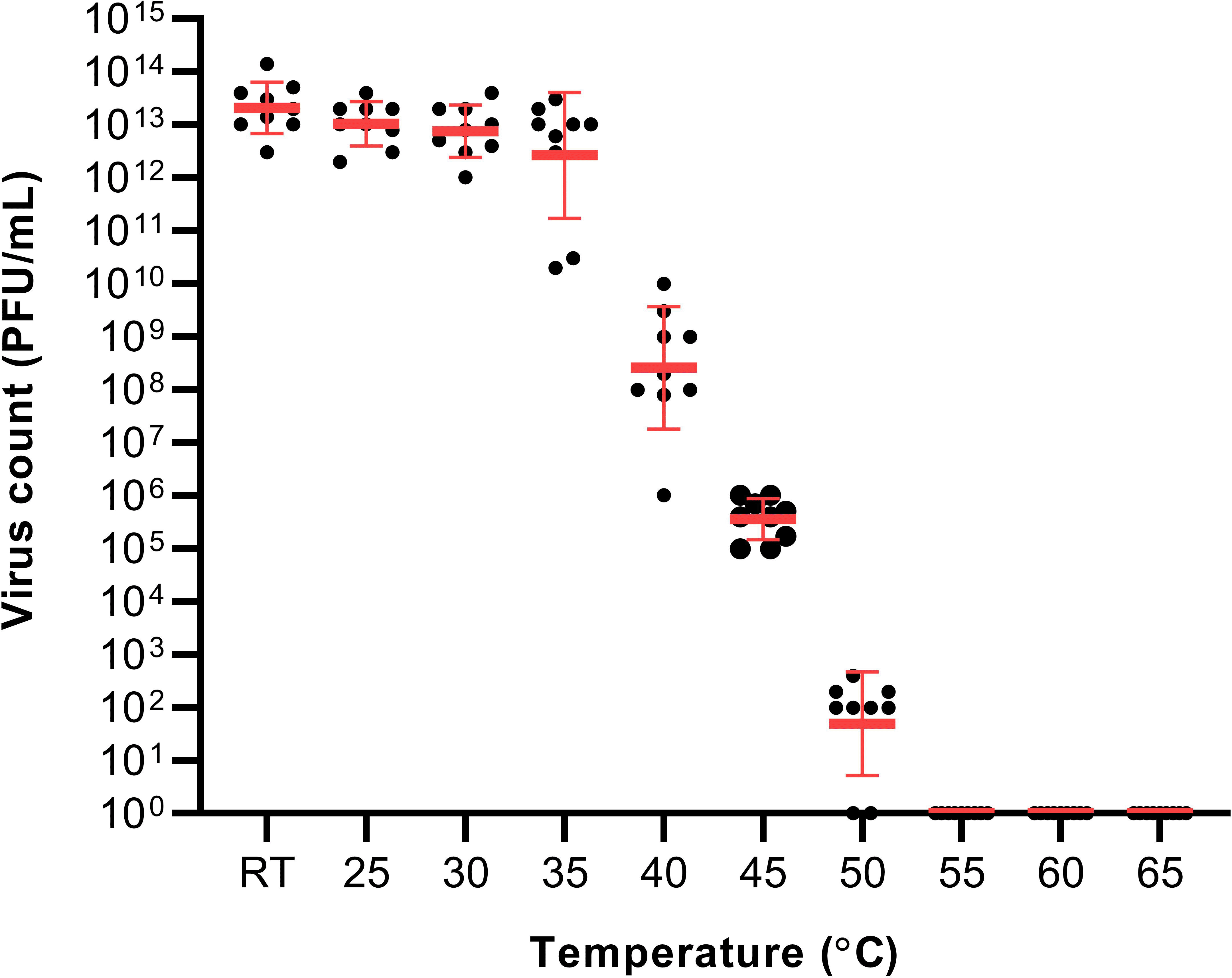
RT – (21-23°C).
Nosocomial transmission of SARS-CoV-2 has been identified to occur via multiple routes in healthcare facilities14 indicating that uncomplicated measures like wearing personal protective equipment along with surface cleaning and decontamination15 could be used effectively to reduce the transmission of infection. Other than that, modification to facilities such as, including isolation chambers with temperature control could help minimize the transmission.11,16 As noted in a previous study,11 temperature affects the stability of the virus in aerosol or on a surface. When the temperature of the room is not elevated, SARS-CoV-2 could remain stable for up to 72 hours on a surface such as plastic and stainless steel and three hours in aerosols.16 Based on the findings of this present study, to reduce the viral viability significantly, a higher room temperature might be important. However, according to a previous report, the average maximum temperature for Indonesian people to still feel comfortable falls between 24 and 29°C17 and to increase the room temperature would probably cause discomfort to the patient.
The administration of heat to the isolation chamber could be conducted prior to patient handling to reduce the likelihood of SARS-CoV-2 transmission. The other possibility is using heat in the isolation chamber as a means of thermotherapy. Thermotherapy is where mild-temperature elevation or hyperthermia (39-42°C) is used as a treatment against SARS-CoV-2 infection.18 Previous studies assessing the indoor temperature and SARS-CoV-2 were associated inactivation of SARS-CoV-2.19–21 Following the increase in temperature, heat-shock proteins (HSPs) are released which downregulates the progression of sepsis-induced acute lung injury.22 However, HSPs could become hosts to several viruses (such as human papillomavirus, adenovirus, and dengue virus) promoting their infectivity.23 In the case of SARS-CoV-2, its infectivity is more likely to be degraded than promoted by the HSPs.18 Therefore, heat administration to the isolation chamber should not be performed on COVID-19 patients with human papillomavirus, adenovirus, or dengue virus co-infections. In addition, it should not be attempted on patients with severe-to-critical COVID-19 as they would be more likely to have an increased risk of mortality following the thermotherapy or heat administration.24
One of the limitations of this study was we did not assess the role of other factors that might influence the SARS-CoV-2 transmission such as humidity. Therefore, the results of our study should be incorporated with data of the other studies assessing the effects of humidity on SARS-CoV-2 viability. In addition, we assessed the effect of the temperature on SARS-CoV-2 in a liquid state only and this might have influenced the results. Assessing the effect of the temperature on different states might could provide better understanding.
This present study also has proven that increasing indoor temperature to 55°C is sufficient to terminate the virus. Further increment to the temperature would not be necessary and only results in higher energy consumption. Similarly, a previous study also reported the inactivation of 90% of SARS-CoV-2 achieved at 54.5°C after 36 minutes.13 However, for use in treatment 55°C, might be too high for patients to tolerate. In that case, we only suggest the use of such temperature to thermally sterilize the isolation chamber prior to its use.
Figshare: Effect of elevated temperature on SARS-CoV-2 viability. DOI: https://doi.org/10.6084/m9.figshare.19243515.v1.25
This project contains the following underlying data:
Data are available under the terms of the Creative Commons Attribution 4.0 International license (CC-BY 4.0).
| Views | Downloads | |
|---|---|---|
| F1000Research | - | - |
|
PubMed Central
Data from PMC are received and updated monthly.
|
- | - |
Provide sufficient details of any financial or non-financial competing interests to enable users to assess whether your comments might lead a reasonable person to question your impartiality. Consider the following examples, but note that this is not an exhaustive list:
Sign up for content alerts and receive a weekly or monthly email with all newly published articles
Already registered? Sign in
The email address should be the one you originally registered with F1000.
You registered with F1000 via Google, so we cannot reset your password.
To sign in, please click here.
If you still need help with your Google account password, please click here.
You registered with F1000 via Facebook, so we cannot reset your password.
To sign in, please click here.
If you still need help with your Facebook account password, please click here.
If your email address is registered with us, we will email you instructions to reset your password.
If you think you should have received this email but it has not arrived, please check your spam filters and/or contact for further assistance.
Comments on this article Comments (0)