Keywords
Sexual dimorphism, Geometric morphometry, Procrustes analysis, Maxillary first premolar, Principle component analysis, Tooth form, Tooth shape
This article is included in the Manipal Academy of Higher Education gateway.
Sexual dimorphism, Geometric morphometry, Procrustes analysis, Maxillary first premolar, Principle component analysis, Tooth form, Tooth shape
In biology, “size” and “shape” are vital to describe an organism or a component of an organism, and expressing these involves the use of morphometry. “Size” is usually represented by linear and angular measurements of an entity. “Shape” on the other hand, is more complex to visualize and involves robust statistical procedures. Shape information is essential for bioarchaeology, anthropology, and forensic sciences to interpret evidence obtained from human remains. Rohlf and Marcus (1993) have reviewed the various procedures utilized in describing shape in biology and have termed the use of geometric morphometric analysis as a revolution in describing the “shape”.1 The geomorphometric analysis involves defining landmarks on the biological structure in two or three dimensions followed by statistical procedures using this data for visualizing the changes in shape. Further, the analysis also graphically represents the landmark variations on transformation grids to identify the deviations seen between species, gender, etc. Forensic anthropologists usually employ these landmark coordinates to define the biological profile.2
Sexual dimorphism in dentition is a well-established detail. Sexual dimorphism in a tooth may be attributed to variations in genetics, epigenetic factors, and the influences of sex hormones. In a study by Guatelli-Steinberg et al. (2008) on seven different populations, they found no significant association of sex hormone concentrations post-birth and tooth patterning.3 However, Ribeiro D et al (2013) have demonstrated a significant role of intrauterine testosterone levels in dental development and size.4 Taking cognizance of the varied reports pertaining to hormonal regulation of tooth size/shape, genetic influence on tooth shape must be considered primary. Genes that influence tooth patterning during odontogenesis are located in the sex chromosomes.
Sexual dimorphism has been researched by numerous morphometric studies involving linear measurements of width, length and diagonal measurements of teeth, measurements of areas of the occlusal surfaces, etc.5-7 Geomorphometric analysis of shape is a relatively new research modality to evaluate shape of the teeth.
The aim of the present present study is to evaluate the geometric morphometric variations of landmarks of the maxillary first premolar and its sexual dimorphism.
This study was conducted on the Dakshina Kannada population of Karnataka, India. The study commenced following the approval by the institutional ethics committee of Manipal College of Dental Sciences, Mangalore, Manipal Academy of Higher Education (vide ref no20018, dated 16th March 2020). Dental study casts of 55 individuals were retrieved from the archives of Department of Orthodontics, Manipal College of Dental Sciences, Mangalore. Broad written consent was taken from the patients during treatment, for use of the plaster casts for research assuring anonymization. Individuals born and brought up in Dakshina Kannada region were included in the study and their study casts were retrieved. The maxillary pre-treatment dental casts (poured in dental stone) of individuals meeting the inclusion criterion;, were retrieved for photography. All models with intact maxillary right first premolar, without any evidence of wear, caries, restorations or crown placement were included in the study. Age, sex and demographic details were noted from the patient management system. The randomization of the orthodontics patient box numbers was done using random numbers generated from www.random.org. The study was a time bound study to be completed in three months’ time. Total 55 random numbers were generated, distributed as 22 Females and 33 Male individuals having a mean age of 18.39±5.07 years (males 20.15±4.97 years and Females 17.33±4.91 years).
The data acquisition process involved photographing the cast, marking the landmarks (using TPS dig and TPS util softwares). The acquired landmark coordinates were then analysed in MorphoJ and PAST softwares. The statistical procedures used were procrustes superimposition, principal component analysis, discriminant function and canonical variate analysis.
Standardised images of the first maxillary premolar’s occlusal surface were taken with a Canon EOS 700D camera (Canon Inc., Japan) using macro mode. Each cast model was placed in the center of the field of focus of the lens with a scale placed adjacent to the cast (positioned at the occlusal surface level). An intermediate value diaphragm was used for an adequately focused photograph of the premolar’s occlusal surface. The Maxillary first premolar was positioned with the cemento-enamel junction (CEJ) perpendicular to the optical axis making it parallel to the camera lens as suggested by Wood and Abbot.8 The photographs were saved in *.tiff format for transfer to the landmark marking software TPSdig.
The landmarks were determined based on the anatomy (anatomic evidence) as well as the geometric contours (geometric evidence) of the tooth. A total of 19 landmarks were identified (11 based on anatomical evidence of cusp, ridges and grooves, and 8 based on geometric evidence of crest of curvature and line angles) as shown in Figure 1.
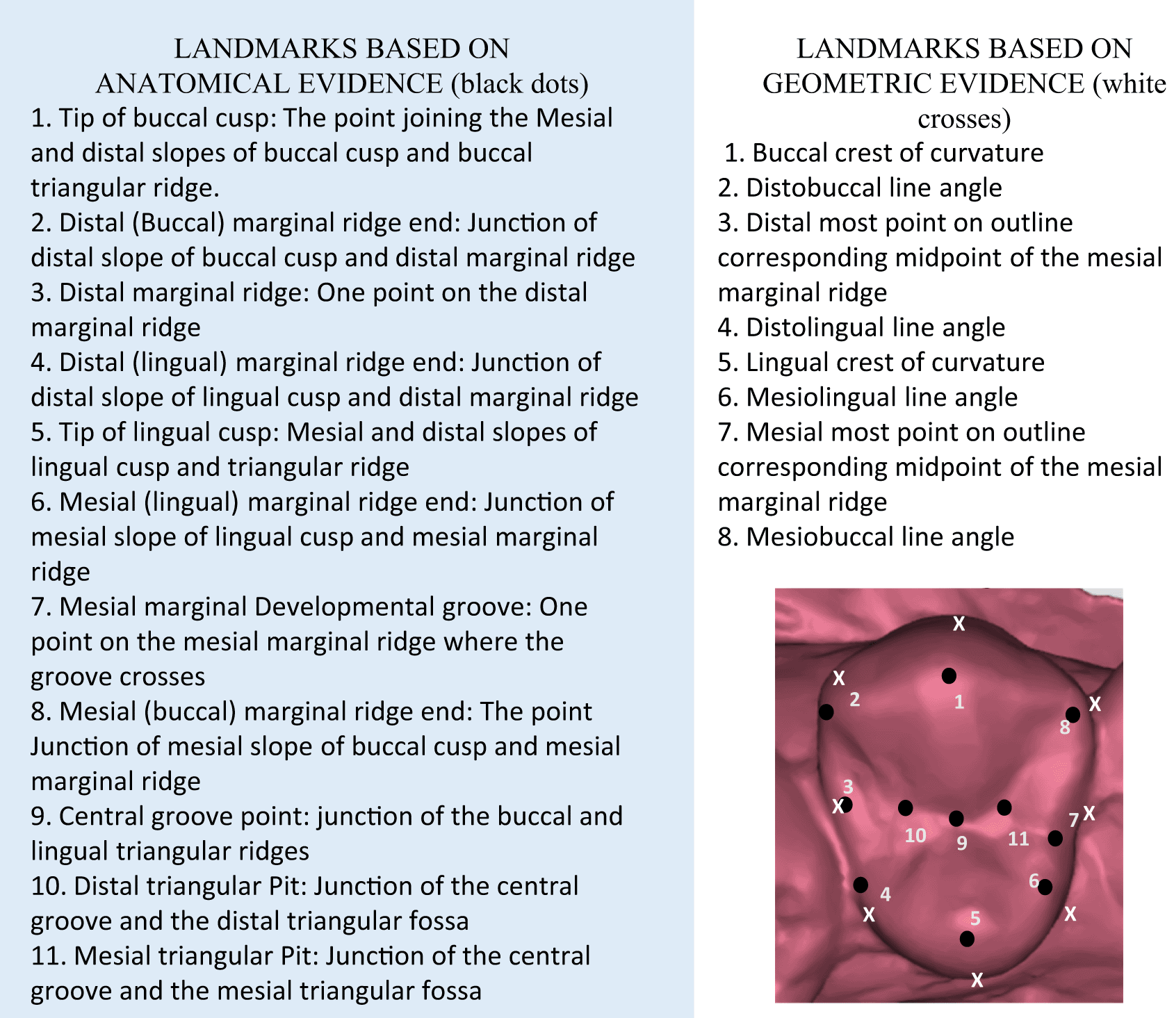
Using the TPSutil software, the *.tps file was generated incorporating 55 photographs of the maxillary casts in high resolution. This was followed by the landmark acquisition using TPSdig2 software. Using the landmark selection tool, the 19 landmarks (as described in Figure 1) were defined for each maxillary right first premolar of the 55 individuals and a *.tps file, (i.e. landmark data file) was generated. This file represented landmarks in two dimensions in “x,y” format. The data was then analyzed using Morpho J and PAST software for performing the principal components analysis, discriminant function analysis, canonical variate analysis, generate transformation grids and graphical representations.
Principal Components Analysis (PCA) was used to analyse differences within the individuals Discriminant Function Analysis (DFA) and Canonical Variate Analysis (CVA) were utilized to compare the data of ‘shape’ between the sexes.
Principal Components Analysis (PCA) showed that the first 11 principal components accounted for 80% of the maxillary first premolar variance, with the first four representing 50% of the variability (Table 1). The scatter was evenly noted on either side of the scatter plot axis, indicating a homogenous distribution of landmarks among individuals. The variability was seen more in males than in females (Figure 2).
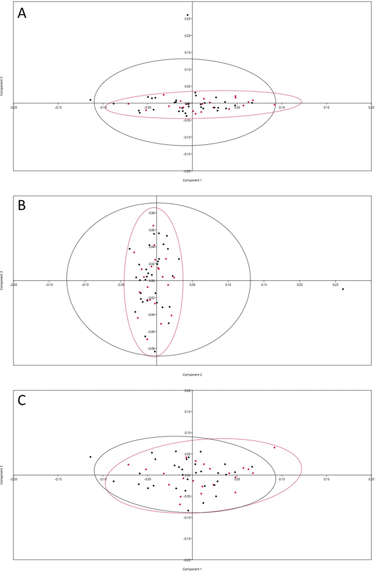
A=PC1 vs PC2; B=PC2 vs PC3 and C=PC1 vs PC3, Crimson dots represent female and black dots represent males.
The deformation graph showed prominent variability in the lingual direction of the buccal and the lingual cusp tips and buccal translation of the buccal as well as lingual crest of curvature. The distal end of the distobuccal cusp ridge and the mesial end of the mesiolingual cusp ridge tends to be shifted more towards the middle of the buccolingual dimension. The mesial marginal developmental groove remains lingual to the groove at all times, but shows some variation buccolingually (Figure 3A).
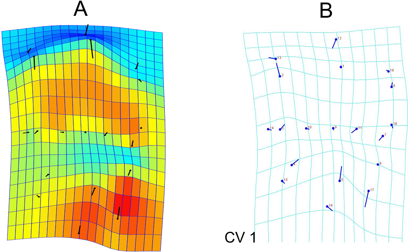
The lollipop graphs show the mean shape of the landmarks as circles and the relative position change of the landmarks is represented by sticks.
Canonical variate analysis was used to evaluate the differences in the landmark positioning between gender. The analysis shows that between gender, the Mahalanobis distances was 2.5676 and Procrustes distances among groups was 0.0303. Transformation grids illustrated that male teeth have more buccally placed distobuccal cusp ridge and lingual cusp tip. Further the buccal crest of curvature, the mesial marginal developmental groove, the mesiolingual cusp ridge end and the lingual crest of curvature are more lingually placed in males compared to females (Figure 3B).
On comparison of the centroid size, females had a mean centroid size of 15.36+1.24 which was marginally larger compared to the male individuals’ centroid size of 15.201+1.33 units. This was however not statistically significant with a p value of 0.606 (t=0.517). Procrustes ANOVA for shape analysis showed a greater variation with an f value of 1.4 and p value of 0.0624, indicating an increased variation in shape of the teeth among gender when compared to size. This result indicated that shape of the premolar was significant at the level of 10% alpha, whereas centroid was not.
Discriminant function analysis was performed based on the procrustes coordinates. There was 90.91 percent accuracy in classification of gender based on the landmark coordinates. The accuracy was 20/22 (90.99%) among females and 30/33 (90.91) among males accounting for 50/55 (90.91) in the total sample (Figure 4).
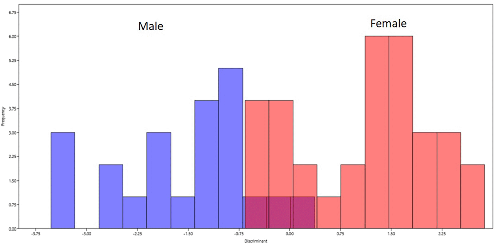
Blue bars on left represents male and red bars on right represents females.
The quantification of an object’s geometric shape by measurement of landmark coordinates is done using geometric morphometric analysis. This method utilizes multivariate statistical procedures that allow preservation of the landmark data in its original geometric shape and enables us to visualize the shape changes in real dimensions.9 There are various methods of evaluation of shape or form of a biological structure. These include Euclidean Distance Matrix analysis,10 Elliptical Fournier analysis11 and the most researched and understood procrustes superimposition method.12
Maxillary first premolar is particularly an essential tooth for taxonomic classification. The tooth has a characteristic asymmetry due to the prominent mesial marginal developmental groove and depression making the mesial outline concave compared to distal outline. Bailey and Lynch (2014) have assessed the shape of the mandibular premolars in Neanderthal and Modern humans and found their classification to be more accurate in modern humans with an accuracy of 98.1% as compared to Neanderthals who had an accuracy of 65%.11 The shape of a tooth is said to be a result of genetic drift rather than environmental factors.11 Genes play a primary role in morphodifferentiation of teeth. MSX, DLX, PAX9 genes are responsible for histo- and morpho-differentiation of tooth germ during odontogenesis. Studies have shown that MSX 1 mutation leads to agenesis of teeth especially the premolar segment.13 SPRY2, GAS1 and RUNX2 are potential candidate genes which influence the formation of secondary dentition including premolars.14 These studies indicate that genes play an important role in formation and morphology of premolar.
In our study, the centroid sizes did not show a significant variation in size of the premolars. This is in line with the other studies in Indian population. Banerjee A et al. (2016) in their odontometric study, showed that the mesiodistal width, buccolingual width and the crown lengths of maxillary first premolar were not significantly different between sexes.15 Yong et al. (2018) have studied the sexual dimorphism of human premolars in the Australian population and found that the centroid size did not show any significant difference by sex. However, Procrustes ANOVA showed significant effects of sex, accounting for 1.1% variation.12 This is in concordance with our present study where we found shape of the tooth to indicate greater sexual dimorphism than the size as seen by procrustes ANOVA.
One of the limitations of our study is the 2 dimensional analysis. The buccolingual inclination of the premolar might affect the landmark visualization in a two dimension. This can be overcome by incorporation of the third dimension of the coordinates, and performing a 3D geomorphometric analysis. This would yield a better discriminating ability of the landmarks. Secondly, a study evaluating the shape variables of all the premolars and molars of the human arch would give an all-inclusive assessment of tooth shape.
For future work, newer mathematical and computational models can be explored for shape analysis of teeth. The newer techniques would be capable in obtaining optimal parameters from the landmark data. In this regard, Choi G et al. (2020) in their recent research have compared area based, procrustes based methods with their new shape analysis technique using quasi-conformational theory. They have demonstrated superior results using their newer conformational theory in delineating gender and ancestry among indigenous and European origin Australian population. They have stated that, procrustes based approach gives satisfactory accuracy in discrimination, however, the Teichmuller distance method used is superior owing to the methodologies incorporating mean and Gaussian curvature analysis.16
2D geomorphometric analysis of the maxillary first premolar was performed utilizing 19 landmarks of geometric and anatomical evidences. The literature shows that size shows minimal variation between gender.12,17 However, the shape using the 19 landmark coordinate data of the premolar teeth, was able to discriminate gender with an accuracy of 90.91% (as demonstrated by discriminant function analysis). Canonical variate analysis showed that the maximum variation was in relation to the positioning of the distobuccal cusp ridge end and the lingual cusp tip, both of which are more buccally placed in males. Such variations play an important role in reproduction of the premolar morphology during restoration and tooth alignment. Further, the shape coordinates can be used to estimate sex of the individual as an adjunct in forensic investigations of skeletonized remains.
Figshare. MAXILLARY Premolar Landmark Data. DOI: https://doi.org/10.6084/m9.figshare.19487717.v118
This project contains the following underlying data:
Data are available under the terms of the Creative Commons Attribution 4.0 International license (CC-BY 4.0).
| Views | Downloads | |
|---|---|---|
| F1000Research | - | - |
|
PubMed Central
Data from PMC are received and updated monthly.
|
- | - |
Provide sufficient details of any financial or non-financial competing interests to enable users to assess whether your comments might lead a reasonable person to question your impartiality. Consider the following examples, but note that this is not an exhaustive list:
Sign up for content alerts and receive a weekly or monthly email with all newly published articles
Already registered? Sign in
The email address should be the one you originally registered with F1000.
You registered with F1000 via Google, so we cannot reset your password.
To sign in, please click here.
If you still need help with your Google account password, please click here.
You registered with F1000 via Facebook, so we cannot reset your password.
To sign in, please click here.
If you still need help with your Facebook account password, please click here.
If your email address is registered with us, we will email you instructions to reset your password.
If you think you should have received this email but it has not arrived, please check your spam filters and/or contact for further assistance.
Comments on this article Comments (0)