Keywords
Radiotherapy, Diagnostic reference levels, Head & Neck, Pelvis, DLP, and CTDIvol
This article is included in the Manipal Academy of Higher Education gateway.
This article is included in the Oncology gateway.
Radiotherapy, Diagnostic reference levels, Head & Neck, Pelvis, DLP, and CTDIvol
Manipal Academy of Higher Education, Manipal, Karnataka, 576104, India was added as an affiliation.
To read any peer review reports and author responses for this article, follow the "read" links in the Open Peer Review table.
Computed tomography (CT) plays a significant part in the radiotherapy treatment process. CT enables personalized radiation treatment delivery with the help of cross-sectional imaging. CT is the only modality used to calculate 3D-dose in radiotherapy.1 Additionally, it facilitates inaccurate delivery of radiotherapy by obtaining digitally reconstructed radiographs for setup verification of the patient on the teletherapy machine before starting treatment.
Despite these advantages, CT carries a significant disadvantage of utilizing ionizing radiation for imaging, with the inevitable risk of stochastic effects. As mentioned in the ALARA principle, these imaging doses, which patient organs will receive during the treatment, should be as low as possible. The “International Commission on Radiological Protection” (ICRP) implemented the “Diagnostic Reference Level” (DRL) used in radiological procedures. A Diagnostic reference level is not a regulatory limit. It is a benchmark that, when exceeded, triggers a review. ICRP recommends that the DRL represent the local practice within a particular geographical area. DRL will not distinguish between good or bad medical practices. The motive of establishing a DRL is to reduce the dose of ionizing radiation that does not contribute to giving any additional clinical information to a medical practitioner. DRL is estimated to improve the radiation dose and image quality.2
DRLs are not applicable in radiotherapy practice, but it is necessary to apply DRL for different imaging techniques used in radiotherapy treatment, imaging for patient position verification, and treatment planning. DRL is the 75th percentile of effective dose distribution, calculated with patient or phantom data. Regional, national and international levels DRLs may be estimated and compared.2 We need to periodically estimate DRLs as there may be a change in the work practice.
Diagnostic reference level for CT procedure is already available, and studies will still establish diagnostic reference levels regionally and nationally. For radiotherapy planning CT, the scanning parameters and the scan length for each region are different. It is essential to establish a DRL for the same. Some studies on establishing DRL for radiotherapy planning CT have already been published.1–3 The objective and need of this study are to establish diagnostic reference levels for head & neck and pelvic CT protocols in RT treatment planning and compare them with other regional, national and international DRL. The established DRL will be used to find further optimized DRL for RT CT.
An image of acceptable quality can be obtained using a minimum radiation dose. The present study was conducted using a phantom to assess the radiographic technical parameters that could lead to the lowest radiation dose and acceptable image quality. The outcomes of this study can be used to improve Head & neck, and pelvis radiographic examinations.
This study was conducted after obtaining approval from the ethical committee of Kasturba (IEC number 925/2018) Hospital. The expected outcome of the current study is “standardization of Radiotherapy Planning CT Head and Neck and Pelvic protocol and optimization of dose can be achieved by using reference DRLs.” By using a convenient sampling technique and Kappa co-efficient, a total of 120 patients with head and neck cancer and 90 patients with pelvic cancer to be treated with radiotherapy (Mean age 53.33 ±8.5, Male 54.16 ± 15 Female 52.5±12.17) were prescribed for RTCT with the age of 18 years and above were taken for the study after obtaining a voluntary written informed consent form from February 2019-August 2019 All the CT simulation images were acquired in the advanced Philips 16 slice big-bore RT CT machine, with a tube voltage of 120 kVp and 300 mAs and used for radiotherapy1 planning. During the prospective study, information such as the patient’s gender, region of interest, Field-Of-View (FOV), type of cancer, DLP, and CTDIvol were noted. The examination protocol and the exposure parameters used to take the scan were also collected. The data collected for this study was anonymized entirely.
In this study, we have included and analysed CT procedures of the Head & Neck, and Pelvis. The CT image acquisition parameters are given in Table 1.
| Location | Acquisition type | Voltage (kV) | Reference (mAs) | Collimation (N×mm) | Rotation Time (sec) | Pitch Slice | Thickness (mm) |
|---|---|---|---|---|---|---|---|
| RT Head & Neck | Helical | 120 | 300 | 16 × 1.5 | 1 | 0.813 | 3 |
| RT Pelvis | Helical | 120 | 300 | 16 × 1.5 | 1 | 0.813 | 5 |
Scanning length per examination varied from one scan to another scan. The considered length for head & neck investigation was from the vertex of the Head to the carina, and for the pelvis, the procedure is from the D10-D12 area to the mid femur. The “American Association of Physicists in Medicine suggests that the scanned volume should extend at least 5 cm superiorly and inferiorly beyond the target area”.4
All RTCT scan images were verified and approved by the oncologist after confirming them to be suitable for volume delineation. The effective dose was arrived at using the Normalized effective dose (k) coefficients and K’s value according to the updated AAPM report 96.
Patient weight 40 – 80 kg mean weight 60 kg ± 5 kg
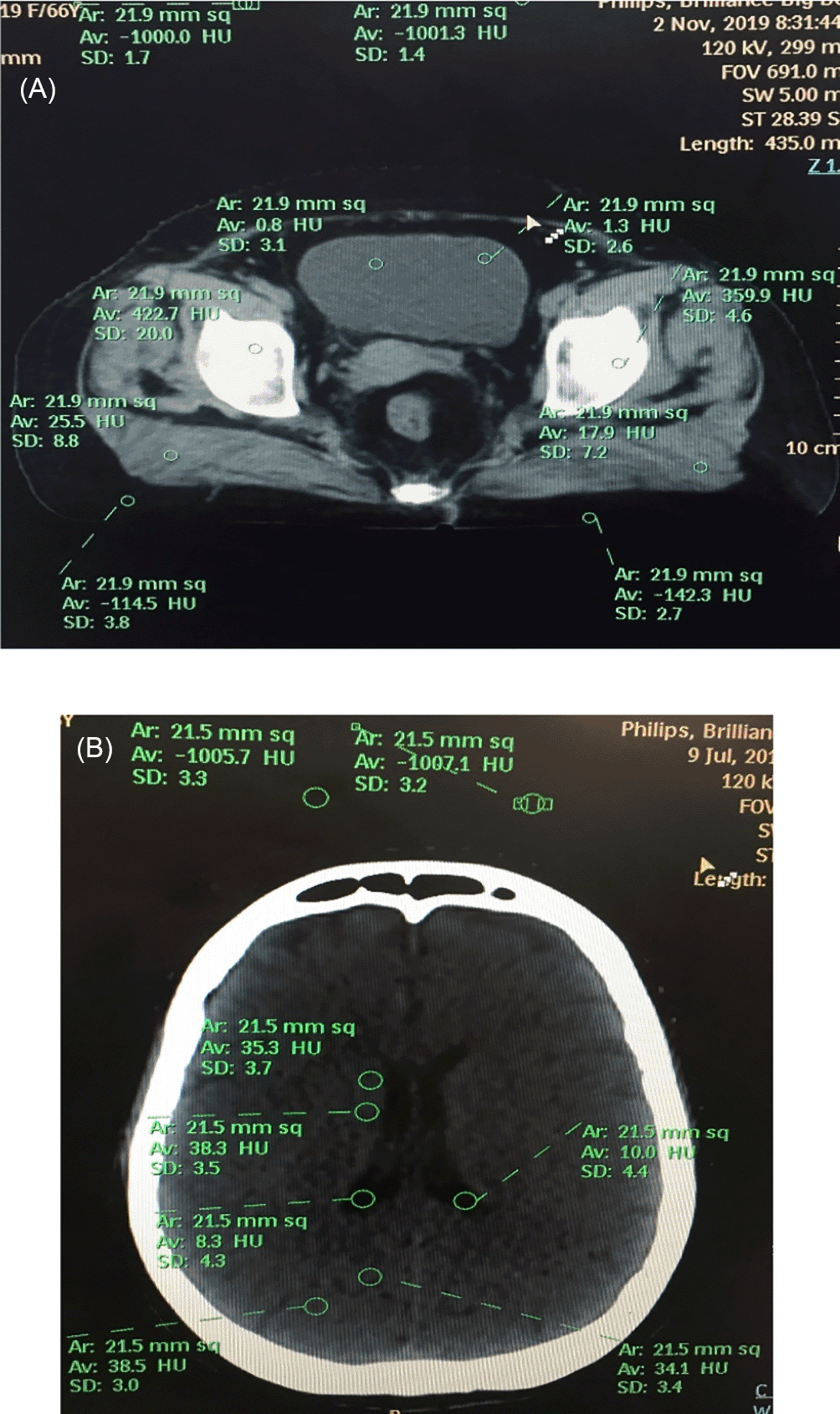
The obtained CT image of a different patient was analyzed for dose and quantitate image quality. The “signal-to-noise” and “contrast-to-noise” ratios for other density regions were analyzed by sketching circular regions of interest (ROIs) of 20-22 mm2 and were manually positioned in the different areas and background to encompass the homogeneity of measured tissues.3
The location of ROIs for quantitative image quality assessment of the pelvis and Head and neck is shown in Figure 1A and Figure 1B, respectively.
Analysis of the SNR, CNR, and FOM ratios for the phantom and the participant population were arrived at by the following equation5:
The statistics were analyzed using IBM SPSS STATISTICS 26.0 software. The descriptive statistical analysis included the average, standard deviation, median, and the third quartile for CTDI vol and total dose length product (DLP), calculated for each procedure.
The maximum crucial part of dose optimization is completed earlier than the patient rests on the Computed tomography table, in the phrases of ensuring the “justification of the clinical indication for CT.” Numerous investigators have discovered different techniques for reducing radiation doses.6–10
Once it is determined that a CT scan is required, the work of reducing radiation dose should confirm that pictures with interpretable indicative data can be achieved with the most reduced feasible radiation dose. A low dose CT scan without the required diagnostic information helps but could lead to further imaging or damage of significant time for producing management choices. “In contrast, a high-dose CT with image quality superior to it needed to get diagnostic information could increase considerations over radiation dose risks.”
To observe a harmony between “image quality and imaging dose, a CatPhan 500 phantom,” which has a 20 cm width, was filtered. “Scans were procured with a rotation time of 0.5 s, slice thickness of 3 mm, the pitch of 0.6, and the greatest accessible, effective tube current”. With the use of reconstruction algorithms, the raw data were reconstructed from the phantom data. “The reconstructed images of the CATPHAN from all diverse tube voltage and effective tube current-time product settings were reviewed by five experienced medical physicists.” “To calculate the contrast-to-noise ratio, circular regions of interest were drawn with the signal ROI inside the 20 mm diameter target in the different density material and the background ROI outside but next to the target. To determine the appropriate tube voltage, the CNR, the increased dose, and penetration were considered”.11 Some of these parameters can optimize the dose and image quality.
Because of the rectilinear relationship between radiation dose & applied tube current, tube current (measured in milliamperes) change is the utmost commonly used scan limit to modify dose.12 Tube current (milliamperes) can be modified manually by selecting fixed or constant milliamperes with a user-required image quality measured to alter tube current based on body shape and body regions. While unnecessarily vigorous tube current reduction causes increased image noise in the mediastinum and chest wall, previous research has shown that tube current can be decreased to 15 to 50 mAs without compromising the diagnosis of lung or mediastinal anomalies.13
Another scan parameter typically tailored for radiation exposure adaptation is tube potential (measured in kilovolts). The variation in radiation dose corresponds to tube current and is generally relative to the square of the adjustment of applied pinnacle kV. (kVp), for example, bringing down the tube potential from 140 to 120 kVp brings about a 35% reduction.
The total radiation dose associated with CT examination is the dose length product (DLP; measured in milli-gray multiplied by centimetres). For helical CT acquisitions, it is calculated as the product of volume CT dose index (CTDIvol, in milli-gray) and scan length (in centimeters). Therefore, a reduction in scan length results in a direct and linear decrease in the DLP. Scan length should always be curtailed to the area of interest.13
This experimental study was conducted in the same setup. The image acquisition was accomplished using a CATPHAN 502 phantom. These scans were used to relatively compare the quality of the obtained images using the technical parameters. Image acquisitions were obtained for the different mAs and kV settings to reduce radiation dose. As with tube current, lowering tube potential also improves image noise, improving image contrast. In some studies, it is mentioned that in common, chest CT can be performed at 80 kV in subjects smaller than 50 to 60 kg and 100 kV for subjects weighing up to 75 to 80 kg.13
The improved image noise with decreased kilovolts does not influence general image quality considerably. Earlier investigations described weight-based streamlining of kilovolts and milliamperes for paediatric chest CT with a considerable decrease in dose.13
A total of 100 images were acquired using the different technical parameters mentioned in Table 2. The acquired images were assessed for objective and subjective image quality acceptability by determining the SNR, CNR, and FOM.
| Imaging parameters | Details |
|---|---|
| kVp | 90, 129, 140 |
| mAs | Started with 100 till 350 with 50 mAs increment |
| FOV | 220 mm |
| Scan length | 183 mm |
| Slice thickness | 3 mm |
Scanning parameters of the phantom study with different kVp and mAs are shown in Figures 2A, 2B, 2C, 2D, 3A, 3B, 3C, and 3D.
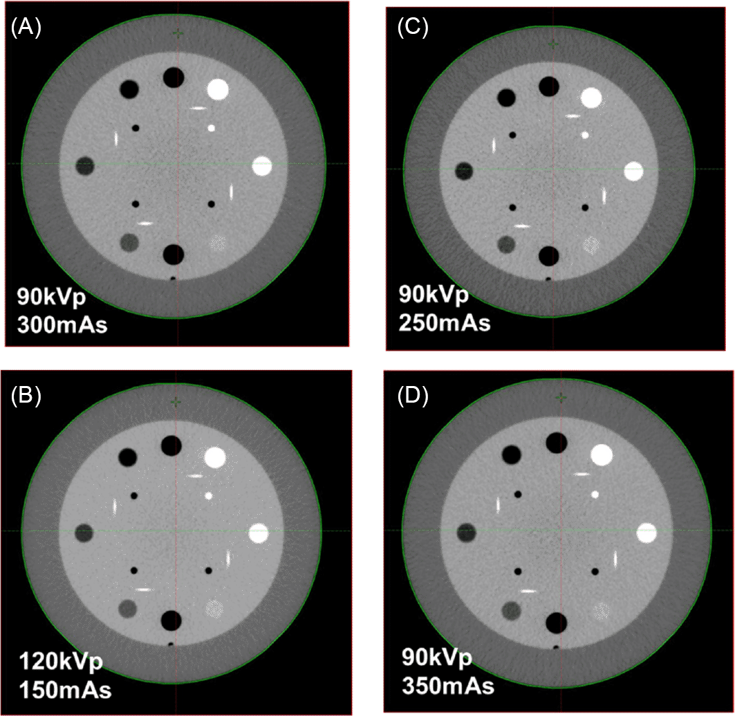
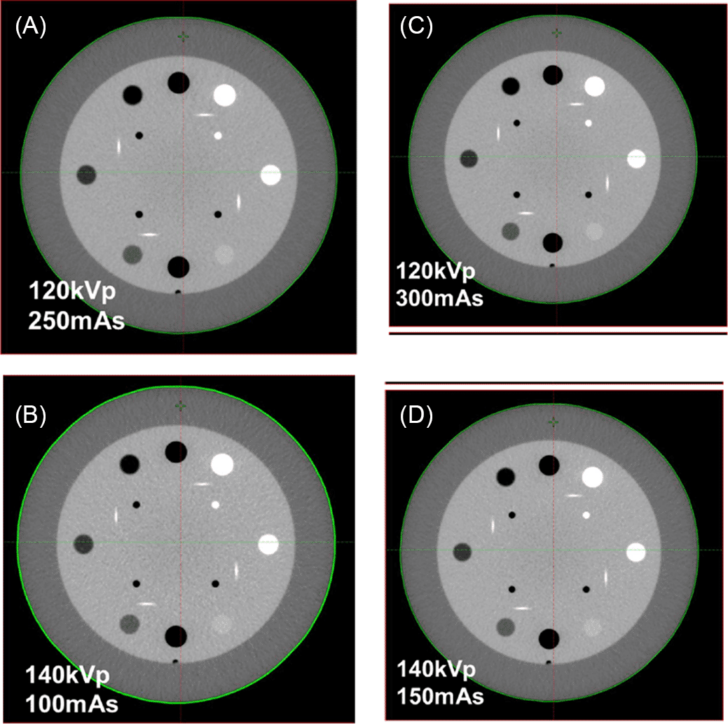
Imaging parameters used in the phantom study are given in Table 2.
In this study, we have used a total of 70 males and 50 female head and neck and 90 female patients that are diagnosed with cancer. The mean, median, effective dose, and third quartile values for DLP, which is the present protocol DRL for the Head and neck and pelvic scans, are summarised in Table 3. All the subjects underwent the scan, and all the participants were eligible for potential reasons. All the 120 participants underwent scans without no missing data.
The mean and standard deviation for image quality (SNR and CNR) and Figure of merit (FOM) for the Head & Neck, and Pelvic scans are summarized in Table 4 and Table 5.
Determination of optimal protocol on phantom data
The summary of the technical parameters selected from the experimental study to be used in the optimization process is presented in Table 6.
The CNR, SNR, and FOM are calculated for ROI created in the phantom scan. The CNR and SNR are compared with the CNR, and SNR is calculated in the patient scans taken in 120 kVp and 300 mAs, selected some combinations of kVp and mAs which was nearer to the patient scan.
In the imaging techniques that use ionizing radiation, involving radiotherapy planning computed tomography scans, to minimize patients’ risk corresponding to the linear no-threshold model, radiation dose needs to be optimized. Diagnostic reference levels are indicators of the typical practice in a country or a region. Because equipment and procedure protocols can vary between different facilities in countries or areas, it is good to establish national or regional diagnostic reference levels.14 The use of DRL is recognized as a dose optimization tool by many professional and regulatory organizations, including ICRP, ACR, IAEA, and AAPM.
The focus of the current research was to establish a diagnostic reference level for present RTCT. This study is the first of its kind RTCT simulation DRLs in India. The Radiotherapy CT scanner and the CT scanner used in radiology have some differences. The CT scanner used for the Radiotherapy scan preferably needs to have a big bore size compared to the radiology CT bore. The reason is to allow some specific fixtures used in radiotherapy CT simulation, which is required to maintain the same position of the patient throughout the radiotherapy treatment and avoid some normal structures from the path of radiation. The literature reports a 10 – 20 mGy more dose delivered with big bore CT than with small-bore size CT scanner because of the variations in mAs settings.15 Comparing Radiotherapy CT DRL with Radiology CT scan procedure DRL is not appropriate because of several differences between the two, including different scanning lengths, protocols, and the requirement of different quality of images.4,16
Clerkin et al.17 projected a “National Irish DRL of 882 mGy cm and CTDIvol of 21 mGy for the Head and neck RT localization CT. Our study’s results comply with this study’s recommendations, with the DLP of 790.65 mGy cm and CTDIvol of 17.76 mGy”.
Nika Zalokar et al.1 projected DRL of 708.2 mGy cm, 663.1 mGy cm, and CTDIvol of 17.7 mGy, 18.3 mGy for pelvic RT localization CT with Philips and Siemens CT, respectively. The output measurement for the pelvis scan was from the “L3-L4” portion to 2 cm under the ischiatic bone. Study results with the Dose length product of 999.7 mGy cm and CTDIvol of 17.76 mGy, and the scan length is D10-D12 region to the mid femur.
Toro et al.18 studied patient exposure concentrations in Computed tomography models by comparing only CTDIvol standards. All CTDIvol values in our study were lower compared to that study. However, wide variations in CTDIvol have been found among various CT units.
The current study has a few limitations. This study calculates SNR, CNR, and FOM. However, the correlation between the dose and quality of the image was not addressed based on BMI and chest circumference. The radiation doses that cancer patients obtain throughout imaging have not been significant because of the treatment doses. The RT CT dose optimization is required because cancer patients might require undergoing many CT scans. In the current study, the variation in CTDIvol, DLP, and scanning protocol in Head and Neck and Pelvis cancer Radiation therapy localization CT imaging in the departments authorize the establishment of DRLs. It may be necessary to consider BMI-specific modified RT CT protocols, and this area warrants further investigation.
The first regional radiation therapy Computed tomography simulation DRLs have been projected and deliver a platform for dose assessment and optimization. Due to the limited number of literature papers on radiation therapy, Computed tomography DRLs. These provide a basis for dose optimization among RT centres and may facilitate the reduction in cumulative radiation exposure. Comparison with previously established RT CT DRLs showed some radiation dose variation, so exposure parameters should be reviewed and optimized.
Harvard Dataverse, V1. Establishment of Diagnostic reference level and Radiation dose variation in Head & Neck and Pelvis treatment planning in RT CT, https://doi.org/10.7910/DVN/GIGDBG.19
This project contains the following underlying data: Slice number of the exact location where we had to draw a Region of Interest (ROI) for obtaining the Hounsfield unit and Standard deviation of the area where the ROI is plotted. The collected values from the region of interest will give the Contrast to Noise Ratio (CNR), Signal to Noise Ratio (SNR), and Figure of Merit (FOM). While calculating the radiation dose levels for an individual part, critical parameters like Dose Length Product (DLP), CTDIvol, and scan length are considered. All the above data has been liked with the link below.
Data are available under the terms of the Creative Commons Zero “No rights reserved” data waiver (CC0 1.0 Public domain dedication).
| Views | Downloads | |
|---|---|---|
| F1000Research | - | - |
|
PubMed Central
Data from PMC are received and updated monthly.
|
- | - |
Provide sufficient details of any financial or non-financial competing interests to enable users to assess whether your comments might lead a reasonable person to question your impartiality. Consider the following examples, but note that this is not an exhaustive list:
Sign up for content alerts and receive a weekly or monthly email with all newly published articles
Already registered? Sign in
The email address should be the one you originally registered with F1000.
You registered with F1000 via Google, so we cannot reset your password.
To sign in, please click here.
If you still need help with your Google account password, please click here.
You registered with F1000 via Facebook, so we cannot reset your password.
To sign in, please click here.
If you still need help with your Facebook account password, please click here.
If your email address is registered with us, we will email you instructions to reset your password.
If you think you should have received this email but it has not arrived, please check your spam filters and/or contact for further assistance.
Comments on this article Comments (0)