Keywords
Breast Abscess, Cholesterol Granuloma, Open Excisional Biopsy
This article is included in the Oncology gateway.
Breast Abscess, Cholesterol Granuloma, Open Excisional Biopsy
We substituted repetitive "such as" words with a similar term and deleted the statement regarding the lymph node due to its average size in the ultrasound image.
See the authors' detailed response to the review by Shoji Oura
See the authors' detailed response to the review by Tommy Supit
Cholesterol granuloma is a chronic inflammatory granulomatous disease caused by cholesterol crystals that have been released into the tissue.1 This disease is mainly found in the middle ear cavity or mastoid process but rarely in the breast.2,3 The incidence rate of cholesterol granuloma in the ear is 0.6 cases per one million population. While the incidence rate of cholesterol granuloma in the breast has never been published, it is estimated to be far less than the incidence rate of cholesterol granuloma in the ear.4
Cholesterol granuloma of the breast is a rare and benign disease. The etiology of this disease in the breast is unclear. However, some reports mentioned the terminal stage of ductal ectasia, which is responsible for the ruptures of the terminal duct, and its lipid-rich material, such as cholesterol crystal escapes the broken luminal structure of the terminal duct. The further inflammatory process surrounds this cholesterol crystal, then forms an encapsulated lesion. The cholesterol crystal is also resistant to resorption by giant cells, creating a problematic situation for the inflammatory process to subside by itself.5,6 However, this theory is still debatable as not all ruptured ductal ectasia will lead to the leak of cholesterol crystals.5
Breast cancer is still a leading cause of newly diagnosed cancer cases in Indonesian women. As most of the patients (80%) are diagnosed in locally advanced stages, breast cancer holds a high mortality rate in Indonesia.7–9 The delay of women seeking medical attention is often the main reason for this highly advanced stage presentation. This is due to the patient’s neglect, inadequate knowledge, and other socio-economic problems.8,9 Sub-urban areas, for instance our current practice location in Tangerang, are no exceptions. Clinicians can easily find new cases of locally advanced breast cancer with a classical clinical presentation, including ulceration, peau d’orange, and multiple regional and distant metastases, with poor prognosis.10 However, in some patients with ambiguous clinical presentation, the clinician needs to consider other differential diagnoses other than breast cancer.
A 43-year-old Javanese housewife woman with a weight of 78 kg, a height of 168 cm, and a body mass index of 27.63 kg/m2 presented to the Surgery Clinic at Siloam General Hospital, Tangerang, Banten, Indonesia, with the primary complaint of a painful mass located in her left breast over the previous week before being admitted to the hospital. The mass was found abruptly in the patient, and she showed severely progressive pain (Visual Analog Score 7/10)11 and a slight fever. Initially, the patient didn’t notice any lumps on either of her breasts. A retracted nipple accompanied this complaint but without any edema and redness on the skin.
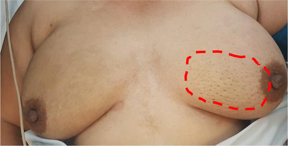
On inspection during the physical examination, peau d’orange and nipple retraction were found prominently (Figure 1). A painful mass of approximately 5 × 7cm was found on palpation. The mass had an irregular border, hard consistency, and no fluctuation. Moreover, the attending clinician noted prominent tenderness and warmth of the skin, which is unusual for such breast cancer cases.

The red line illustrates the location of the mass. The skin of the left breast was consistent with peau d’orange, the classical sign of locally advanced breast cancer.
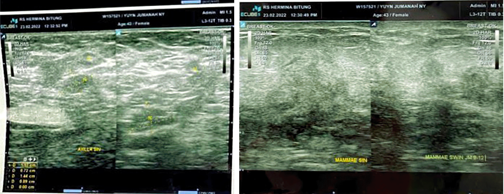
Preliminary ultrasonography was conducted to further investigate the diagnosis, which showed a solid mass at the left breast with an irregular border and multiple enlarged lymph nodes in the left axillary region (Figure 2). These findings suggest a malignant mass at the left breast with regional metastasis to the left axillary region. The laboratory exam was unremarkable, without elevation in systemic inflammatory markers for example leucocytes and neutrophils. The BIRADS score of this patient was five points which is highly suggestive of malignancy.

A solid mass with an irregular border is shown in the left picture. The right image shows multiple enlarged lymph nodes in the left axillary region.
Since neglected breast carcinoma is prevalent in our daily clinical practice, the primary working diagnosis was breast cancer with a secondary differential diagnosis of breast abscess. These differential diagnoses were made due to the abrupt incidence of the mass, accompanied by severe pain and warmth of the surrounding skin of the affected breast, which is unusual for patients with breast cancer. Typically, breast cancer requires a core biopsy as a further diagnostic procedure. However, the clinician felt the urge for an open biopsy because the breast abscess was the possible secondary diagnosis.
Open biopsy and drainage were performed under general anesthesia. An abscess cavity was found within the left breast, with size and location in accordance with the mass previously palpated in pre-operative clinical examination. There was approximately 15cc purulent material found with much necrotic tissue and a hard, solid abscess wall (Figure 3). The surgeon decided to do a biopsy of the abscess wall, followed by debridement of the abscess cavity, and left the wound open, thus permitting secondary wound healing while awaiting the biopsy result. Culture of the purulent material was done, but no bacteria were found, further proving that this is a “sterile abscess”.
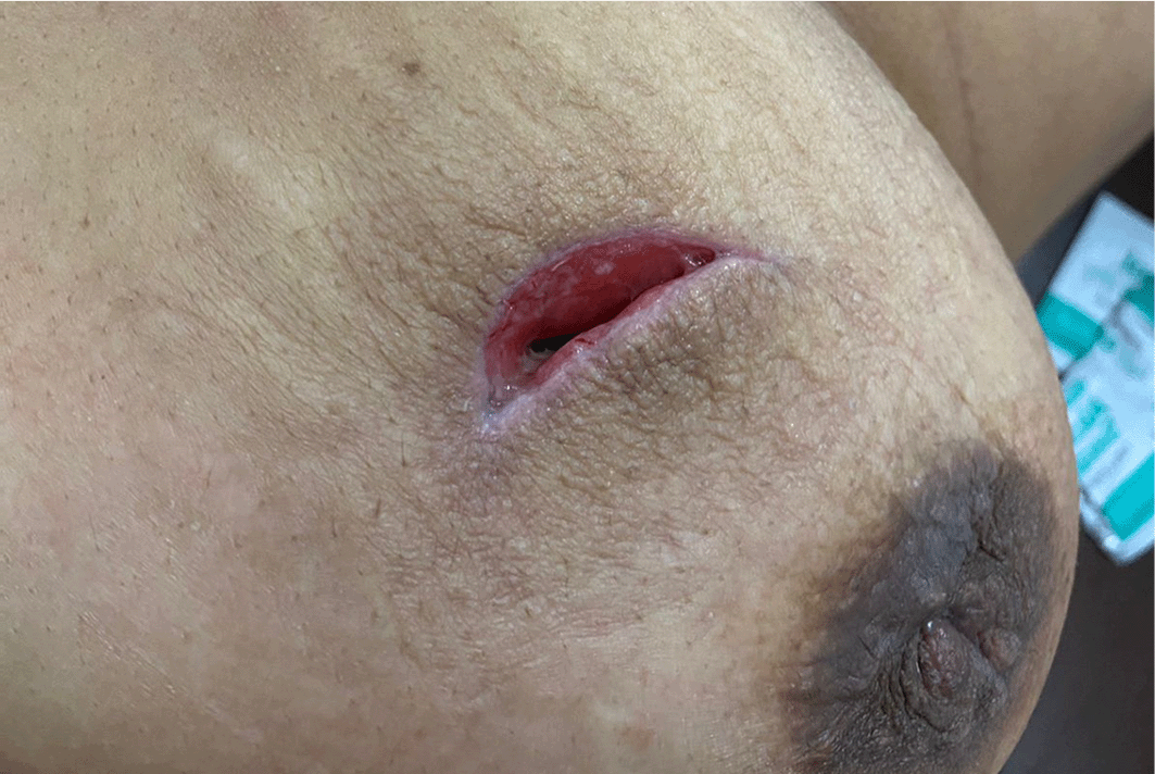
The patient was given Ceftriaxone 2 g twice daily intravenously from preoperative to postoperative day one. Following the operation, the patient’s pain scores were notably reduced to only 3/10. This low pain level was easily managed by oral analgesics such as paracetamol (500 mg per oral solution). The patient was discharged on the first day after the operation. Furthermore, Clindamycin 300 mg twice daily per os continued until postoperative day seven. A follow-up meeting was scheduled two weeks, four weeks, and six months after the surgery. The patient did not complain of any pain. The scar healed entirely within four weeks and was observed for six months with satisfactory results (Figure 4).

Peau d’orange and necrotic tissue or purulent material were absent. The scar was partially healed.
At one-month post-surgery, a follow-up with the patient was scheduled. At this follow-up, the patient reported no complaints at her surgical site. The pain score was reduced to zero points, and the wound showed an improvement without any secondary infection (Figure 5).
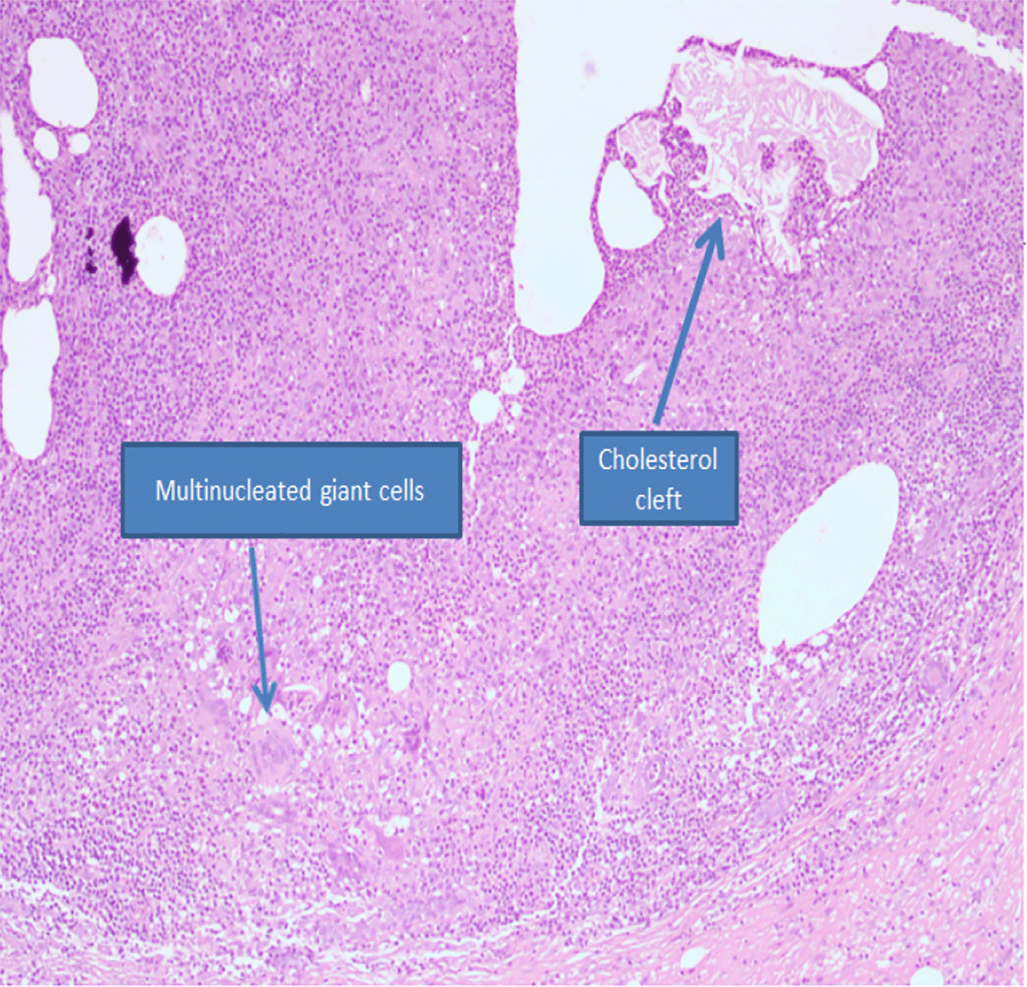
Biopsy of the abscess wall showed dilated duct lumen containing needle-like cholesterol crystal, with surrounding chronic inflammatory infiltrate and foreign body type multinucleated giant cells (Figure 6). Some areas showed cholesterol crystals being engulfed by multinucleated giant cells (Figure 7). Thus, the biopsy confirmed cholesterol granuloma of the breast as the diagnosis, and a malignant lesion of the breast was not found.

A dilated duct lumen contains needle-like cholesterol crystals, with surrounding chronic inflammatory infiltrate and foreign body type multinucleated giant cells (H&E, magnification ×10). H&E, hematoxylin and eosin.
The management of breast carcinoma and breast abscess are entirely different. Typically, the difference is prominent in clinical pictures and imaging, so clinicians will not consider these two entities as differential diagnoses.12
The primary diagnostic assessments for breast cancer are clinical examination, radiology, and core biopsy of the mass.9,13 However, the experience of both the clinician and radiologist is also a dominant factor in determining whether a mass is benign or malignant.13 A report of two cases of cholesterol granuloma on the breast showed that both mammography and ultrasonography were suggestive of carcinoma of the breast, but open biopsy showed cholesterol granuloma.14 While some reports find this is an accurate tool to get a definitive diagnosis for invasive breast cancer with sensitivity reported 90-99%, reports in Indonesia show its sensitivity is only 78% in early breast cancer.15–17 The last fact, combined with the plausibility of breast abscess as the diagnosis in this case report, led the surgeon to choose open biopsy instead of core biopsy for the patient.
Due to its rarity and similarity to breast cancer signs and symptoms, treatment guidelines for breast abscess due to cholesterol granuloma have not yet been established. A report by Jeong et al., 2016 indicated that core biopsy was sufficient to diagnose breast abscess, and afterward, the follow-up showed a decrease in size.18 But serial cases from Nam et al., 2019 of 12 cases of cholesterol granuloma of the breast showed that the more suitable treatment for breast abscess was an excisional biopsy.5 Reports from Osada & Kitayama, 2002 and Fujii et al., 2013 stated that open and excisional biopsies provided satisfying results and no recurrence.6,19 In most cases, diagnosis based on microscopy is precise, but some conditions may mimic malignancy in fragmented core biopsy samples. Conversely, some malignancies can also simulate benign inflammatory or reactive conditions.20
Due to its consideration as a breast abscess, open biopsy and drainage were preferable, which acted as diagnostic and curative procedures. On the other hand, core biopsy simply is less suitable in many cases because of cholesterol granuloma present in other body regions, including the ovarium, middle ear cavity, brain, and testis, which also require resection of the affected tissue and removal of the inflammatory tissue.1,2,21,22
This case report has many strengths; the physical, laboratory, and radiography examinations were done thoroughly, which support the diagnosis, and definitive treatment of surgery was performed with a satisfactory result with no complications and complete scar healing. Moreover, the complete patient follow-up during the hospitalization and post-hospitalization were comprehensively collected. In addition, most previously reported cases didn’t report or evaluate post-procedure pain scores. However, in this case report, the pain score was dramatically reduced.
Although it could be very similar in presentation, an experienced clinician should be wary and consider some entities that could mimic carcinoma of the breast, for instance cholesterol granuloma. An excisional biopsy is essentially required to eradicate inflammatory tissue to ensure the healing process.
Written informed consent for publication of their clinical details and clinical images was obtained from the patient.
FH worked on the Conceptualization, Data Curation, Formal Analysis, Funding Acquisition, Investigation, Methodology, Resources, Software, Visualization, Writing – Original Draft Preparation. RVH was involved in Conceptualization, Formal Analysis, Methodology, Software, Visualization, Writing-Review & Editing. EJW was involved in Project Administration, Supervision, Writing – Review & Editing. FH wrote the draft of the article, RVH and EJW helped with the final manuscript preparation. All figures are original to this manuscript and permission from the patient to publish such an image is obtained. All authors read and approved the final manuscript.
The authors would like to thank Patricia Diana MD from the Faculty of Medicine, Pelita Harapan University for her detailed explanation of the pathology result and picture.
| Views | Downloads | |
|---|---|---|
| F1000Research | - | - |
|
PubMed Central
Data from PMC are received and updated monthly.
|
- | - |
Competing Interests: No competing interests were disclosed.
Is the background of the case’s history and progression described in sufficient detail?
No
Are enough details provided of any physical examination and diagnostic tests, treatment given and outcomes?
No
Is sufficient discussion included of the importance of the findings and their relevance to future understanding of disease processes, diagnosis or treatment?
No
Is the case presented with sufficient detail to be useful for other practitioners?
No
Competing Interests: No competing interests were disclosed.
Is the background of the case’s history and progression described in sufficient detail?
Partly
Are enough details provided of any physical examination and diagnostic tests, treatment given and outcomes?
Partly
Is sufficient discussion included of the importance of the findings and their relevance to future understanding of disease processes, diagnosis or treatment?
Yes
Is the case presented with sufficient detail to be useful for other practitioners?
Yes
Competing Interests: No competing interests were disclosed.
Reviewer Expertise: General surgery, biomolecular aspects of wound healing
Alongside their report, reviewers assign a status to the article:
| Invited Reviewers | ||
|---|---|---|
| 1 | 2 | |
|
Version 3 (revision) 11 Jul 22 |
read | |
|
Version 2 (revision) 14 Jun 22 |
read | |
|
Version 1 12 May 22 |
read | |
Provide sufficient details of any financial or non-financial competing interests to enable users to assess whether your comments might lead a reasonable person to question your impartiality. Consider the following examples, but note that this is not an exhaustive list:
Sign up for content alerts and receive a weekly or monthly email with all newly published articles
Already registered? Sign in
The email address should be the one you originally registered with F1000.
You registered with F1000 via Google, so we cannot reset your password.
To sign in, please click here.
If you still need help with your Google account password, please click here.
You registered with F1000 via Facebook, so we cannot reset your password.
To sign in, please click here.
If you still need help with your Facebook account password, please click here.
If your email address is registered with us, we will email you instructions to reset your password.
If you think you should have received this email but it has not arrived, please check your spam filters and/or contact for further assistance.
Comments on this article Comments (0)