Keywords
COVID-19, Anti-coagulant, mortality, length of stay, severity, health
This article is included in the Emerging Diseases and Outbreaks gateway.
COVID-19, Anti-coagulant, mortality, length of stay, severity, health
Coronavirus disease 2019 (COVID-19) infection has caused widespread novel COVID-19 pneumonia, causing respiratory problems.1 The World Health Organization (WHO) declared this as a global pandemic affecting many aspects of life.2 A severe degree of COVID-19 pneumonia is defined as a condition in patients complaining of difficulty in breathing, plus one of: respiratory rate >30 times per minute; severe respiratory distress; or oxygen saturation (SpO2) <93% in room air or a PaO2/FiO2 ratio (PF Ratio) <300. In children, it is defined as having a cough or difficulty breathing, plus at least one of: central cyanosis or SpO2 <90%; severe respiratory distress (such as snoring, heavy chest wall traction); signs of severe pneumonia, namely inability to suckle/drink; lethargy or decreased consciousness, or seizures. Other signs of pneumonia are chest retractions, rapid breathing >60×/minute in children aged <2 months, >50×/minute in children aged 2-11 months, >40×/minute in children aged 1-5 years, or >30×/minute in children aged >5 years.2
Severe SARS-CoV2 infection promotes a syndrome related to prothrombotic conditions, in which blood clots easily. This condition is characterized by several specific abnormal laboratory values, such as mild thrombocytopenia, increased fibrin, degradation of fibrin products, fibrinogen, and D-dimers. Increased D-dimers are strongly correlated with worsened clinical conditions and increased risk of death in COVID-19.3,5 Based on reports from several studies, there is a growing incidence of several thromboembolic states in patients with COVID-19 admitted to the intensive care unit (ICU), one of which is pulmonary embolism. COVID-19 patients suspected of experiencing a thromboembolic event should be given anticoagulant therapy when radiological imaging is difficult to perform.3–5
The pathophysiology of hypercoagulation in COVID-19 patients is still currently being explored. A case series presenting three cases of COVID-19 with antiphospholipid syndrome has recently been reported by The New England Journal of Medicine.5 SARS-CoV-2 infection is associated with antiphospholipid antibodies, which predispose to hypercoagulation. The study of Campbell et al. reported severe COVID-19 patients with increased levels of lactate dehyrdrogenase (LDH), D-Dimer, bilirubin, decreased platelets, mild anemia, heart and kidney injury, and diffused thrombotic micro-angiopathy.5–7
Patients with severe COVID-19 experience complications of coagulopathy in the form of disseminated intravascular coagulation (DIC) which might result in death. Severe COVID-19 patients experience respiratory problems and increased virulence, according to the criteria of the Third International Consensus Definitions for Sepsis (Sepsis-3). Severe COVID-19 patients are also at risk of venous thromboembolism (VTE) due to prolonged bed rest. The International Society of Thrombosis and Haemostasis (ISTH) provides a new category to identify the early phase of sepsis-associated DIC which is also called sepsis-induced coagulopathy (SIC). COVID-19 patients who fit the diagnostic criteria for SIC can be also given anticoagulant therapy.5–10
Anticoagulant therapy in COVID-19 patients is administered to those who show signs of thrombosis, such as elevated inflammatory factors and D-dimers within 7-14 days, with threefold D-Dimer value. The option would be to use low molecular weight heparin (LMWH) at a dose of 100 IU/ kg twice a day, for 3-5 days. The European Society of Cardiology also includes anticoagulants in the COVID-19 therapy algorithm. For patients admitted to the ICU, the parenteral heparin drip protocol must be strictly controlled and the time value of the active prothromblastin time is 60-85 seconds. For non-ICU patients, the subcutaneous dose of enoxaprin is started at 1mg/kg twice daily. After all the completed researches, further study on how to use anticoagulants is still being analysed to promote the best prognosis.3,11,12
Approval was issued by the research ethics board Universitas Airlangga Hospital (No: 179/KEP/2020) on 2nd October 2020. The consent to participate was not applicable since our data were obtained from medical records.
We performed a retrospective cohort study with consecutive sampling among COVID-19 adult patients admitted to two referral hospitals for COVID-19, Universitas Airlangga Hospital (UAH) and Husada Utama Hospital, Surabaya, East Java, Indonesia, from March 15th 2020 to August 31st 2020. The patients included were ones in moderate, severe, or critical condition. We assessed patients referring to WHO guidelines and Indonesian Ministry of Health guidelines, and tested for SARS-CoV-2 Polymerase Chain Reaction (PCR) through oropharyngeal and nasopharyngeal swabs. Sequential chest x-ray and laboratory inflammatory marker evaluation were performed, then we did three timeframes of assessment. The first evaluation was done at the admission, followed by a second evaluation on the sixth day, and lastly evaluated before discharge or death (mean time 13 days).
In October 2020, we collected the data from medical records of those admitted to UAH and Husada Utama hospital between March 15th, 2020 to August 31st, 2020. We had 331 patients with moderate to critically ill COVID-19. Incomplete medical records were excluded. From this selection, we organized patient records into 110 moderate cases, 140 severe cases, and 81 critically ill cases with ventilator (see Figure 1).
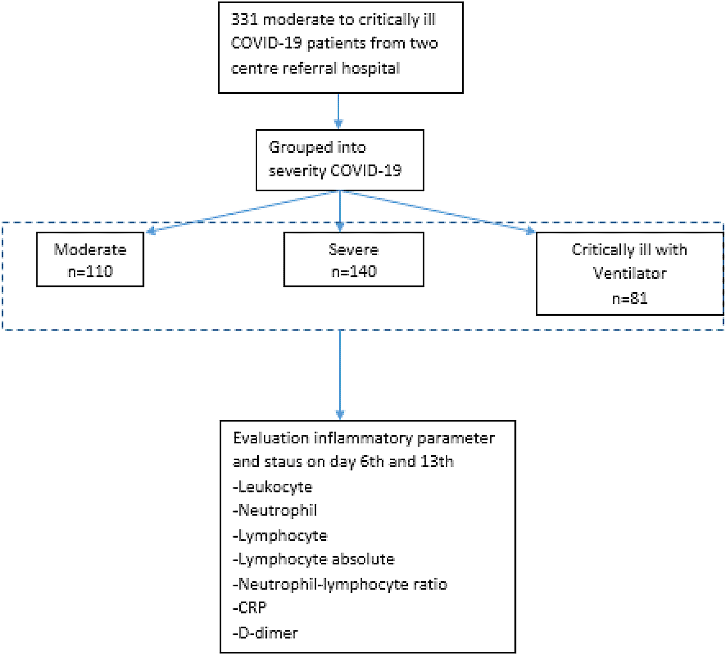
Patients were categorized as a moderate case if they had either or both of 1) signs of pneumonia; 2) O2 sat ≥93% free air. Meanwhile, patients were classified into severe cases if there were clinical signs of pneumonia, with one of the following: 1) respiratory rate (RR) >30 times per minutes; 2) severe respiratory distress; 3) oxygen saturation < 93% free air. They were grouped into critically ill if they suffered from acute respiratory distress syndrome (ARDS), sepsis, and septic shock.2
The criteria of inflammatory markers were: 1) white blood cell (WBC) >6.16x10³ cells/μL;13 2) neutrophil-lymphocyte ratio (NLR) >6.5;14 3) absolute lymphocyte count ALC < 1.0x10³ cells/μL;15 4) c-reactive protein (CRP) >41.8 mg/L;16 and 5) Procalcitonin > 0,07 ng/mL.16
We examined the data using SPSS version 24.0 (Chicago, IL, USA). Descriptive analysis incorporated categorical variables reported as number (percentage) and continuous variables as mean (standard deviation). We displayed categorical variables as number (%) and continuous variables as mean (standard deviation) or median (range), depending on whether the data are normally distributed. Means of Chi square or McNemar was used to assess statistical significance for dichotomous variables, while paired t-test or Wilcoxon test were used to examine continuous variables, depending on the data distribution. We compared the mean/median difference of the first to the second evaluation and the second to the third evaluations of the laboratory results, length of stay, days of death, and mortality rate according to severity of COVID-19 using ANOVA or Kruskal Wallis. Bivariate analysis between inflammatory state and mortality was conducted using Spearman. We also determined the relationship between inflammatory state after anti-coagulant with patients’ mortality, along with some variables, which were age, gender, disease severity, and comorbidity.
Of 331 patients enrolled, 200 were male and 131 were female. Patients were grouped based on the category of severity of disease and resulted in 110 moderate patients, 140 severe patients, and 81 critically ill patients with ventilator support. The average age of patients with severe COVID-19 was 57.45 ± 13.5 years. In the anticoagulant group, the most frequent comorbid factors found were diabetes mellitus (DM) (52.86%), hypertension (HT) (46.91%), and geriatric age (47.14%) while others had history of heart disease (5.45%), stroke (3.7%), and chronic kidney disease (1.82%). We evaluated inflammatory markers as baseline study data. There were significant differences in laboratory markers between each severity group (p value <0.05). The highest white blood count was 8.53 ± 5.41 × 10³/L, neutrophils 83.75 ± 9.51%, lymphocyte 17.1 ± 9.9%, lymphocyte absolute 1.1 ± 0.57 × 10³/L, neutrophil lymphocyte ratio (NLR) 8.38 ± 12.5, the C-reactive protein (CRP) 40.1 ± 42.43 mg/L and d-dimer 1.2 ± 8.53 mcg/L. The outcome analysis showed significant differences in the mean length of stay (p value <0.05) (see Table 1).
| Severity of COVID-19 | |||||||
|---|---|---|---|---|---|---|---|
| Moderate | Severe | Critically ill with ventilator support | Total | ||||
| 110 | 140 | 81 | 331 | ||||
| Sex | |||||||
| Male (n,%) | 64 | 58,18 | 82 | 58,57 | 54 | 66,67 | 200 |
| Female (n,%) | 46 | 41,82 | 58 | 41,43 | 27 | 33,33 | 131 |
| Age (mean/SD) | 53,62 | 13,67 | 57,45 | 13,5 | 56,11 | 13,63 | 167,18 |
| Comorbid | |||||||
| Geriatric age (age>60 years old) (n,%) | 40 | 36,36 | 66 | 47,14 | 29 | 35,80 | 135 |
| DM (n,%) | 33 | 30,00 | 74 | 52,86 | 23 | 28,40 | 130 |
| HT (n,%) | 40 | 36,36 | 54 | 38,57 | 38 | 46,91 | 132 |
| Heart Disease (n,%) | 6 | 5,45 | 7 | 5,00 | 4 | 4,94 | 17 |
| Chronic Kidney Disease (n,%) | 2 | 1,82 | 2 | 1,43 | 1 | 1,23 | 5 |
| Stroke (n,%) | 1 | 0,91 | 4 | 2,86 | 3 | 3,70 | 8 |
| Laboratory | p Value | ||||||
| Leucocyte (103/uL; median, SD) | 6,49 | 3,55 | 7,166 | 5,89 | 8,53 | 5,41 | <0.05* |
| Neutrophil (%; median, SD) | 72,4 | 15,7 | 78 | 21,41 | 83,75 | 9,51 | <0.05* |
| Lymphocyte (%, median, SD) | 17,1 | 9,9 | 12,15 | 10,42 | 9,5 | 7,01 | <0.05* |
| Lymphocyte absolute (103/uL, median, SD) | 1,1 | 0,57 | 0,915 | 1,31 | 0,84 | 0,51 | <0.05* |
| Neutrophil-Lymphocyte Ratio (NLR) (median, SD) | 4,18 | 5,3 | 6 | 5,61 | 8,38 | 12,5 | <0.05* |
| C-Reactive Protein (mg/L; median, SD) | 18,7 | 39,43 | 40,1 | 42,43 | 34,1 | 49,28 | <0.05* |
| D-Dimer (mcg/l; median, SD) | 0,62 | 3,2 | 0,9 | 7,48 | 1,2 | 8,53 | <0.05* |
| Outcome | |||||||
| Length of Stay (mean, SD) | 18,46 | 9,54 | 19,54 | 10,63 | 14,26 | 11,5 | <0.05* |
| Discharge (n,%) | 108 | 98,18 | 104 | 74,29 | 11 | 13,58 | |
| Death (n,%) | 2 | 1,82 | 36 | 25,71 | 70 | 86,42 | |
We evaluated and compared inflammatory markers up to three times. To analyze the relationship between the administration of anticoagulants in each severity of COVID-19 cases, we calculated the decrease or increase in these inflammatory markers.
Based on the first and second laboratory evaluations, there were differences in decreasing leukocyte count and D-dimer in severe cases (p 0.03; p 0.026), decreasing neutrophils neutrophil-lymphocyte ratio and CRP in moderate cases, (p 0.004; p 0.028; and p<0.05), and increasing lymphocyte in critically ill cases (p<0.05). In this initial evaluation, there were no differences in inflammatory status between each degree of COVID-19 cases.
In contrast to the initial evaluation, the comparison of the inflammatory status between the second and third examinations found that there were differences in each severity group (moderate (p<0.05); severe (p<0.05) and critically ill (p 0.001)). Inflammation marker examination found decreasing leukocytes in moderate and severe cases (p<0.05; p 0.001), decreasing neutrophils in moderate, severe and critically ill cases (p<0.05; p<0.05; p 0.007), increasing lymphocyte count in moderate cases (p<0.05); increasing absolute lymphocyte in moderate and severe cases (p<0.05; p<0.05), decreasing NLR in moderate, severe and critically ill cases (p<0.05; p<0.05; p<0.05), decreasing CRP in moderate and severe cases (p<0.05; p< 0.05), and decreasing d-dimer in all case groups (p<0.05; p<0.05; p<0.05).
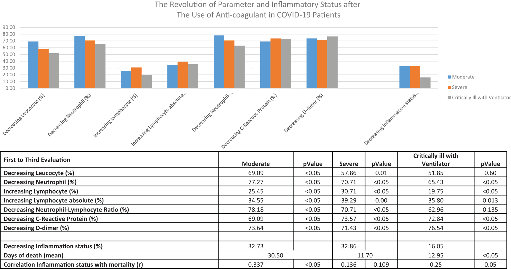
With these significant results, we evaluated the inflammatory parameters from the initial to the last examination. In moderate group, there was an improvement in inflammation parameters in all variables (p<0.05). In severe cases, significant improvements in inflammatory parameters were recorded, including decreasing leukocytes, decreasing neutrophils, decreasing lymphocytes, increasing absolute lymphocytes, decreasing NLR, decreasing CRP and d-dimer (p 0.01; p<0.05; p<0.05; p<0.05; p 0.0; p<0.05; p<0.05 p<0.05).
In the critically ill with ventilator support group, significant results were also obtained on each inflammatory marker variable except for decreasing leukocytes (p 0.6). From the clinical aspect, analysis of the use of anticoagulants on day of death found significant differences among groups of severity (p<0.05). Significant correlation of inflammatory status on the thirteenth day with mortality based on patient comorbidities was seen in moderate and critical cases on a ventilator (r=0.337; p<0.05 and r=0.25; p 0.05) (see Table 2 and Figure 2).
| First to second evaluation | Moderate | Severe | Critically Ill with ventilator | ||||||||||||
|---|---|---|---|---|---|---|---|---|---|---|---|---|---|---|---|
| n=110 | P value | n=140 | P Value | n=81 | P Value | ||||||||||
| Inflammatory status | 1st | 40 | 36.36 | 0,003* | 73 | 52.14 | 0,142 | 41 | 50.62 | 0.32 | |||||
| 2nd | 21 | 19.09 | 61 | 43.57 | 48 | 59.26 | |||||||||
| Laboratory evaluation | |||||||||||||||
| Decreasing leucocyte (103/uL; median, SD) (n,%) | 7.3 | 5.74 | 51 | 46.364 | 0.71 | 8.46 | 5.7 | 58 | 41.43 | 0,03* | 10.31 | 7.06 | 31 | 38.27 | 0.173 |
| Decreasing neutrophil (%; median, SD) (n,%) | 67.85 | 26.25 | 69 | 62.727 | 0.004* | 77.25 | 28.22 | 69 | 49.29 | 0.486 | 83 | 37.75 | 43 | 53.09 | 0.091 |
| Increasing lymphocyte (%, median, SD) (n%) | 16.85 | 12.05 | 55 | 50 | 0.866 | 9.65 | 13.35 | 58 | 41.43 | 0.07 | 5 | 9.48 | 20 | 24.69 | < 0.05* |
| Increasing lymphocyte absolute (103/uL, median, SD) (n,%) | 1.2 | 0.83 | 64 | 58.182 | 0.16 | 0.925 | 1.11 | 73 | 52.14 | 0.544 | 0.706 | 0.865 | 35 | 43.21 | 0.056 |
| Decreasing neutrophil-lymphocyte ratio (NLR) (median, SD) (n,%) | 3.12 | 7.98 | 68 | 61.818 | 0.028* | 5.67 | 9.43 | 65 | 46.43 | 0.74 | 9 | 11.77 | 40 | 49.38 | 0.897 |
| Decreasing C-reactive protein (mg/L; median, SD) (n,%) | 17.26 | 31.429 | 63 | 57.273 | <0.05* | 23.64 | 70.22 | 94 | 67.14 | <0.05 | 13.8 | 40.35 | 29 | 35.80 | 0.324 |
| Decreasing D-dimer (mcg/l;median, SD) (n,%) | 0.51 | 3.19 | 58 | 52.727 | 0.598 | 1.362 | 2.32 | 79 | 56.43 | 0.026 | 1.08 | 11.85 | 31 | 38.27 | 0.427 |
| Second to third evaluation | Moderate | Severe | Critically Ill with ventilator | ||||||||||||
|---|---|---|---|---|---|---|---|---|---|---|---|---|---|---|---|
| n=110 | P value | n=140 | P value | n=81 | P Value | ||||||||||
| Inflammatory status | 2nd | 21 | 19.091 | <0.05* | 61 | 43.57 | <0.05* | 48 | 59.26 | 0.001* | |||||
| 3rd | 4 | 3.6364 | 27 | 19.29 | 28 | 34.57 | |||||||||
| Laboratory evaluation | |||||||||||||||
| Decreasing leucocyte (103/uL; median, SD) (n,%) | 3.73 | 4.76 | 67 | 60.909 | <0.05* | 5.57 | 11.9 | 78 | 55.71 | 0.001* | 8.7 | 14.73 | 36 | 44.44 | 0.349 |
| Decreasing neutrophil (%; median, SD) (n,%) | 31,1 | 35,19 | 73 | 66,364 | <0,05* | 49.05 | 39.51 | 89 | 63.57 | <0.05* | 69.6 | 42.61 | 41 | 50.62 | 0.00*7 |
| Increasing lymphocyte (%, median, SD) (n%) | 10 | 14.58 | 31 | 28.182 | <0.05* | 3.7 | 10.77 | 42 | 30.00 | <0.05* | 2.4 | 8.9 | 24 | 29.63 | 0.078 |
| Increasing lymphocyte absolute (103/uL, median, SD) (n,%) | 0.757 | 1,067 | 34 | 30,909 | <0,05* | 0.564 | 2.14 | 47 | 33.57 | <0.05* | 0,373 | 4.29 | 28 | 34.57 | 0.218 |
| Decreasing neutrophil-lymphocyte ratio (NLR) (median, SD) (n,%) | 2.23 | 4,37 | 77 | 70 | <0,05* | 5.02 | 7,93 | 88 | 62.86 | <0.05* | 3.3 | 14.82 | 39 | 48.15 | <0.05* |
| Decreasing C-reactive protein (mg/L; median, SD) (n,%) | 2,71 | 7,83 | 64 | 58,182 | <0,05* | 6.37 | 15.9 | 75 | 53.57 | <0.05* | 7.8 | 19.9 | 43 | 53.09 | 0.221 |
| Decreasing D-dimer (mcg/l;median, SD) (n,%) | 0.39 | 0.822 | 62 | 56.364 | <0,05* | 1.078 | 3.68 | 70 | 50.00 | <0,05* | 0.847 | 2.1 | 35 | 43.21 | < 0.05* |
| First to third evaluation | Moderate | P Value | Severe | P Value | Critically ill with ventilator | P Value |
|---|---|---|---|---|---|---|
| Decreasing leucocyte (%) | 69.09 | <0.05* | 57.86 | 0.01* | 51.85 | 0.60 |
| Decreasing neutrophil (%) | 77.27 | <0.05* | 70.71 | <0.05* | 65.43 | <0.05* |
| Increasing lymphocyte (%) | 25.45 | <0.05* | 30.71 | <0.05* | 19.75 | <0.05* |
| Increasing lymphocyte absolute (%) | 34.55 | <0.05* | 39.29 | 0.00* | 35.80 | 0.013 |
| Decreasing neutrophil-lymphocyte ratio (%) | 78.18 | <0.05* | 70.71 | <0.05* | 62.96 | 0.135 |
| Decreasing C-Reactive protein (%) | 69.09 | <0.05* | 73.57 | <0.05* | 72.84 | <0.05 |
| Decreasing D-dimer (%) | 73.64 | <0.05* | 71.43 | <0.05* | 76.54 | <0.05* |
| Decreasing inflammation status (%) | 32.73 | 32.86 | 16.05 | |||
| Days of death (mean) | 30.50 | 11.70 | 12.95 | <0.05* | ||
| Correlation of inflammation status with mortality (r) | 0.337 | <0.05** | 0.136 | 0.109 | 0.25 | 0.05** |
Univariate and multivariate analysis using regression logistic revealed that no decline of inflammatory profile both on day 6 (adjusted odds ratio [aOR] = 2.36; 95% CI: 1.46-3.83, p<0.05) and day 13 (adjusted odds ratio [aOR]=4.15; 95% CI: 2.33-7.42, p<0.05) was related to the mortality event (see Table 3).
| Status inflammation | Outcome death | |||||
|---|---|---|---|---|---|---|
| Unadjusted OR | 95%CI | P Value | Adjusted OR | 95%CI | P value | |
| Inflammation 1st day | 1.23 | 0.98-1.55 | 0.091 | 1.29 | 0.76-2.18 | 0.34 |
| Inflammation 6th day | 1.59 | 1.24-2.06 | 0.001* | 2.36 | 1.46-3.83 | <0.05** |
| Inflammation 13th day | 1.67 | 1.48-1.89 | <0.05* | 4.15 | 2.33-7.42 | <0.05** |
COVID-19 has been linked to coagulation disorders which cause various complications. An increase in coagulation parameters, such as D-dimers, is an independent risk factor for death. Patients with D-dimers of more than 1000 ng/mL have a 20 times greater risk of death due to infection.17 Although the pathogenesis of coagulopathy in COVID-19 cannot be fully explained, the mechanism may resemble septic coagulopathy in bacterial infections. Overabundant pro-inflammatory cytokines increases the level of damage-associated molecular patterns (DAMP). Activation of coagulation factors due to cell and endothelial damage is the most common mechanism of infection. Both the pathogen and DAMP from damaged tissue activate monocytes. The activated monocytes release pro-inflammatory cytokines (IL-1, IL-6, IL-10, TNF-α) and chemokines that animate neutrophils, lymphocytes, platelets, and vascular endothelial cells. The coagulation cascade is commenced by tissue factor and phosphatidylserine on the cells’ surface. Healthy endothelial cells retain anti-thrombogenetic properties by expressing glycocalyx and binding with anti-thrombin proteins. Damaged endothelial cells change their nature to become more procoagulant due to glycocalyx disorders and loss of anticoagulant proteins.7,12,18,19 Markers of hypercoagulation and high levels of inflammatory mediators are consistent with poor outcome in patients having acute respiratory distress syndrome (ARDS) and sepsis. These observations have led to several studies focused on inflammation and coagulation pathways in acute lung injury, ARDS or sepsis.17,20,21
The routine use of anticoagulant regimens in COVID-19 patients requiring hospitalization has been recommended, after several studies found an association between viral inflammation and coagulation disorders.22 Several guidelines for the management of COVID-19 have included anticoagulant regimens regarding COVID-19-related coagulation disorders. Recommendations issued by the Anticoagulation Forum (ACF) and the American College of Chest Physicians (ACCP) selected Low Molecular Weight Heparin (LMWH) over Unfractioned Heparin (UFH) to minimize laboratory evaluation. ACCP also recommends fondaparinux over UFH in patients with a high risk of bleeding, kidney problems, and any plans for procedures in the near future. UFH is more recommended by the ACF for patients with renal impairment with creatinine clearance <15-30 mL/min. The American Society of Hematology (ASH) states that LMWH or UFH is the therapy of choice over oral anticoagulants due to the potential for drug interactions and short half-lives.23,24 In our study, the population involved was a group of patients requiring hospitalization due to SARS-CoV2 infection ranging from moderate to critically ill. At all three severity groups, patients receiving anticoagulant therapy had increased D-dimer values. This increase in D-dimers expresses clusters of fibrin lysis and thrombus in the pulmonary vessels. Previously, Guan et al. (2020) found that 46% of 1099 COVID-19 patients had an increase in D-dimer and only 5% experienced a decrease in platelet count.19,25
The relationship between coagulation function and indicators of inflammation and infection has been previously analyzed by Long et al. (2020). D-dimers were positively correlated with CRP (r=0.36, p=0.0007) and procalcitonine (r=0.45, p<0.001).26 Increased CRP was also found in patients with coagulation disorders by Friedrich et al. (2020) to a mean level of 131 ± 106 mg/l.27 In our study, there was an increase in CRP by a mean of 40.1 ± 42.43/l in the severe group population receiving anticoagulants that met the criteria for anticoagulant according to The International Society of Thrombosis Haemostasis (ISTH) or due to an increase in D-dimer. A meta-analysis regarding dosing of anticoagulant therapy on the COVID-19 mortality rate found that there was a slight decrease in the mortality rate in COVID-19 patients in need of a ventilator.28 Several studies have shown different results on the mortality outcome of COVID-19 patients receiving anticoagulant therapy. In theory, the coagulation cascade is active when inflammation is present due to SARS-CoV2 infection. Therefore, it may be possible to obtain beneficial effects from the use of anticoagulants as anti-inflammatory agents.
According to the theory of hypercoagulation disorders, there is a two-way relationship between the immune system and thrombin formation, where inhibition of thrombin formation may reduce the inflammatory response.29 The positive effects of using anticoagulants on mortality were reported by Nadkarni et al.; compared to non-users, recipients of anticoagulants for both therapy and prophylaxis had reduced mortality (adjusted hazard ratio [aHR] = 0.53; 95% CI: 0.45-0.62, and aHR = 0.50; 95% CI.: 0.45-0.57, respectively), and intubation rates (aHR 0.69; 95% CI: 0.51-0.94, and aHR 0.72; 95% CI: 0.58-0.89, respectively).30 Our study revealed that no reduction in inflammatory markers was significantly correlated with the mortality.
Several other studies have classified the use of anticoagulants according to their intended use for therapeutical and prophylaxis purpose. Klok et al. (2020) found that 15% of the population needed ICU while Helms et al. (2020) revealed 25 of their patients had pulmonary embolism despite receiving anticoagulant therapy.22 Therefore, anticoagulant prophylaxis is rational. In addition, the prophylactic use of apixaban (odds ratio [OR] 0.46, p=0.001) and enoxaparin (OR=0.49, p=0.001) exhibited a significant decrease in mortality. There was also an association between therapeutic apixaban and decreased mortality rates (OR 0.57, p=0.006). Pawlowski et al. (2020) compared the effects of enoxaparin and heparin, showing that patients receiving heparin had a higher risk of death and higher ICU admission than ones in enoxaparin group (risk ratio: 6.76; 95% C.I: [3.39, 12.7]; adjusted p-value <0.0001); (risk ratio of ICU admission: 1.51; 95% C.I.: [1.12, 2.03]; adjusted p-value 0.01). Additionally, ICU and hospital length of stay were shorter in the enoxaparin population (mean ICU duration: 0.9 days [standard deviation: 2.5], mean hospital duration: 5.4 days [standard deviation: 4.3]).31
In this study, we found favorable changes in inflammatory markers such as white blood cells, neutrophils, lymphocytes, CRP, and D-dimers after the use of anticoagulants. The analysis of the results of this study has been divided based on the subgroup of the severity of COVID-19 disease. From the clinical aspect, analysis of the use of anticoagulants on day of death found significant differences among groups of severity (p<0.05). There was also a significant correlation between inflammatory status on the thirteenth day and mortality based on patient comorbidities obtained from moderate and critical cases on a ventilator (r=0.337; p< 0.05 and r=0.25; p 0.05). Univariate and multivariate analysis exhibited that no reduction in inflammatory profile on day 6 (adjusted odds ratio [aOR]=2.36; 95% CI: 1.46-3.83, p value < 0.05) and day 13 (adjusted odds ratio [aOR]=4.15; 95% CI: 2.33-7.42, p value < 0.05) were linked to the patients’ mortality. This study is able to explain that the values of inflammatory and hypercoagulable markers go hand in hand with the severity of the patient. Although not all inflammatory markers improve after anticoagulant treatment, inflammatory variables mostly manifested good results. This study did not distinguish one regimen of anticoagulant from another; thus, it could be a confounder. Conclusively, it is necessary to carry out further subgroup analysis of the types of anticoagulants and the comparison of therapeutical effects and prophylactic use of anticoagulant towards the mortality outcome and length of stay.
Administration of anticoagulants to COVID-19 patients with moderate to critical presentation promoted significant outcomes of inflammatory markers which ultimately showed a statistical difference in mortality. Most of the inflammatory markers in patients improved after anticoagulant administration. Therefore, our findings confirm that the administration of anticoagulants can be optimized since they are able to work as anti-inflammatories.
figshare: Data of Inflammatory Parameters after Anti-coagulant among Moderate, Severe, and Critically Ill COVID-19 Patients. https://doi.org/10.6084/m9.figshare.16910905.v2
This project contains the following files:
Data are available under the terms of the Creative Commons Attribution 4.0 International license (CC-BY 4.0).
Pradana Zaky Romadhon: Conceptualization, Formal Analysis, Investigation, Methodology, Software, Supervision, Validation, Writing – Original Draft Preparation, Writing – Review & Editing
Siprianus Ugroseno Yudho Bintoro: Conceptualization, Formal Analysis, Investigation, Methodology, Supervision, Validation, Writing – Original Draft Preparation, Writing – Review & Editing
Satriyo Dwi Suryantoro: Conceptualization, Formal Analysis, Investigation, Methodology, Software, Supervision, Validation, Writing – Original Draft Preparation, Writing – Review & Editing
Merlyna Savitri: Conceptualization, Formal Analysis, Investigation, Methodology, Software, Supervision, Validation, Writing – Original Draft Preparation, Writing – Review & Editing
Putu Niken Ayu Amrita: Conceptualization, Formal Analysis, Investigation, Methodology, Software, Supervision, Validation, Writing – Original Draft Preparation, Writing – Review & Editing
Muhammad Noor Diansyah: Conceptualization, Formal Analysis, Investigation, Methodology, Software, Supervision, Validation, Writing – Original Draft Preparation, Writing – Review & Editing
Ami Ashriati Prayoga: Conceptualization, Formal Analysis, Investigation, Methodology, Supervision, Validation, Writing – Original Draft Preparation, Writing – Review & Editing
Choirina Windradi: Data Curation, Investigation, Methodology, Resources, Software, Visualization; Writing – Original Draft Preparation
Bagus Aulia Mahdi: Conceptualization, Data Curation Formal Analysis, Investigation, Methodology, Project Administration,Resources, Software, Validation, Writing – Original Draft Preparation
Krisnina Nurul Widiyastuti: Data Curation, Investigation, Methodology, Resources, Software, Visualization; Writing – Original Draft Preparation
Dwiki Novendrianto:: Data Curation, Investigation, Methodology, Resources, Software, Visualization; Writing – Original Draft Preparation
Esthiningrum Dewi Agustin:: Data Curation, Investigation, Methodology, Resources, Software, Visualization; Writing – Original Draft Preparation
Firas Farisi Alkaff: Data Curation, Investigation, Methodology, Resources, Software, Visualization; Writing – Original Draft Preparation
Kartika Prahasanti: Data Curation, Investigation, Methodology, Resources, Software, Visualization; Writing – Original Draft Preparation
Didi Darmahadi Dewanto: Formal Analysis, Investigation, Methodology, Project Administration, Supervision, Validation, Writing – Original Draft Preparation
| Views | Downloads | |
|---|---|---|
| F1000Research | - | - |
|
PubMed Central
Data from PMC are received and updated monthly.
|
- | - |
Provide sufficient details of any financial or non-financial competing interests to enable users to assess whether your comments might lead a reasonable person to question your impartiality. Consider the following examples, but note that this is not an exhaustive list:
Sign up for content alerts and receive a weekly or monthly email with all newly published articles
Already registered? Sign in
The email address should be the one you originally registered with F1000.
You registered with F1000 via Google, so we cannot reset your password.
To sign in, please click here.
If you still need help with your Google account password, please click here.
You registered with F1000 via Facebook, so we cannot reset your password.
To sign in, please click here.
If you still need help with your Facebook account password, please click here.
If your email address is registered with us, we will email you instructions to reset your password.
If you think you should have received this email but it has not arrived, please check your spam filters and/or contact for further assistance.
Comments on this article Comments (0)