Keywords
Angiosarcoma, gastrointestinal tract, gastrointestinal bleeding, cutaneous metastasis, case report
This article is included in the Oncology gateway.
Angiosarcoma, gastrointestinal tract, gastrointestinal bleeding, cutaneous metastasis, case report
The new version is not very different from the first one. We corrected some spelling mistakes and removed the CT-scan figure as it was not of good quality. For the endoscopy figure, I think it is good to keep it to know the endoscopic aspect of this rare pathology.
See the authors' detailed response to the review by Meriam Sabbah
Angiosarcoma is a malignant tumor that has morphological and immunohistochemical characteristics that resemble those of endothelial cells. It is a rare soft tissue sarcoma. It mainly occurs in the skin, breast, heart and liver but it can also be found in any soft tissue structure or viscus.1 It rarely involves the gastrointestinal (GI)2 tract and only individual case reports and small series have been reported in the literature.2 It can be both primary, seated in the GI tract, or secondary to the direct extension of a retroperitoneal tumor. A metastatic origin is just as possible.3 There are few cases where synchronous intestinal and subcutaneous angiosarcoma have been reported.4,5 The diagnosis of GI angiosarcoma is often delayed as it usually presents with non-specific signs such as GI bleeding and anemia. The diagnosis is based on pathological study and immunohistochemistry. The prognosis of angiosarcoma is generally poor due to delayed diagnosis and insufficient therapeutic management.1
In this paper, we report the first African case of small intestine epithelioid angiosarcoma revealed by melena and low hemoglobin level, which also presented with a similar neoplasm arising from multifocal subcutaneous tissues.
A 66-year-old Tunisian man, a retired taxi driver, with a medical history of hypertension and who stopped treatment two weeks before, presented in February 2020 with a one-month history of chest pain, dyspnea, melena and fatigue.
He was first admitted in the visceral and digestive surgery department of Mohamed Taher Maamouri Hospital.
On admission, physical examination of the patient showed pale skin. His blood pressure was 130/90 mmHg and his pulse 88 bpm. We also noted the presence of two subcutaneous masses on the right and left flanks measuring 3 cm in diameter. Hemoglobin level was 5.7 g/dL. Mean corpuscular volume level was 88 fL. The white blood cells count was 8,100 per μL and platelet count was 229,000 platelets per μL of blood. Plasma creatinine level was 213 μmol/L (Normal value <90 μmol/L).
Esophagogastroduodenoscopy and duodenoscopy revealed multiple purpuric and hemorrhagic nodules of the ampullary region, the duodenal bulb and the duodenum, with a size ranging from 5 mm to 10 mm in diameter (Figure 1). A subsequent jejunoscopy showed multiple other erythematous and purpuric nodules and masses, some of which were bleeding. A colonoscopy was performed with no exceptional features noted.

Pathological study of the biopsies of the ampullary formation concluded in the presence of a mixed tumor with solid and vascular architecture in the form of anastomosed slits infiltrating the chorion and sparing the submucosa. The cells bordering the vascular slits had an abundance of eosinophilic cytoplasm, vesicular nuclei, and prominent nucleoli. Immunohistochemical analysis revealed a tumor positivity for vimentin and erythroblast transformation specific related gene (ERG) (Figure 2). The tumor was negative to other markers both vascular (CD31 and CD34) and non-vascular (AE1AE3, CD45, MelanA, HMB45, Dog-1, CD138, PS100, actin, desmin and HHV8). TFE3 was not expressed. Therefore, the pathological study concluded to the diagnosis of epithelioid angiosarcoma.
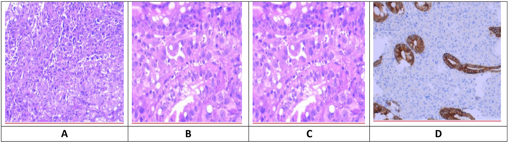
(A) Vascular slits. (B) Epithelioid cells. (C) Nuclear expression of erythroblast transformation specific related gene. (D) Negative for epithelial marker (AE1AE3).
Biopsy of a subcutaneous nodule and pathological study confirmed a cutaneous and muscular tumoral lesion characteristic for epithelioid angiosarcoma (Figure 2).
A computed tomography (CT) scan, performed as part of the extension assessment, showed the presence of a left apical sub-pleural nodule of 3 mm diameter, subcutaneous nodules on the right and left flanks and on the right posterior chest wall measuring 28 * 22 mm, 36 * 18 mm and 18 * 9 mm, respectively. They were associated to masses with heterogeneous contrast enhancement of the right gluteus maximus and left gluteus medius muscles.
The patient was transfused with a unit of red blood cells daily during a week due to a continued drop in hemoglobin. One week after his hospitalization, he underwent interventional endoscopy to control active bleeding by argon plasma coagulation. No surgery has been performed. A drop in hemoglobin persisted and the patient died within 10 days following uncontrolled hemorrhagic shock.
Angiosarcomas are a subtype of soft-tissue sarcoma with vascular or lymphatic differentiation. They are rare, representing 1–2% of soft tissue sarcomas.1 Angiosarcomas can develop at any age but are more common in patients during their sixth decade.1,6 Although they are usually found in skin and subcutaneous tissues, they can arise in any soft-tissue structure or viscera due the ubiquity of blood vessels and lymphatics.1 Thus, internal organs may be affected such as the liver, spleen, heart and uncommonly the GI tract.7 Within the GI tract, the small intestine is the most frequent location.2 Still, it is hard to distinguish between primary and metastatic angiosarcoma.4 A retrospective study of 66 cases of angiosarcoma of the small intestine showed that angiosarcoma of cutaneous or aortic origin often metastasized to the small intestine.8 In our case, it was difficult to ascertain whether small intestinal angiosarcoma was primary or metastatic spread from cutaneous and muscular masses as angiosarcoma was multifocal at outset.
The predisposing factors of small intestine angiosarcoma are still unclear. Studies have shown that a history of exposure to radiation, environmental toxins or foreign bodies as well as certain familial syndromes such as neurofibromatosis may be predisposing factors.1 Our patient did not have any of these predisposing factors.
Clinical presentation of small intestine angiosarcoma consists of non-specific symptoms. It includes mainly abdominal pain, anemia, GI bleeding, fatigue, weight loss and occasionally abdominal obstruction and perforation. Therefore, delayed diagnosis often occurs after substantial searches for a source of bleeding using endoscopy, enteroscopy or capsule endoscopy. On endoscopy, GI lesions may appear as a nodule, hemorrhagic mass, purpuric nodules, or submucosal mass.9 The extent of angiosarcoma can be assessed by CT, magnetic resonance imaging, or positron emission tomography scans.9 The definitive diagnosis is confirmed by pathological and immunohistochemical examinations.
Angiosarcoma is histologically classified into three subtypes: a) spindle-shaped endothelial cells; b) epithelioid with large, round or polygonal cells; c) pleomorphic cells.10 It is difficult to distinguish such tumors from poorly differentiated carcinoma, lymphoma or malignant melanoma. Hence, immunohistochemical study is essential to establish a definitive diagnosis of angiosarcoma, by showing the expression of vascular markers such as CD31, CD34, factor VIII, vimentin, endothelin-1, vascular endothelial growth factor receptor, ERG and ulex europaeus agglutinin-1.1 In our case, the tumor was only positive for vimentin and ERG. ERG appears as an important marker to consider in the diagnosis of undifferentiated tumors of the digestive tract.
The prognosis of angiosarcoma is very poor with a survival ranging from 10 days11 to 33 months.3 Our patient died 10 days after diagnosis despite interventional endoscopy by argon plasma coagulation to control active bleeding. Surgical resection is considered the treatment of choice for small intestine angiosarcoma.12 However, microscopically margin-negative resection is almost impossible in disseminated angiosarcomas. Adjuvant treatment with chemotherapy could prolong survival in metastatic angiosarcoma.13 In our case, the patient died before any chemotherapy could be considered.
This is the first African case of small intestine associated with synchronous subcutaneous angiosarcoma. Its non-specific clinical signs can lead to delayed diagnosis and worsen the prognosis. In such poorly differentiated and confusing tumor, ERG appears as an important marker to consider in the antibody panel of pathological diagnosis. Interventional endoscopy to control bleeding can be considered in localized forms, but no evidence has been reported in the literature discussed. In our case, it was of little help. The prognosis remains poor due to delayed diagnosis and insufficient therapeutic management although chemotherapy may help to prolong survival in metastatic and disseminated angiosarcoma. Further studies should be conducted to improve the prognosis.
| Views | Downloads | |
|---|---|---|
| F1000Research | - | - |
|
PubMed Central
Data from PMC are received and updated monthly.
|
- | - |
Provide sufficient details of any financial or non-financial competing interests to enable users to assess whether your comments might lead a reasonable person to question your impartiality. Consider the following examples, but note that this is not an exhaustive list:
Sign up for content alerts and receive a weekly or monthly email with all newly published articles
Already registered? Sign in
The email address should be the one you originally registered with F1000.
You registered with F1000 via Google, so we cannot reset your password.
To sign in, please click here.
If you still need help with your Google account password, please click here.
You registered with F1000 via Facebook, so we cannot reset your password.
To sign in, please click here.
If you still need help with your Facebook account password, please click here.
If your email address is registered with us, we will email you instructions to reset your password.
If you think you should have received this email but it has not arrived, please check your spam filters and/or contact for further assistance.
Comments on this article Comments (0)