Keywords
Chemotherapy, Imaging, Nasopharyngeal Carcinoma, Orbital Metastases, Radiotherapy.
This article is included in the Oncology gateway.
Chemotherapy, Imaging, Nasopharyngeal Carcinoma, Orbital Metastases, Radiotherapy.
Each year, around 129,000 new instances of nasopharyngeal carcinoma (NPC) are discovered around the world,1 a number of cases have been found in Asia.2 Heritability, environmental exposures, and Epstein-Barr virus (EBV) infection are all potential causes for NPC (EBV).3 The prevalence of Epstein-Barr virus (EBV)-associated nasopharyngeal carcinoma (NPC) in endemic Epstein-Barr virus (EBV)-infected people varies widely around the world.4 NPC affects one out of every 100,000 people worldwide.5 There was only one case of nasopharyngeal cancer incidence (0–14 years) and four cases (15–19 years) by diagnosis period in Bangladesh from 2001 to 2014.6 The common sites of distant metastasis of NPC are the bones, lungs, liver, and retroperitoneal lymph nodes.7 Nasopharyngeal carcinoma (NPC) is a tumour that develops from the nasopharyngeal epithelial cells. The nasal chamber, nasopharynx, pterygopalatine fossa, and apex of the orbital could all be affected by NPC.8 Ocular metastasis, on the other hand, is uncommon. Patients having NPC possess 1-year and 5-year overall survival of 92% and 70%, correspondingly, based on current screening and diagnostic procedures, and 20% to 25% of survivors will demonstrate metastatic diseases.9 In this case, we gathered information from patients with NPC eye metastasis (EM) and analyzed blood concentrations of EBV potential variables to determine their predictive usefulness for identifying NPC. Merely very few cases of paediatric nasopharyngeal carcinoma (NPC) disseminated to the ocular have been reported to our knowledge.
A 16-year-old child was admitted to the hospital with nasopharyngeal carcinoma metastasis to the eye. The patient had no known significant medical, family cancer or smoking history. In 2019, a 14-year-old child had a growing lump in his right cervical region below the right ear. He had gradually developed pain on that swelling. Then his family member obtained homoeopathy medicine treatment because of these concerns. One week later, he developed significant bleeding from the mouth and nose. As his complaints were not cured, his family went to ENT Head & Neck Specialist, Anwar khan modern hospital, Dhaka. Examination revealed mental wellbeing and several lymphadenopathies of which the largest was seen in the right cervical region. The subject’s entire system evaluations were also normal. Then Fiber Optic Laryngoscopy (FOL) was done on 8 July, 2019 was normal. Later CT of Nasopharynx done on 22 July 2019 revealing soft tissue inflammatory mass at the nasopharynx and suspected for carcinoma (Table 1). Excisional biopsy was recommended by an ENT Head and Neck Specialist resulting in Metastatic carcinoma, nasopharyngeal type (Table 1). In mid-August 2019, blood test for EBV ab (Epstein Barr Virus Antibody) revealed IgG- 112 (Positive) IgM- <5.0 (Negative) and a Biochemistry of the blood was performed (Table 2). From 20 August 2019, He had a minor hearing impairment, so an audiogram was performed, but the results were normal. On 31 August 2019, he went to National Institute of Cancer Research & Hospital (NICRH) and started chemotherapy (Table 3). Next cycle he went to Bangabandhu Sheikh Mujib Medical University (BSMMU) and Radiation treatment on NICRH (Table 3).
The patient was diagnosed with nasopharyngeal cancer in July 2019, and treatment began in August 2019 with cisplatin, Fluorouracil (5FU), Paclitaxel (PTX), Granulocyte colony-stimulating factor (G-CSF or GCSF), Gemcitabine and radiotherapy for seven sessions (Table 3). Previously regarded was used in two further cycles of adjuvant chemotherapy. After two courses of palliative chemotherapy, a partial response was observed. Complete blood count, peripheral blood smear and biochemical examinations were normal until December 2019.
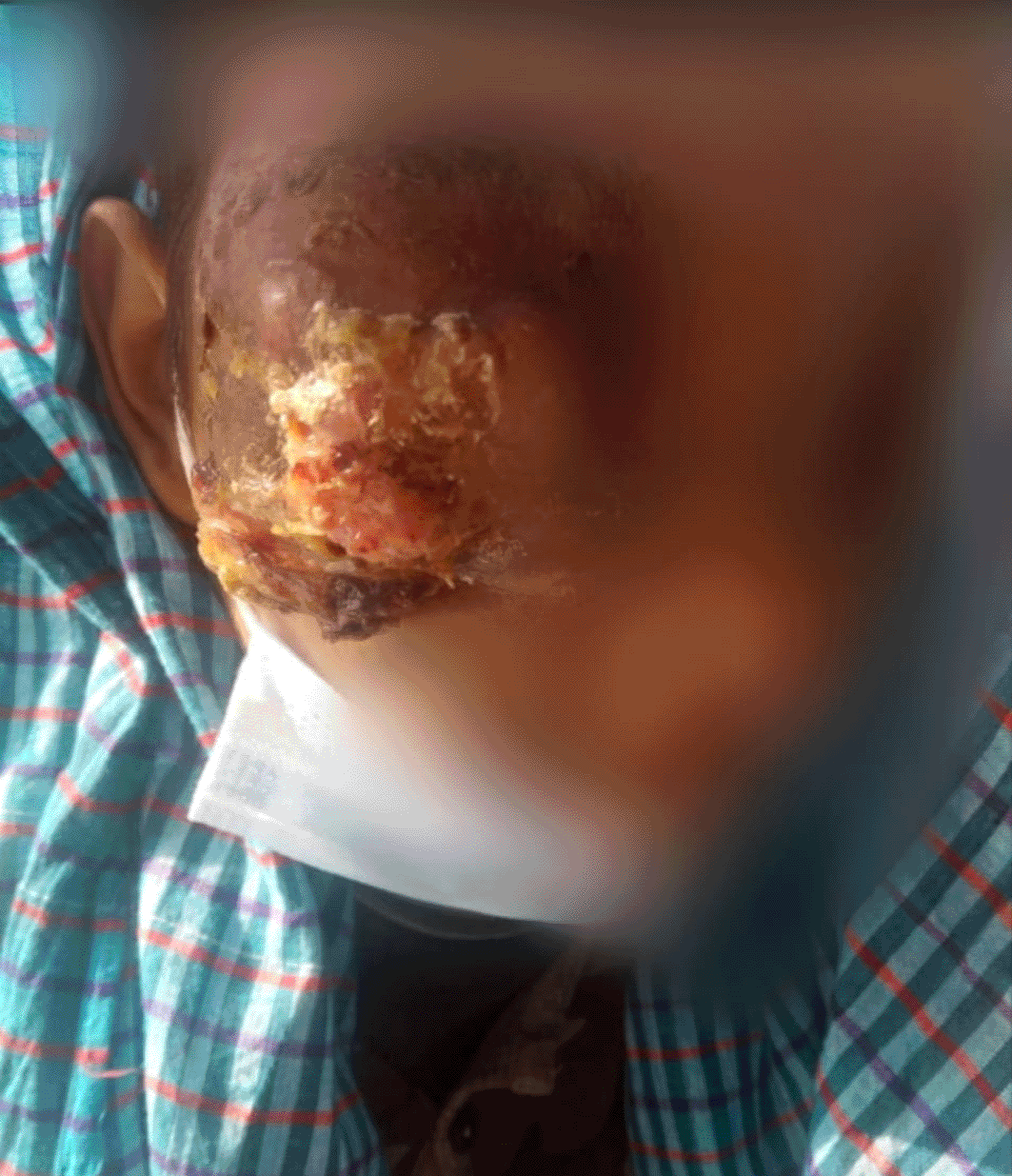
Unilateral uncomfortable palpable globs in the medial canthal area were noticed four months after the chemotherapy was finished in December 2019. He had history of eye bleeding, diplopia, and headaches. He arrived at the emergency room with a severe red eye and edema on his right side. He had noticed uncomfortable eyelid redness and swelling in his right eye, which was quickly obscuring his vision. He described involuntary tear production and complete visual impairment in his right eye, but no purulent discharges the patient had reported a decrease of appetite and weight loss in the prior three months. The right conjunctiva exhibited a vast, raised, and rough lesion that completely obscured the right eye on examination (Figure 1). The ulcerated tumor was painful.
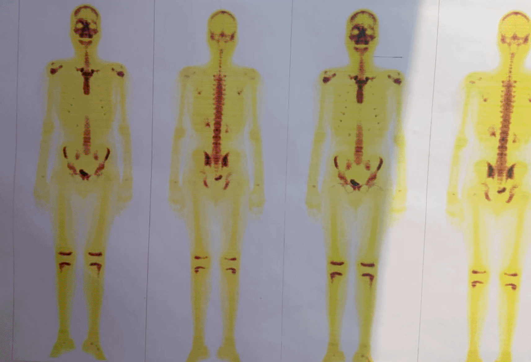
An MRI of Brain & PNS with the base of the skull with contrast, Bone Scintigraphy (Figure 2), Histopathology (Sinonasal mass frontal right), positron emission tomography (PET)/CT scan, Echocardiography, Chest X-ray were all performed, which revealed in the (Table 1).
However, his health deteriorated for four months from August to December 2021, with complete loss of right eye vision, low nutritional intake, lethargy, cachexia, and weight loss. In the right eye, ocular motion revealed unilaterally limited movement and no light perception. According to a CT scan and histology, the nasopharyngeal mass had increased, and there were metastases to the eye. A continuation of chemoradiotherapy cycle was started for him. He had a history of considerable fresh blood loss from the eye without any contact or trauma from August to December 2021, for which he received four blood transfusions because his Hb was 4.2 mg/dl at the time. Patient is now residing at home and receiving chemotherapy, blood transfusions, and other essential investigations in the hospital on an out-of-hospital basis.
NPC is rare cancer that affects just a small percentage of Bangladeshis. In terms of disease presentation, it is complex and one-of-a-kind. Neck swelling is the most prevalent presenting complaint (57.2%), followed by nasal symptoms (19.5%), headache (14.3%), and auditory symptoms (14.3%), aural symptoms (7.2%) and about 5.4% of people had ophthalmic manifestations.10 NPC has the highest tendency for rapid distant metastases among all head and neck cancers, with the probability increasing with progressive disease and recurring tumors, particularly in lymph node involvement of the primary tumor, which is consistent with our case.11 Orbital metastases in children is considerably more uncommon. The most prevalent primary tumors of these paediatric age group are sarcomas and neural embryonal tumors like neuroblastoma.12 According to the most advanced N classification, individuals with lymphatic metastases and a high probability of metastatic disease had the worst outcome.13 The most common cause of therapy failure in people with NPC is distant metastases. After completing radiation and chemotherapy, around 15% to 30% of patients with NPC develop metastases.14 The lungs, liver, bones, and retroperitoneal lymph nodes are frequent locations for distant metastases.15 The median duration of survival from the discovery of distant metastasis has been estimated to be 11.2 months for bone metastasis, 16.3 months for lung metastasis, and 3.2 months for liver metastasis.7 There were no metastases in the common areas in the case report mentioned here. On the other side, orbit intervention, may arise by direct extension through various paths, based on the surface area of the initial tumor. The pterygopalatine fossa and infratemporal fossa are the most common, followed by the inferior orbital fissure, in which most posterior section corresponds to the orbital apex. These ocular metastases have a wide range of clinical manifestations.11 Practically, orbital tumors and orbital metastases often present with an early onset of rapidly progressing symptoms. Most metastases entered intra/extraconal orbital soft tissues unilaterally, leading to globe dislocation with proptosis, diplopia, and reduced eye movement. Direct compression of the optic nerve, most usually at the ocular apex, resulted in Relative Afferent Pupillary Defect (RAPD) and vision loss, both of which significantly influenced on the victims’ clinical status.16 Nasal endoscopy is used to detect the presence of a mass, followed by cross-sectional imaging such as a CT scan, MRI, or PET for TNM staging. When detecting distant metastases, PET is more sensitive and accurate than MRI. Biopsy offers the necessary histopathological diagnostic for therapy planning. The results of radiological and histological tests are used to guide treatment decisions. The preferred treatment for NPC and associated regional nodal metastases is radiotherapy. Chemotherapy is used as an adjuvant for cancers that have progressed. Biopsy is the most common reason for surgery.17 Almost all metastases will emerge within three years, even with adequate treatment. The major component of NPC death is tumor metastasis.18 Radiotherapy is still the most common treatment for NPC. Because of the tumor’s deep anatomical position, a substantial cumulative amount of radiation is required to provide good therapeutic benefits. However, because the tumor is close to the eyes, radiation may affect the structure and function of the eyes.19 Eye discomfort caused by EM is indistinguishable from radiation-induced eye issues in people with NPC. As a result, efficient biological indicators with high specificity and sensitivity would be beneficial in determining whether radiation or NPC EM caused the eye symptoms.
Chemotherapy is one of the treatments for nasopharyngeal cancer that has progressed. The medications used in cancer treatment act by harming, inhibiting, or stopping the spread of cancer cells that are rapidly growing. On the other hand, chemotherapy medications damage both cancer cells and healthy tissue. They can harm the oral and gastrointestinal mucosa, hair follicles, reproductive system, and erythroid system in high enough doses. Malnutrition in cancer patients has many causes that are complex and multifactorial. Tumour cells secrete the chemicals serotonin and bombesin, which decrease appetite and cause anorexia. Nasopharyngeal cancer can also lead to inflammation of the oral mucosa and digestive mucous membranes, discomfort, decreased salivary gland secretion, psychologic distress, and tooth decay. Reduced oral intake can lead to a loss of strength, infection, and malnutrition. The tumour on NPC is weak, with enhanced neovascularization and a high risk of bleeding. Anaemia may develop as a result of chronic bleeding.20 In patients with nasopharyngeal cancer, anaemia is recognized as one of the predictive variables that affect numerous markers of life expectancy. A study in China discovered a substantial difference in overall survival between patients with Hb less than 11 g/dl and those with more than 11 g/dl, with 70% and 78%, respectively. Another Chinese study discovered a substantial difference in response to intensity-modulated radiation therapy among patients with and without anaemia. Anaemia patients had a complete response of 69.8% and a partial response of 30.2%, respectively, whereas non-anaemia patients had a complete response of 85.7% and partial response 14.3%. This was a statistically significant difference (p=0.02).20 In our patient’s case, he had a history of blood loss while touching the mass, even though it was a modest fresh quantity of blood loss, and he had a history of a decrease in food taste and losing weight while undergoing chemotherapy. He might develop anaemia due to external blood loss and chemotherapy, for which he had received blood transfusions multiple times.
The radiographic examination is necessary for the diagnosis of the disease as well as the evaluation of local bone and extra-bony expansion. The most common methods of examining suspected ocular lesions are computed tomography (CT) and magnetic resonance imaging (MRI). MRI provides the best resolution of orbital soft tissues, despite CT being frequently the primary choice in examining the orbit. A CT scan may be more effective in patients with suspected bone involvement. The anterior region of the orbit is where metastatic lesions are most frequent. These lesions are distinguished by their local aggressiveness, as evidenced by extraocular muscles and bone invasion.12
The uncommon occurrence of NPC metastasis in the eye poses a significant challenge to doctors, and at its early stage, it can be fatal.21 Due to a lack of specifics on radiation dosimetry, this study has a limitation. More detailed research into this area of interest is needed to help manage this severe consequence and offer the best and safest method for ocular metastatic surveillance. In most cases, histological proof is not required after a thorough radiological examination. In fact, in patients with advanced disease, a biopsy is rarely required to demonstrate metastasis. Biopsy may be required in some cases to make therapy decisions. This may interest in newly metastatic breast cancer, especially if ocular biopsy is proven to be more accessible, similar to executing novel targeted therapy by giving genetic studies of the tumour. In light of our patient’s unilaterality, we regarded his orbital involvement as metastatic after of the extension assessment.
A comprehensive history, a careful ophthalmological examination, and an overall physical assessment are required when orbital or ocular metastases is anticipated. It is vital to refer patients with no known history of cancer to an oncologist for a simultaneous broad extension screening to investigation of the main tumor. The goal of orbital metastasis treatment is to improve the patient’s quality of life while preserving their functional prognosis. Radiotherapy and chemotherapy may be used. Since NPC is recognized for its radio sensitivity, the most common treatment for orbital metastases from NPC is radiotherapy. However, chemotherapy is still used in certain individuals. Limitation, the presence of cranial nerve palsy is not evaluated in our study.
Nasopharyngeal cancer can cause a wide range of symptoms which are not always associated to the nose. One of the rare early presentations is orbital metastases with vision problems. However, it is important to keep in mind that tumor recurrence and eye metastasis are also conceivable. Orbit is a unique location. Nasopharyngeal carcinoma seldom invades the orbit. Ophthalmic symptoms from orbital invasion, on the other hand, can be the first sign of NPC. As a result, it is critical to distinguish this disease entity, particularly in younger individuals, where malignancy is often overlooked as a possible differential diagnosis. The prognosis is generally dangerous and varies depending on the source malignancy. Furthermore, orbital metastases frequently arise in advanced stages of disease and in the scenario of many metastases. In terms of improving the function and quality of life, palliative radiation and chemotherapy are used. Despite recent therapy advancements, these individuals’ expectancy remains limited.
The patient’s written informed consent for publishing of this case report, as well as images, was acquired.
Mohammad Ashraful Amin: Conceptualization, Writing- Original draft preparation: Mohammad Ashraful Amin, Sabrina Nahin: Visualization and Supervision.: Mohammad Delwer Hossain Hawlader, Mohammad Ashraful Amin, Sabrina Nahin, Atia Sharmin Bonna, Mohammad Delwer Hossain Hawlader: Writing- Reviewing and Editing.
Data can be shared based on the reader’s reasonable request and priority base and some restrictions will apply. All data related to the case are available; Amin, Mohammad Ashraful (2022), “Nasopharyngeal carcinoma has metastasis to the orbit, consequences in anaemia: A paediatric unusual case of Bangladesh.”, Mendeley Data, V1, doi: https://doi.org/10.17632/kw4fdmdgy8.1.
| Views | Downloads | |
|---|---|---|
| F1000Research | - | - |
|
PubMed Central
Data from PMC are received and updated monthly.
|
- | - |
Provide sufficient details of any financial or non-financial competing interests to enable users to assess whether your comments might lead a reasonable person to question your impartiality. Consider the following examples, but note that this is not an exhaustive list:
Sign up for content alerts and receive a weekly or monthly email with all newly published articles
Already registered? Sign in
The email address should be the one you originally registered with F1000.
You registered with F1000 via Google, so we cannot reset your password.
To sign in, please click here.
If you still need help with your Google account password, please click here.
You registered with F1000 via Facebook, so we cannot reset your password.
To sign in, please click here.
If you still need help with your Facebook account password, please click here.
If your email address is registered with us, we will email you instructions to reset your password.
If you think you should have received this email but it has not arrived, please check your spam filters and/or contact for further assistance.
Comments on this article Comments (0)