Keywords
Ascites, fibroma, oophorectomy, pleural effusion
This article is included in the Oncology gateway.
Ascites, fibroma, oophorectomy, pleural effusion
Meigs syndrome is defined as the triad of benign ovarian tumor, especially ovarian fibroma with ascites and pleural effusion that resolves after tumor resection.1 It occurs as a result of increased capillary permeability thought to be a result of vascular endothelial growth factor (VEGF) production. Pleural effusions are usually right-sided because the transdiaphragmatic lymphatic channels are larger in diameter on the right.2 The pleural effusion as well as accompanying ascites are typically transudative. Meigs syndrome is also seen in cases like large, cystic leiomyomas or other benign ovarian tumors, thecoma, cystadenoma or granulosa cell tumor.3 Ovarian fibromas constitute 2 to 5% of all ovarian tumors and Meigs syndrome occurs in just 1% of these tumors indicating rarity of this clinical condition.4 Though diagnosis is possible preoperatively with ultrasound and magnetic resonance imaging (MRI), a high index of suspicion may be important as it radiologically and clinically mimics ovarian malignancy.5 This case is described for its rarity in presentation and clinical confusion in diagnosis.
This was a case of 61-year-old, post-menopausal, Asian housewife, para four living three woman, presenting with abdominal distension for two to three months along with decreased appetite and constipation. She had a history of abdominal mass suspected of being ovarian malignancy for two years prior, for which she had not undergone any treatment due to personal difficulties. She experienced no vomiting and abdominal pain. She was former smoker and a known case of diabetes mellitus.
On examination, general condition was fair with no pallor, edema, lymphadenopathy or any signs of dehydration. Vitals were stable. On respiratory examination, decreased air entry was observed on the right side. Cardiovascular examination was normal. On abdomen examination, an irregular firm mass could be felt which was mobile and extending from the upper border of symphysis pubis to just above the umbilicus. The mass was mobile and non tender on palpation. Speculum examination showed normal cervix with rectocele. Vaginal examination showed an anteverted uterus with fullness felt in all the fornices separate from uterus.
Chest X-ray showed blunting of right costophrenic angle suggesting pleural effusion (Figure 1). Ultrasound of abdomen and pelvis revealed gross ascitis, huge complex right ovarian cyst (Figures 2, 3), confirmed by CT abdomen and pelvis as malignant ovarian tumor (Figures 4, 5). Ascitic tapping showed transudative nature of fluid, cancer antigen 125 (CA-125) was 84.6 U/ml and carcinoembryonic antigen (CEA) and lactate dehydrogenase (LDH) were normal.
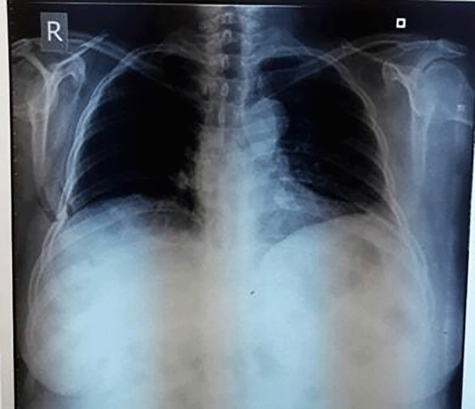
Chest X-ray showing right sided pleural effusion.
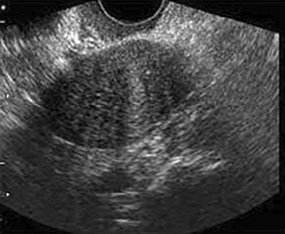
Ultrasound of the abdomen and pelvis showing ovarian cyst and ascites.
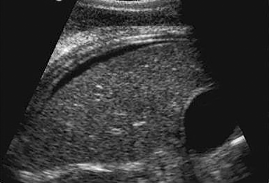
Ultrasound of the abdomen and pelvis showing ovarian cyst and ascites.
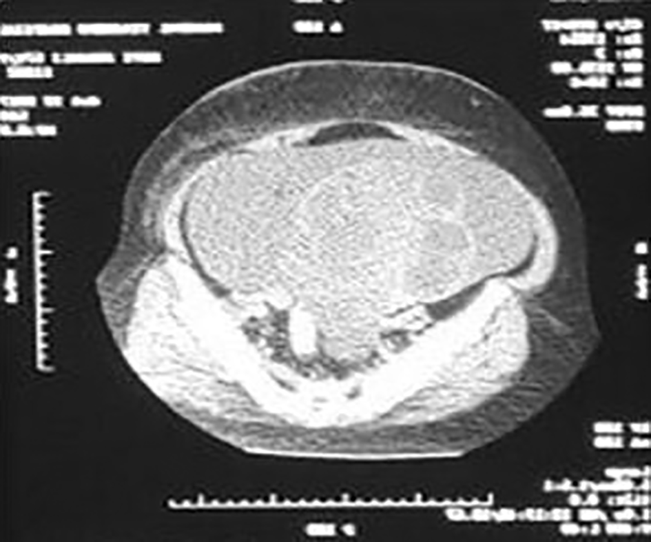
CT scan of the abdomen and pelvis showing the complex ovarian cysts.
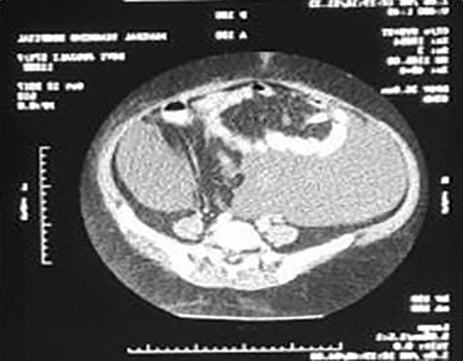
CT scan of the abdomen and pelvis showing the complex ovarian cyst.
She underwent staging laparotomy with total abdominal hysterectomy, bilateral salphingo-oophorectomy and omental resection for biopsy. There were six litres of straw-colored ascitic fluid with right ovarian hard mass with irregular surface of 15 × 10 cm on laparotomy (Figures 6, 7). Uterus, left ovary and cervix were normal. Omentum, bowel, liver, and peritoneal surface were normal. She was diagnosed with stage I C ovarian tumor. Her post-operative period after laparotomy with total abdominal hysterectomy and bilateral salphingo-oopherectomy was uneventful Biopsy report showed right ovarian fibroma. She recovered well and was living a comfortable life on follow up.
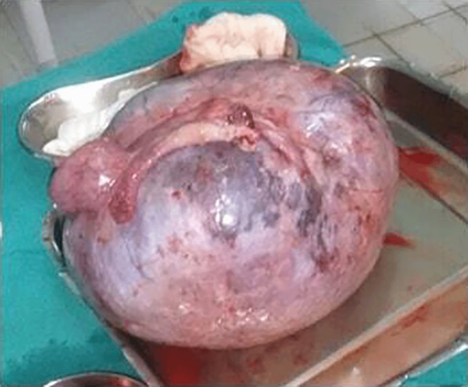
Gross surgical specimen showing right ovarian hard mass with irregular surface of 15 × 10 cm2.
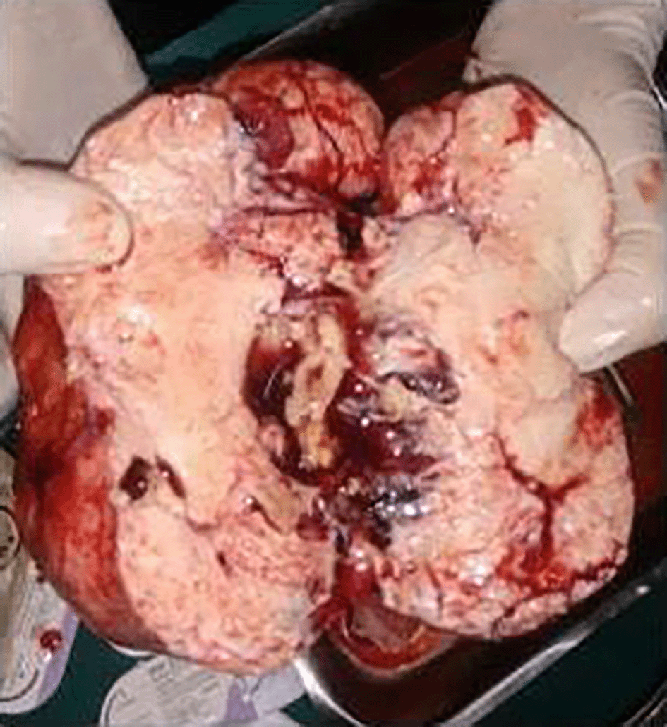
Gross surgical specimen showing right ovarian hard mass with irregular surface of 15 × 10 cm2.
Ovarian fibromas are the most common hormonally inactive sex cord stromal tumor variants that usually occur in perimenopausal and menopausal women.6 They represent only 4% of all ovarian neoplasms and are the least common major subtype of ovarian cancer. Meigs’s Syndrome occurs in just 1% of these cases. Although Meigs’s Syndrome is extremely rare, it is known to produce pleural effusion and ascites. Because several conditions are linked to the development of these common indications, the correct diagnosis and treatment are frequently missed.7 The cause of Meigs’s condition is still unknown. Ascites are a common symptom of ovarian tumors, and numerous causes have been proposed, including tumor torsion and restriction of venous drainage. According to laboratory investigations, the fluid collected in most but not all cases is transudate. The chest and abdomen fluids are identical in all patients.8 Peritoneal cytology, tumour markers, and other signs of malignant pathology may be confusing. Hence, laparotomy is essential for the correct identification of ovarian tumours.9 Due to the rarity of this condition, this is a diagnosis of exclusion but should be considered as soon as ovarian malignancy is excluded. The presentation in our case was similar where a long-standing ovarian mass presented with ascitis and pleural effusion.
Ovarian fibromas are uncommon sex cord stromal tumor commonly seen in post menopausal women. Ovarian fibroma could be a possibility in cases of ovarian tumors with ascitis and pleural effusion, especially when longstanding.
| Views | Downloads | |
|---|---|---|
| F1000Research | - | - |
|
PubMed Central
Data from PMC are received and updated monthly.
|
- | - |
Provide sufficient details of any financial or non-financial competing interests to enable users to assess whether your comments might lead a reasonable person to question your impartiality. Consider the following examples, but note that this is not an exhaustive list:
Sign up for content alerts and receive a weekly or monthly email with all newly published articles
Already registered? Sign in
The email address should be the one you originally registered with F1000.
You registered with F1000 via Google, so we cannot reset your password.
To sign in, please click here.
If you still need help with your Google account password, please click here.
You registered with F1000 via Facebook, so we cannot reset your password.
To sign in, please click here.
If you still need help with your Facebook account password, please click here.
If your email address is registered with us, we will email you instructions to reset your password.
If you think you should have received this email but it has not arrived, please check your spam filters and/or contact for further assistance.
Comments on this article Comments (0)