Keywords
Superficial retinal capillary plexus, Deep retinal capillary plexus, Ischemic cascade, Radiation retinopathy, OCT angiography
This article is included in the Eye Health gateway.
Superficial retinal capillary plexus, Deep retinal capillary plexus, Ischemic cascade, Radiation retinopathy, OCT angiography
Optic coherence tomography angiography (OCT-A) is a non-invasive imaging technique that allows the retina to be 'scanned' and its vessels to be studied, providing images of retinal anatomy in 3 dimensions. Unlike conventional imaging techniques such as fluorescein angiography or indocyanine green angiography, OCT-A does not require intravenous injections of contrast agents and avoids serious allergic reactions that can occur. Compared with OCT, it shows the various chorioretinal vascular structures, in particular the areas of the superficial vascular plexus and the deep capillary plexus.
We report a case of radiation retinopathy, a complication of the treatment of nasopharyngeal cancer by local radiotherapy, a rare and serious pathology that irreversibly compromises visual prognosis. The aim of this case report is to demonstrate the contribution of A-OCT in the diagnosis of radiation retinopathy and to confirm the ischemic origin of retinal vascular damage.
In this new version, we have just changed the structure of the abstract to make readers more aware of the value of A-OCT in radiation retinopathy, and we have minimized the use of abbreviations to make the text easier to understand. The figures and references have not changed. The reviewers suggested these changes.
See the authors' detailed response to the review by Helmi Ben Saad
See the authors' detailed response to the review by David Solá-Del Valle
Locoregional radiotherapy is the gold standard treatment against nasopharyngeal carcinoma (NPC).1 The orbital proximity may lead to severe damage.2 However, late-onset, sight-threatening ocular complications may occur, including cataract, optic neuropathy, radiation retinopathy (RR), and ocular surface disease.2 The early diagnosis of these lesions allowed for better prognosis.1 The optical coherence tomography angiography (OCT-A) allowed us to investigate neovascular alteration for patients suffering from RR even before the onset of loss of vision.1
We reported a case of RR in a 31-year-old female with NPC, treated by locoregional radiotherapy. The patient presented all post-radiotherapy ocular complications with a late diagnosis of RR and a poor prognosis.
The purpose of this case report was to analyze the findings and the usefulness of OCT-A.
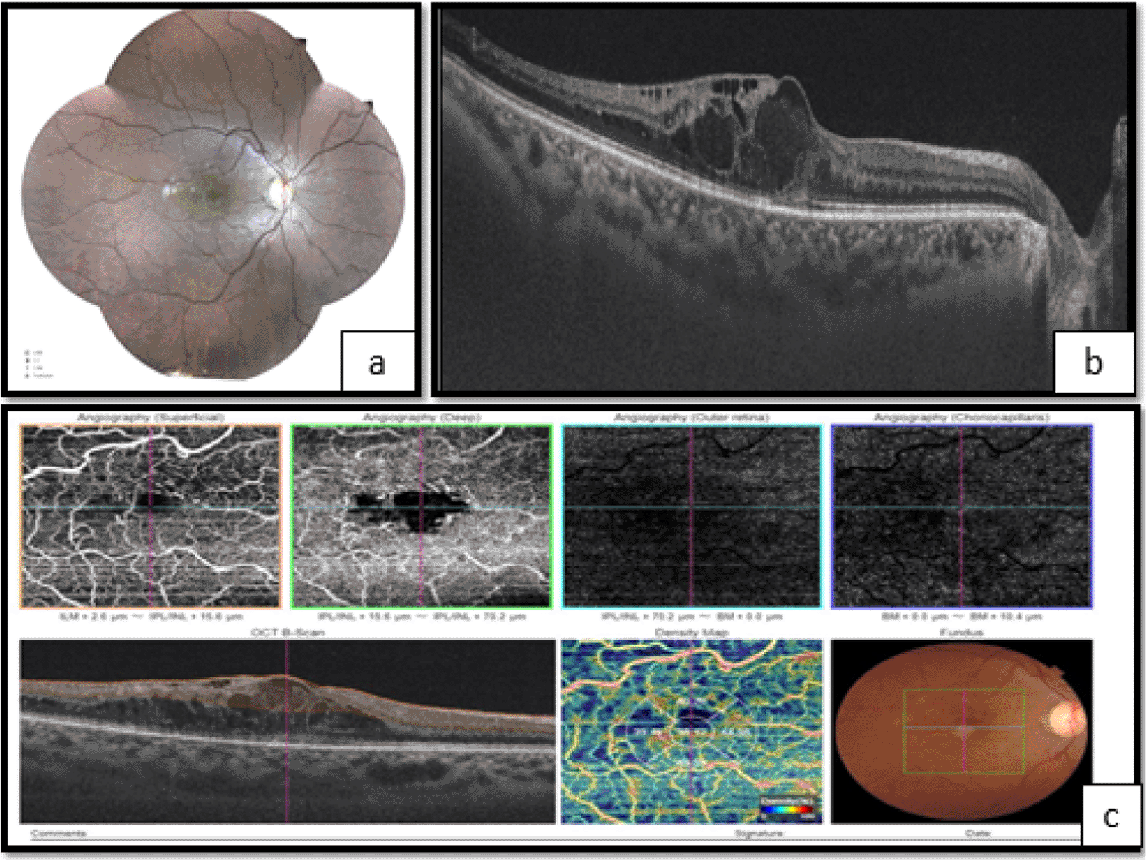
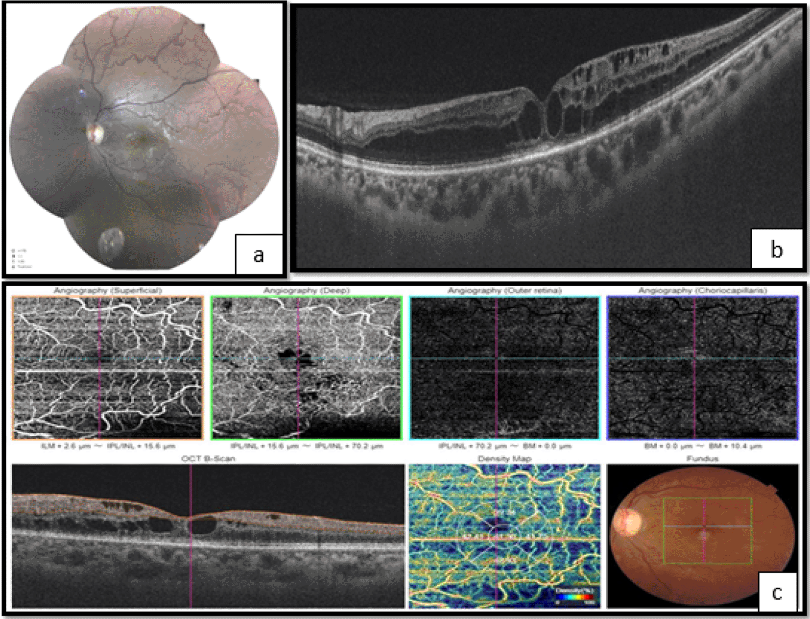
A 31-year-old Tunisian, unemployed female diagnosed with NPC in 2009 was treated by locoregional radiotherapy. The overall administered dose was about 75 Gray. The patient presented with adrenal insufficiency, hypothyroidism, and osteonecrosis as side effects of the treatment. She complained of progressive painless loss of vision in both eyes. On examination, her best-corrected visual acuity was 20/40 in both eyes. The ocular motility was full, and no afferent pupillary defect was noted. Symmetrical subcapsular cataract was noted. The rest of the anterior segment examination was unremarkable. No vitreous cells were noted. Fundoscopy showed microvascular changes mainly marked by vascular tortuosity and microaneurysms, optic disc pallor, and decreased foveal reflex. Fluorescein angiography was not performed because the patient was allergic to fluorescein. The optic coherence tomography (OCT) showed bilateral macular edema with a central macular thickness of 532 μm in the right eye and 406 μm in the left eye. The OCT-A disclosed enlargement of the central avascular zone, and hypoperfusion of both superficial and deep retinal capillary networks (Figures 1 and 2). The vessel density was reduced to 38.12 % in the inferior macular area of the right eye, and to 39.34 % in the superior macular area of the left eye. A systemic workup was performed to rule out other causes of ischemic retinopathy such as diabetes mellitus, blood dyscrasias, and carotid insufficiency. Based on medical history, ocular findings, and negative systemic workup the diagnosis of RR was finally established. After informed consent, and a negative pregnancy test, the patient underwent three monthly intravitreal bevacizumab injections at 1.25 mg. The improvement of visual acuity was poor.

a. Fundoscopy showing vascular tortuosity and dilation of peripheral retinal vessels, the disappearance of foveolar reflection, and mild pallor of the optic disc.
b. Optical coherence tomography (OCT) showed macular edema.
c. OCT angiography (OCT-A) showing enlargement of the centralavascular zone, and hypoperfusion of both superficial and deep retinal capillary network.

a. Fundoscopy showing vascular tortuosity and dilation of peripheral retinal vessels, the disappearance of foveolar reflection, and mild pallor of the optic disc.
b. The optical coherence tomography (OCT) shows macular edema.
c. OCT angiography (OCT-A) showing enlargement of the central avascular zone, and hypoperfusion of both superficial and deep retinal capillary network.
RR was first described in 1933 by Stallard,3 as a predictable complication of radiation exposure. It most commonly occurs between six months and three years after irradiation.4 In this case, the diagnosis was later, twelve years after irradiation. A higher total radiation dose is the highest risk factor, as the incidence of RR increases at doses greater than 45 Gray. Our patient received 75 Gray. Histopathological studies have illustrated a vasculopathy with the destruction of the endothelial cells followed by vascular occlusion and capillary dropout.3,4 The microvascular alterations are associated with a reduction of retinal oxygenation, blood flow, and ischemia.2–4 Contrast sensitivity decrease and visual field impairment were notified in patients treated with radiotherapy.1 Our patient had gradually decreased bilateral visual acuity, as well as cataract and optic neuropathy. The clinical appearance mimics many lesions of diabetic retinopathy such as microaneurysms, macular edema, cotton-wool spots, retinal neovascularization, vitreous hemorrhage, and tractional retinal detachment.4 The main tests usually performed on patients are fluorescein fundus angiography and OCT. The first exam hallmarks are capillary dilatation and microaneurysms, frequently in combination with ischemia or macular edema.5 On OCT images, we found a disappearance of the macular depression with macular edema, and a significant thinning of the inner plexiform, inner nuclear, and outer plexiform layers.5 The fluorescein angiography is an invasive diagnostic technique. Intravenous dye injection may cause severe anaphylaxis, particularly in immunocompromised patients. It was not performed on our patient. Besides, OCT cannot capture vessel network status. Recently, OCT-A has been reported to be a safe and non-invasive examination that combines traditional OCT and Doppler shift. It can provide high-resolution images of each layer of the retina and quantify the retinal microvascular networks without the use of exogenous dyes. It is based on calculating the difference between signals, of moving structures, from two sequential OCT scans at the same position.6 OCT-A has been introduced for the detection of subtle microvascular changes in RR.7,8 Vascular abnormalities are manifested by an enlargement of the central avascular zone and a reduction of vessel density in the deep vascular plexus of the foveal area. Whereas it is less reduced in superficial layers. The susceptibility of the deep layer can be explained by the direct connection of the superficial capillary plexus to the retinal arterioles with greater perfusion and oxygen supply.3 This change in structure can be explained by direct compression of the retinal vascular network, deep in the first place, by intra-retinal fluid cysts. Zijing et al.1 reported that OCT-A detects early vascular alterations of the retina in patients with normal-ranged visual acuity. OCT-A provided a quantitative measurement of retinal capillary changes which may predict future development of radiation-induced retinal toxicity.5 Zijing et al.1 suggested the implementation of OCT-A for the early detection and consistent monitoring of RR. In this sense, a grading system was proposed based on clinical findings in OCT-A, increased central macular thickness, evident cysts, and ophthalmoscopy findings.5 The disadvantage is the presence of several artifacts, especially after treatment. The treatment is inspired by diabetic retinopathy because of clinical and pathophysiological similarities.9
Initially, treatments were based on the use of retinal laser.9 Sector photocoagulation improves clinical signs, but the visual outcome is poor.9 Intravitreal injection of anti-vascular endothelial growth factor (VEG) or corticosteroids has been shown to improve visual acuity, reduce cystoid macular edema, and the risk of the development of RR.3,9 The visual acuity of our patient didn’t change, probably because she presented with several complications of local radiotherapy, such as cataract and optic neuropathy, and ischemia affecting deep layers. Continuous treatment is necessary to maintain acuity improvement8; this requires good patient adherence.9 The optimal regimen for anti-VEGF therapy is not yet identified.8 There have been recent preventive efforts to avoid signs that radiation damage has already occurred, particularly since there is still no curative treatment.10
The OCT-A findings of RR are an enlargement of the foveolar avascular zone and a rarefaction of the vascular network at the level of the deep and vascular networks, even in eyes without clinical evidence of RR.
The patient has consented to the submission of the case report for submission to the journal.
| Views | Downloads | |
|---|---|---|
| F1000Research | - | - |
|
PubMed Central
Data from PMC are received and updated monthly.
|
- | - |
Competing Interests: No competing interests were disclosed.
Reviewer Expertise: Ophthalmology and glaucoma
Competing Interests: No competing interests were disclosed.
Reviewer Expertise: Medical writing skills; physiology
References
1. Gagnier JJ, Kienle G, Altman DG, Moher D, et al.: The CARE Guidelines: Consensus-based Clinical Case Reporting Guideline Development.Glob Adv Health Med. 2013; 2 (5): 38-43 PubMed Abstract | Publisher Full TextCompeting Interests: No competing interests were disclosed.
Reviewer Expertise: Medical writing skills; physiology
Is the background of the case’s history and progression described in sufficient detail?
Yes
Are enough details provided of any physical examination and diagnostic tests, treatment given and outcomes?
Yes
Is sufficient discussion included of the importance of the findings and their relevance to future understanding of disease processes, diagnosis or treatment?
Yes
Is the case presented with sufficient detail to be useful for other practitioners?
Yes
Competing Interests: No competing interests were disclosed.
Reviewer Expertise: Glaucoma/Ophthalmology
Is the background of the case’s history and progression described in sufficient detail?
Yes
Are enough details provided of any physical examination and diagnostic tests, treatment given and outcomes?
Yes
Is sufficient discussion included of the importance of the findings and their relevance to future understanding of disease processes, diagnosis or treatment?
Yes
Is the case presented with sufficient detail to be useful for other practitioners?
Yes
References
1. Gagnier JJ, Kienle G, Altman DG, Moher D, et al.: The CARE Guidelines: Consensus-based Clinical Case Reporting Guideline Development.Glob Adv Health Med. 2013; 2 (5): 38-43 PubMed Abstract | Publisher Full TextCompeting Interests: No competing interests were disclosed.
Reviewer Expertise: Medical writing skills; physiology
Alongside their report, reviewers assign a status to the article:
| Invited Reviewers | ||
|---|---|---|
| 1 | 2 | |
|
Version 3 (revision) 25 Sep 23 |
read | read |
|
Version 2 (revision) 19 Jul 23 |
read | |
|
Version 1 22 Aug 22 |
read | read |
Provide sufficient details of any financial or non-financial competing interests to enable users to assess whether your comments might lead a reasonable person to question your impartiality. Consider the following examples, but note that this is not an exhaustive list:
Sign up for content alerts and receive a weekly or monthly email with all newly published articles
Already registered? Sign in
The email address should be the one you originally registered with F1000.
You registered with F1000 via Google, so we cannot reset your password.
To sign in, please click here.
If you still need help with your Google account password, please click here.
You registered with F1000 via Facebook, so we cannot reset your password.
To sign in, please click here.
If you still need help with your Facebook account password, please click here.
If your email address is registered with us, we will email you instructions to reset your password.
If you think you should have received this email but it has not arrived, please check your spam filters and/or contact for further assistance.
Comments on this article Comments (0)