Keywords
Basaloid squamous cell carcinoma, dimorphic pattern, basaloid cells, comedo necrosis
This article is included in the Oncology gateway.
This article is included in the Datta Meghe Institute of Higher Education and Research collection.
The upper aerodigestive tract is where basaloid squamous cell carcinoma (BSCC), a rare variation of conventional SCC, is most frequently found. The hypopharynx, tonsil, supraglottic larynx, tongue (base), and head-neck regions are particularly susceptible to BSCC. Clinically, the presentation of BSCC is similar to that of conventional SCC, but it has a poorer prognosis than conventional SCC. BSCC is distinguished histopathologically by a dimorphic pattern, a distinctive basal cell component paired with a squamous component, and a squamous component. However, its similar features to conventional SCC make it difficult to diagnose. Therefore, histopathology and immunohistochemistry play a crucial role in diagnosing such tumors. Here we present the case of a 70-year-old male diagnosed with BSCC involving the tongue.
Basaloid squamous cell carcinoma, dimorphic pattern, basaloid cells, comedo necrosis
In the present version we have reframed the discussion part as suggested by the reviewer and also we have added the conclusion in the manuscript.
The title has also been amended.
See the authors' detailed response to the review by Muhammad Kashif
The aggressive squamous cell carcinoma of oral cavity (OSCC) form known as basaloid squamous cell carcinoma (BSCC) is rare. Wain et al.1 were the first to report the existence of BSCC, which was later proved to be a high-grade variety of SCC that is most common in the head and neck.2 Males over the age of 50 are more likely to develop BSCC. It is regarded as a high-grade tumor with a higher risk of nodal metastasis (64%) and distant metastasis (44%), compared to conventional SCC.3 The larynx and hypopharynx, which are parts of the upper-aerodigestive-tract, are often impacted. The tongue (base) is most frequently affected (61%), and BSCC is more common in the oral cavity to the rest of the body. The palate, the retromolar trigone, the gingival-mucosa, and the floor-of-the-mouth (30%) are other affected locations.4,5 In terms of histopathology, the presence of solid epithelial cells with malignant characteristics and a basaloid appearance distinguishes BSCC the most.6 The invading tumor exhibits a variation of development forms including cords and nests, trabeculae, cysts and glands.7 Based on histopathologic and immunohistochemical findings, BSCC is distinct from conventional SCC. In addition, BSCC exhibits a different clinical behavior and prognosis than conventional SCC.8 Compared to conventional SCC, the prognosis for BSCC is worse. Despite having different histological characteristics, BSCC is frequently misdiagnosed as neuroendocrine tumors, small cell carcinoma, adenosquamous carcinoma, and adenoid cystic carcinoma.9,10 Here, we describe a case of BSCC in a 70-year-old man that affected the right lateral border of the tongue.
A male patient aged 70 was referred to our Institute with a painful ulcer over the right lateral border of the tongue for two years. He also had pain that was dull type, continuous in nature, and non-radiating. He also complained of a burning sensation when eating spicy food. He was experiencing difficulty in mastication and deglutition, and the tongue movements were restricted. He experienced weight loss, loss of appetite, decreased salivation, and hoarseness of voice. The patient had a habit of kharra (smokeless tobacco) chewing two-three times a day for two years. He was also a chronic bidi smoker, from 20-25 years (two-three times per day). He claimed to have quit the habit ten years before.
Extra-oral findings revealed bilateral submandibular LN (lymph nodes), which were tender and palpable, measuring approximately 3 × 4 cm along its maximum dimension [Figure 1].
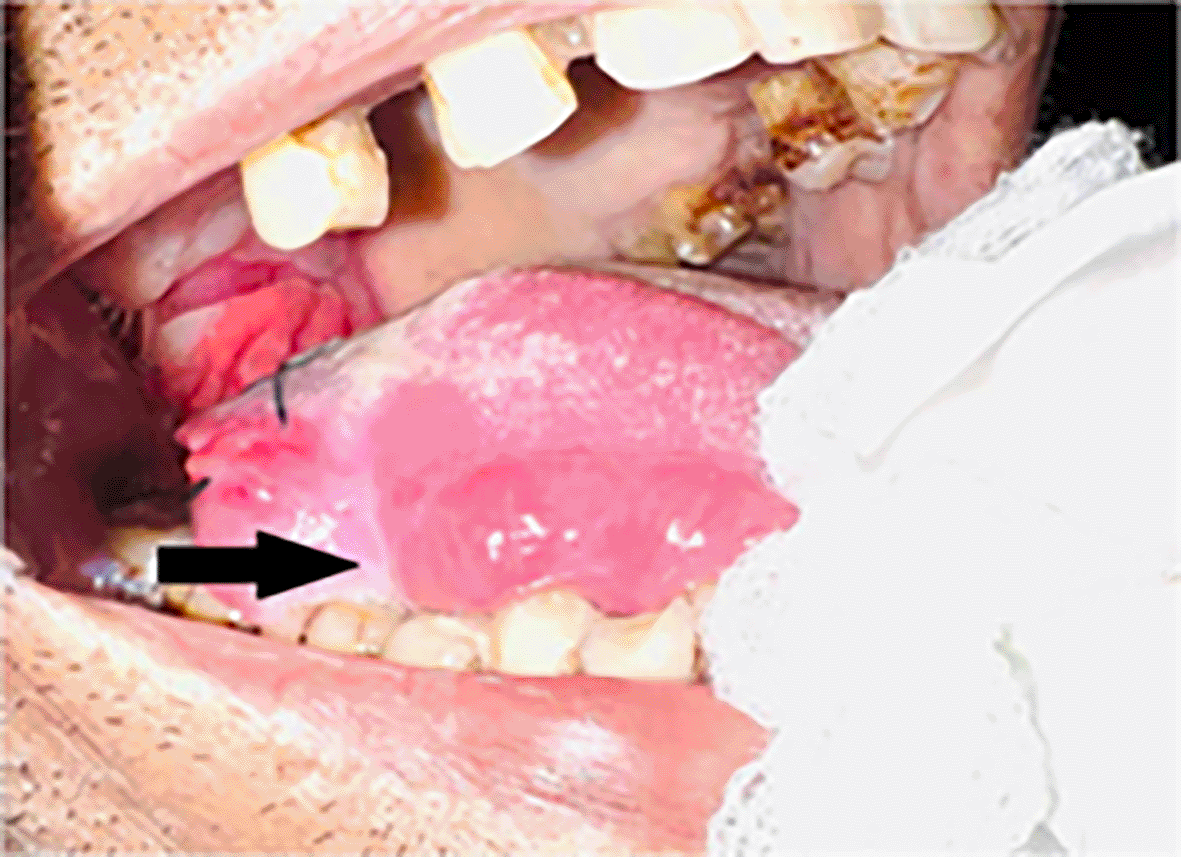
Intraoral examination revealed an ulceroproliferative lesion of approximately 2 × 3 cm on the lateral border of the tongue (right side) [Figure 2], which was extending supero-inferiorly from the dorsal surface to the ventral surface of the tongue, anteroposteriorly from 46 region to the retromolar trigone (RMT) and involving soft palate. The lesion showed characteristic malignant features. The margins were everted and induration was present on palpation. A provisional diagnosis was made of malignancy of the tongue.

Further, a tongue MRI with contrast was performed, which showed a heterogeneously enhancing mass lesion on the tongue (right side) with areas of necrosis within, abutting the lingual septum medially and extending into the infratemporal fossa laterally measuring approximately 7.6 × 4.5 × 4.3 cm. There was evidence of multiple subcentimetric to centimetric heterogeneously enhancing lymph nodes in the submental, bilateral submandibular, and jugulodigastric region, the largest being 4.3 × 3.2 cm in size in the right submandibular region with necrotic areas within. Impression of the tongue MRI revealed the abovementioned characteristics, suggesting tongue carcinoma with lymphadenopathy [Figure 3].
An incisional biopsy was done at our institute. The details of the biopsy report are mentioned below.
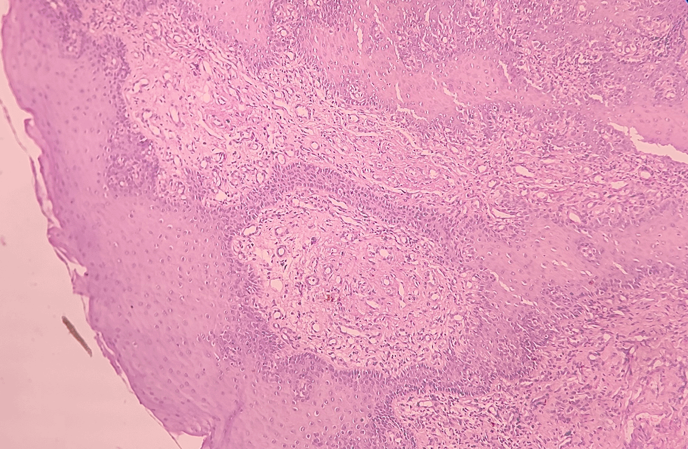
Haematoxylin and eosin-stained tissue section revealed an overlying hyperplastic parakeratinizing stratified squamous epithelium and underlying fibro cellular connective tissue (CT) stroma [Figure 4].

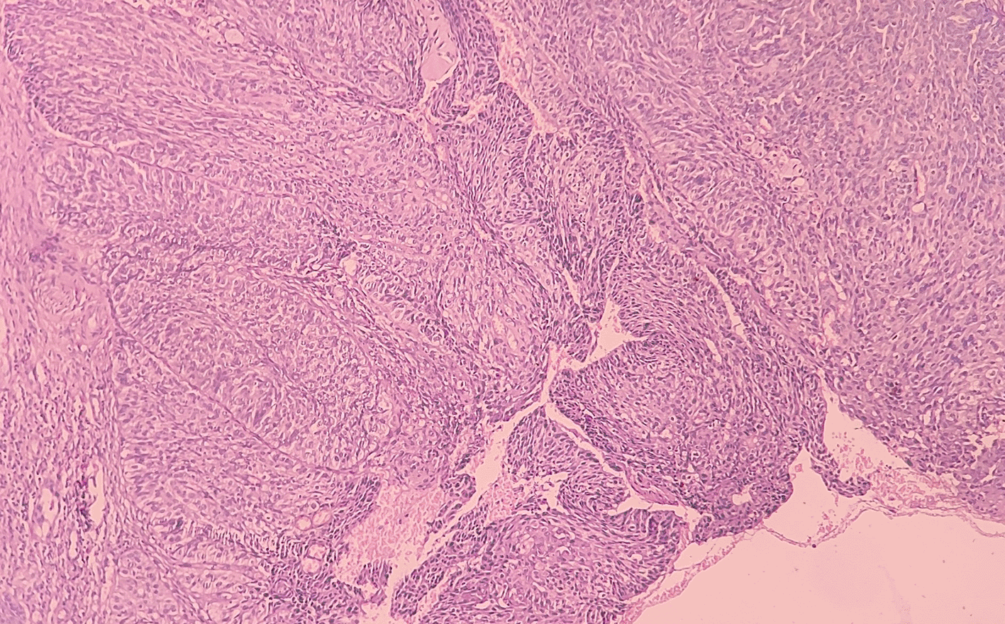
At low-power view [Figure 5], it was evident that the epithelial cells invaded the CT in the form of islands. Some of these islands consisted of basaloid and squamous cells. These islands showed cystic spaces with a central area of comedo-necrosis. There was presence of malignant epithelial cells arranged in an organoid pattern displaying lobules of neoplastic epithelial cells. Fibrous CT septa separated these cells. The tumor cells were compactly arranged and showed cellular pleomorphism. The CT were comprised of collagen fibers and a few fibroblasts. Numerous endothelial cells-lined blood vessels with intravasated and extravasated red blood cells (RBCs) were seen. Moderate to chronic inflammatory cell infiltrates were also seen.

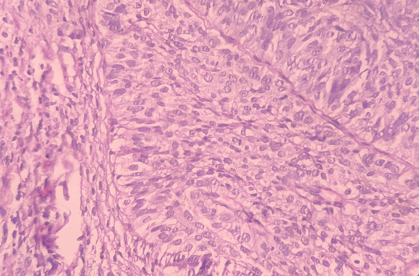
Under the high-power view [Figures 6, 7], all findings of the low power view were confirmed. The periphery of neoplastic islands showed cuboidal to low-columnar basaloid cells with palisading nuclei. The nuclei were ovoid-shaped, showing nuclear hyperchromatism and scant cytoplasm. The neoplastic cells showed characteristics like cellular pleomorphism, nuclear pleomorphism and hyperchromatism; there was increase in the nuclear-cytoplasmic ratio, and abnormal mitosis was also evident. There was presence of chronic inflammatory cell infiltrate chiefly comprising of lymphocytes.

BSCC, a distinct variation of conventional SCC, more commonly affects males in the age group of 60 to 70.11 Wain et al.1 originally identified BSCC as an uncommon, histologically different, and extremely aggressive subtype of SCC in 1986. These four main histologic characteristics were used to make the diagnosis of BSCC: (a) cells present in solid groups in a lobular arrangement, close to the surface mucosa; (b) small, densely packed cells with scant cytoplasm; (c) dark/hyperchromatic nuclei without nucleoli; and (d) small, cystic spaces consisting of mucin-like material.12 BSCC was initially noted in the oral cavity by Cadier and others. The literature has described 45 instances of BSCC affecting the oral cavity, with the base of the tongue (61%) and floor of the mouth (30%) showing a strong preference.6,13
Similar to our case, BSCC is said to be more common in older age groups.14 However, compared to conventional SCC, several investigations have indicated an equal frequency in both sexes.15 In terms of etiology and pathology, basaloid SCC is comparable to conventional SCC. The majority of BSCC patients are seen to have a long history of alcohol and tobacco consumption.
Similar to conventional SCC, BSCC has a painless irregular mass that is hard, verrucous or smooth,15 and may or may not be ulcerative.14,16–18 Because of this, it is quite challenging to distinguish it from conventional SCC. Therefore, the histopathologic and immunohistochemical characteristics play a major role in the diagnosis.8 It can be difficult for a pathologist to diagnose BSCC using an incisional biopsy since BSCC shares many histological characteristics with other neoplasms that have a similar microscopic appearance.
Basal-cell-carcinoma, adenoid-cystic carcinoma (solid variety), adeno-squamous carcinoma, basal-cell adenocarcinoma, salivary-duct carcinoma, and neuro-endocrine carcinoma are all included in the differential diagnosis for BSCC.
The solid type of adenoid cystic carcinoma (ACC) is characterized by clusters of cuboidal cells with black nuclei.19 Most ACCs respond with antibodies to CD117; squamous differentiation, cytologic atypia, and nuclear atypia are absent from ACCs.20 CD117 is used to distinguish between ACCs (solid variant) and BSCCs since ACCs show a positive CD117 test whereas BSCCs do not. Basaloid, columnar, or mucin-secreting cells line the glandular structures seen in adenosquamous carcinomas. Mucicarmine staining demonstrates intracytoplasmic mucin.19
Small round cells and giant polygonal cells are the two cell types commonly mixed together in basal cell adenocarcinomas. In order to diagnose cancer, more than four to five mitotic figures per ten high-power fields are required.19
In cases of basal cell ameloblastoma, homogeneous basaloid-appearing cells surround core islands of odontogenic epithelium, which are periphery surrounded by cuboidal cells. Both the squamous component and central comedo necrosis are absent.19
Tumour islands with extensive centre cystic gaps and comedo necrosis are observed in salivary duct carcinoma. The peripheral tumour cells are cuboidal/polygonal and contain a modest proportion of eosinophilic cytoplasm. The tumour cells are several cell layers thick.19
When compared to the others, adenoid cystic carcinoma (ACC) most closely resembles BSCC. According to Klijanienko et al., it can be challenging or impossible to distinguish between BSCC and ACC, particularly in incisional biopsies. Clinically, BSCC is thought to be more destructive than conventional SCC.10 In comparison with conventional SCC, BSCC has a worse prognosis and survival rate. Less than half as many BSCC patients survive compared to those with conventional SCC.18
Positive staining of cyto-keratin 13 (CK-13) in the well-differentiated-squamous cells distinguishes SCC from BSCC, however the majority of basaloid cells in BSCC does not exhibit immunoreactivity.8 In a study by Ricardo et al.,17 it was discovered that BSCC has higher levels of the PCNA (proliferating-cell nuclear-antigen), AgNOR (argyrophilic-nucleolar-organizing region), and p53 protein than SCC did. Additionally, matrix megalloproteins (MMP-1, 2 and 9) expression levels were observed to be higher in BSCC than in SCC, stating that BSCC exhibits a more destructive/aggressive behavior than SCC.6
While distant metastasis is around six times higher in BSCC than in conventional SCC, local recurrences are less common.5 In contrast to just 13% of conventional SCC, Winzenburg et al. reported 52% distant metastasis of BSCC.21
There isn’t a definite treatment consensus. The majority of the literature has recommended radiotherapy combined with surgery to remove the tumour and lymph nodes as the initial course of treatment.22 Concurrent chemoradiotherapy (CCRT) was used as the main form of treatment in our situation.
Although there is still a lot of disagreement on how to compare the clinical trajectory and prognosis of BSCC and conventional SCC. It has been established that BSCC is a worse version of conventional SCC. Compared to SCC, it has a worse prognosis and a higher recurrence rate. In order to better understand and distinguish unusual lesions like BSCC from conventional SCC and improve therapy and prognosis, it is necessary to report them.
BSCC is a rare and aggressive variant of squamous cell carcinoma. Because of its aggressive nature, it is crucial to identify the disease at an early stage. Histopathology and immunohistochemistry play an important role in the diagnosis of BSCC and to differentiate it from conventional SCC. Appropriate treatment should be followed after the diagnosis considering its aggressiveness and high rate of metastasis.
Prior written informed consent was obtained from the patient and other individuals involved in the study.
All data underlying the results are available as part of the article and no additional source data are required.
Zenodo: Figure 4, https://doi.org/10.5281/zenodo.7944356. 23
Zenodo: Figure 5, https://doi.org/10.5281/zenodo.7944418. 24
Zenodo: Figure 6, https://doi.org/10.5281/zenodo.7944429. 25
Zenodo: Figure 7, https://doi.org/10.5281/zenodo.7944441. 26
Zenodo: CARE checklist for Case Report: Basaloid Squamous Cell Carcinoma, https://doi.org/10.5281/zenodo.7902239. 27
Data are available under the terms of the Creative Commons Attribution 4.0 International license (CC-BY 4.0).
I am thankful to all the participants for their contribution and support in this study.
| Views | Downloads | |
|---|---|---|
| F1000Research | - | - |
|
PubMed Central
Data from PMC are received and updated monthly.
|
- | - |
Is the background of the case’s history and progression described in sufficient detail?
Yes
Are enough details provided of any physical examination and diagnostic tests, treatment given and outcomes?
Yes
Is sufficient discussion included of the importance of the findings and their relevance to future understanding of disease processes, diagnosis or treatment?
Yes
Is the case presented with sufficient detail to be useful for other practitioners?
Yes
Competing Interests: No competing interests were disclosed.
Reviewer Expertise: oral pathology, molecular biology oral cancer
Competing Interests: No competing interests were disclosed.
Reviewer Expertise: HNSCC, Oral Pathology, Oral Immunology, Tumour Immunology
Is the background of the case’s history and progression described in sufficient detail?
Partly
Are enough details provided of any physical examination and diagnostic tests, treatment given and outcomes?
Yes
Is sufficient discussion included of the importance of the findings and their relevance to future understanding of disease processes, diagnosis or treatment?
Yes
Is the case presented with sufficient detail to be useful for other practitioners?
Yes
Competing Interests: No competing interests were disclosed.
Reviewer Expertise: Salivary Biochemistry and Salivomics Cancer Biology Histopathology and Cytology Immunohistochemistry Genomics, RNAomics, and Proteomics Diagnostic Appliances
Is the background of the case’s history and progression described in sufficient detail?
Yes
Are enough details provided of any physical examination and diagnostic tests, treatment given and outcomes?
Partly
Is sufficient discussion included of the importance of the findings and their relevance to future understanding of disease processes, diagnosis or treatment?
No
Is the case presented with sufficient detail to be useful for other practitioners?
Partly
Competing Interests: No competing interests were disclosed.
Reviewer Expertise: HNSCC, Oral Pathology, Oral Immunology, Tumour Immunology
Alongside their report, reviewers assign a status to the article:
| Invited Reviewers | ||||
|---|---|---|---|---|
| 1 | 2 | 3 | 4 | |
|
Version 3 (revision) 26 Mar 24 |
read | |||
|
Version 2 (revision) 29 Nov 23 |
read | read | ||
|
Version 1 21 Aug 23 |
read | read | ||
Provide sufficient details of any financial or non-financial competing interests to enable users to assess whether your comments might lead a reasonable person to question your impartiality. Consider the following examples, but note that this is not an exhaustive list:
Sign up for content alerts and receive a weekly or monthly email with all newly published articles
Already registered? Sign in
The email address should be the one you originally registered with F1000.
You registered with F1000 via Google, so we cannot reset your password.
To sign in, please click here.
If you still need help with your Google account password, please click here.
You registered with F1000 via Facebook, so we cannot reset your password.
To sign in, please click here.
If you still need help with your Facebook account password, please click here.
If your email address is registered with us, we will email you instructions to reset your password.
If you think you should have received this email but it has not arrived, please check your spam filters and/or contact for further assistance.
Comments on this article Comments (0)