Keywords
Colon- adenocarcinoma-GIST- C-Kit-synchronous
This article is included in the Oncology gateway.
We have little knowledge about the synchronous occurrence of gastrointestinal stromal tumors (GISTs) and other types of histologic tumors. This association is very rare.
We describe a case of synchronous stromal tumor and adenocarcinoma of the left side colonic localization. Immunohistochemistry identified c-Kit expression. The discovery of colonic adenocarcinoma was on operative specimen after histologic examination. The patient underwent left oncologic colectomy with stoma. Follow-up at one year postoperatively did not detect tumor recurrence.
Clinical implications of the association between these two neoplasms are not clearly described. Treatment depends on the the most aggressive histologic type or obviously highest stage. Knowledge of the genetic data of this association offers opportunity of treatment with the new targeted-therapy molecules. Surgical resection, may remain the curative treatment.
Synchronous adenocarcinoma and GIST has been more commonly described in the stomach. The pathogeneses of tumorigenesis may not be the same for the two tumors. More studies seem be necessary to clarify a potential role of different genes in the development of adenocarcinomas. And therefore, above all their therapeutic implications.
Colon- adenocarcinoma-GIST- C-Kit-synchronous
These revisions significantly enhance the manuscript's scientific and translational value by deepening the analysis of the molecular pathology and providing explicit, evidence-based clinical recommendations.
The Discussion section is expanded to move beyond the concept of incidental co-occurrence, detailing the potential convergence of oncogenic signaling at the RAS/MAPK pathway as a shared functional dependency that may facilitate co-tumorigenesis, despite the tumors having distinct initial drivers (KIT/PDGFRA in GIST vs. APC/KRAS in CRC). Furthermore, following reviewer input, the manuscript now includes concrete, evidence-based recommendations for the multidisciplinary management of this synchronous disease. This guidance emphasizes that the Very High-Risk GIST component (pT4 with high mitotic rate and capsular rupture) dictates the adjuvant protocol (mandatory Imatinib), and stresses the imperative for exhaustive molecular profiling (KIT/PDGFRA and KRAS/BRAF) of both lesions to maximize therapeutic foresight.
See the authors' detailed response to the review by Shamus R. Carr
See the authors' detailed response to the review by Nafiza Martini
Stromal tumors are the most common mesenchymal tumors of the gastrointestinal tract. They derive from the interstitial cells of Cajal.1 The coexistence of gastrointestinal stromal tumors (GIST) and colorectal adenocarcinomas is unusual. This association has been rather more described in the stomach. Most of them were discovered during surgical intervention for primary gastrointestinal adenocarcinoma.1,2 The synchronous occurrence of primary colonic adenocarcinoma and stromal tumors brings us to think about the possibility of similar origin and carcinogenetic process, and the possibility of similar systemic drugs specially target therapy. Furthermore, the association of specific tumors often leads to the discovery of novel genetic pathways to carcinogenesis that could be important for the development of oncologic therapeutics protocols.
A 79-year-old White retired school-teacher male was admitted complaining of asthenia and diffuse abdominal pain. The patient had no notable pathological history and had never been operated on. The patient also had no familial pathologic history notably no cancer history. The physical examination revealed a large, solid pelvic mass extending to the epigastrium, which was responsible for abdominal pain and a feeling of tightness ( Figure 1).
A colonoscopy was performed but was incomplete due to the presence of an impassable stenosis at the sigmoid, which seemed to be extrinsic. An abdominal-pelvic CT scan was performed and described a large abdominal-pelvic mass of 25 cm in length, which was enhanced after injection of contrast product and seemed to have a digestive origin ( Figure 2).
Laparotomy confirmed the presence of a voluminous mass of the sigmoid adhering and invading the bladder dome extended to the upper rectum. This mass was friable, necrotic in places and centered by a liquefied hematoma ( Figure 3).
There was no evidence of metastatic disease. The patient had undergone extensive resection with lymph node curage according to oncological requirements and the bladder dome was partially resected. In fact, a bladder bezel that was adhering to the tumor was removed. Given the hemorrhagic nature and the precarious nutritional state of the patient. We decided to postpone an anastomosis and perform a Hartmann stoma. Surgical follow-up was favorable, and the patient was discharged on the eighth day of the post-operative period.
The surgical resection piece was sent to the department of Pathology, macroscopically, the specimen corresponded to the left colon extended to the rectum measuring 20 cm in length, 3.5 cm at the colonic border and 2.5 cm at the rectal border. The wall was the site of a shredded tumor lesion extending 15 cm in height ( Figure 4).

On opening, the colonic lumen was partially obstructed by a 4 cm high protrusion of the colonic mucosa, under which there was a whitish tumor proliferation with two macroscopic aspects, whitish fasciculated in the submucosa ulcerating the mucosa ( Figure 4).
This aspect is partially separated by the muscularis propria from the other aspect of the tumor, which shows necrotic and hemorrhagic remodeling, and extends towards the serosa, where there is a capsular rupture.
At 1.5 cm from this tumor there was an intraluminal polypoid lesion measuring 1.5 cm long.
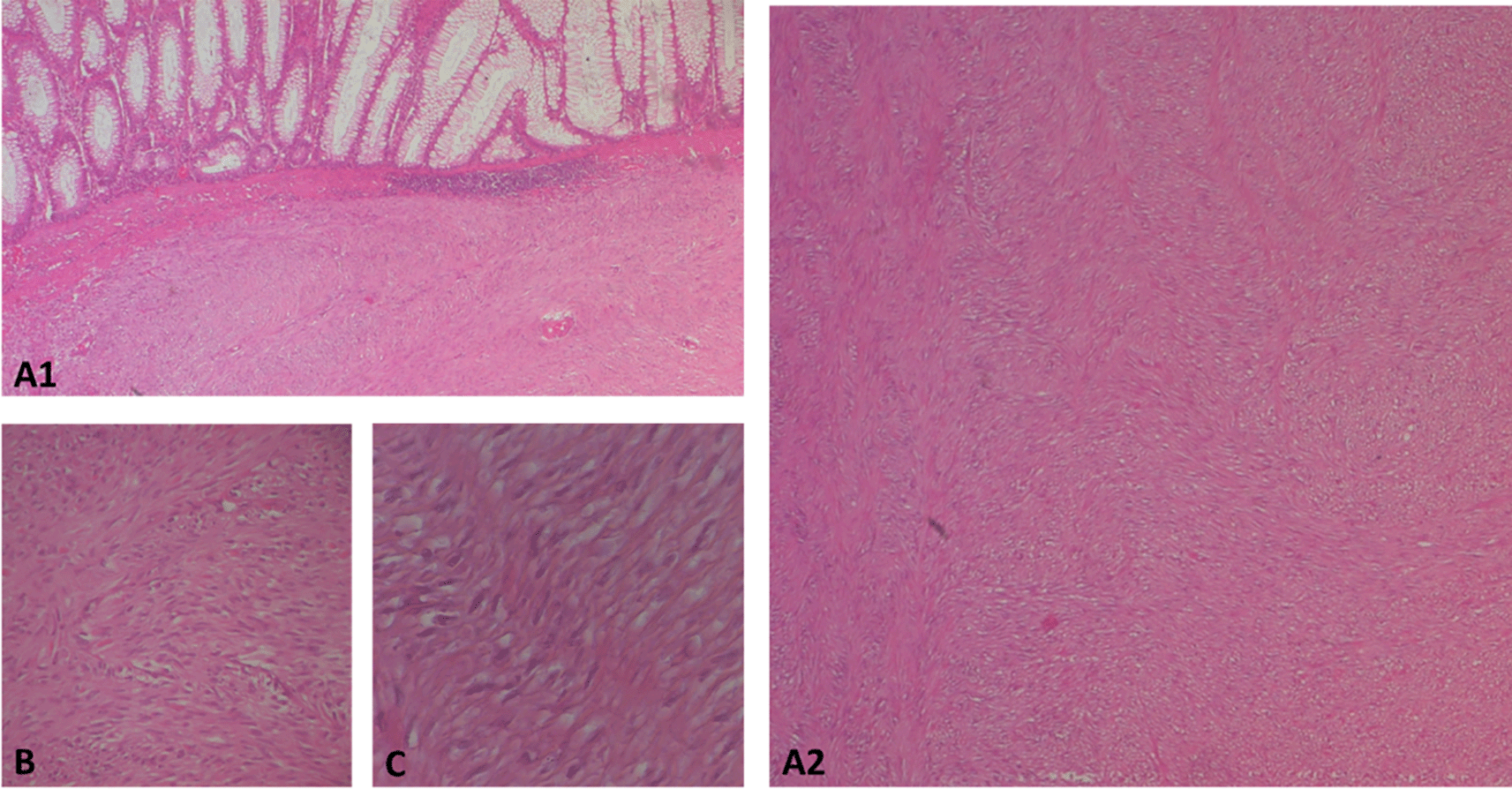
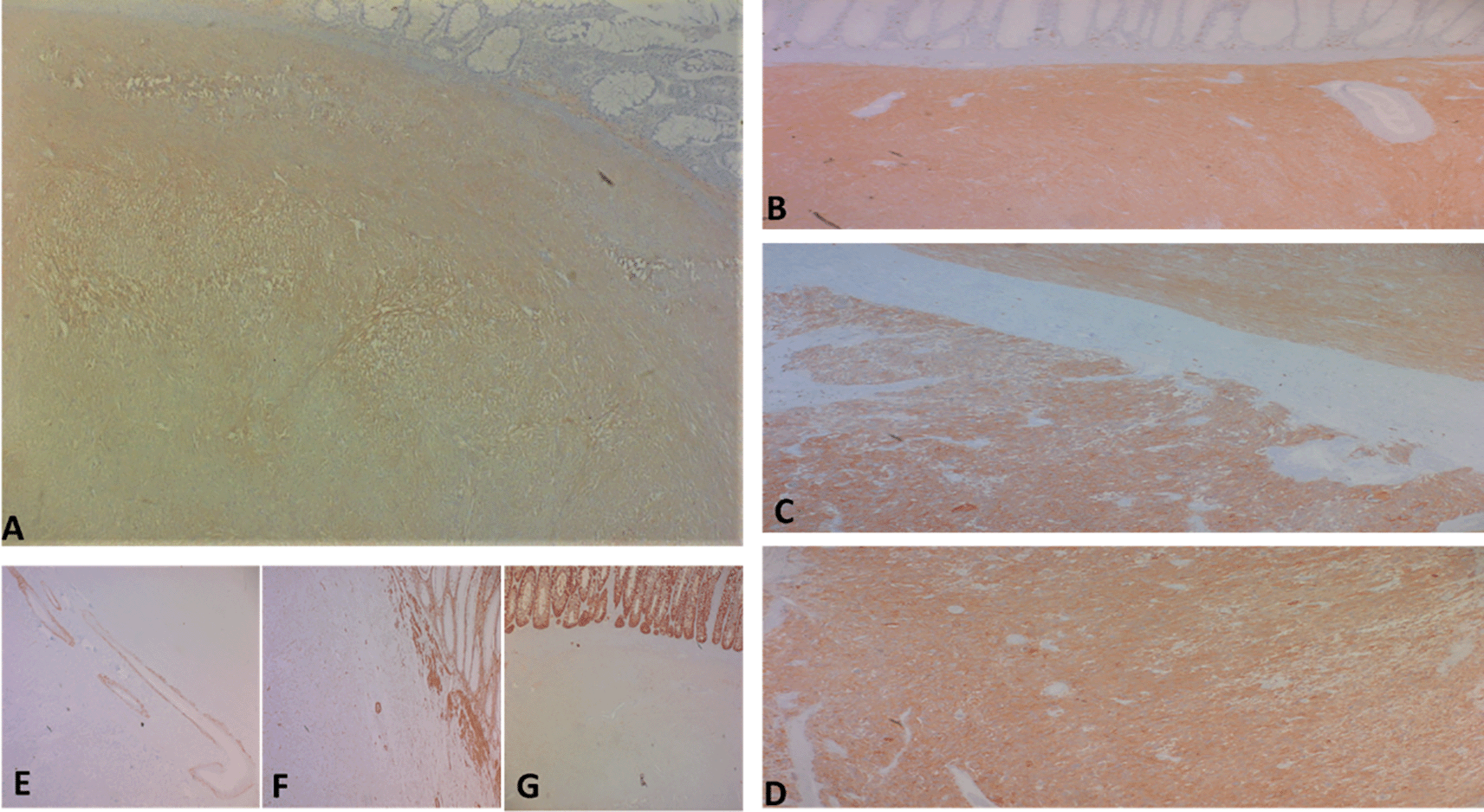
Regarding histology, the main tumor was a mixed gastrointestinal stromal tumor (GIST), with spindle cells in the submucosa and epithelial cells in the outer layers of the colonic wall, with a high risk of recurrence due to the innumerable mitoses, which exceeded 100 mitoses per 50 fields at high magnification, and the capsular rupture, according to the Miettinen and Joensuu classification ( Figure 5) and was classified pT4 according to TNM 2017 in the eighth edition.3

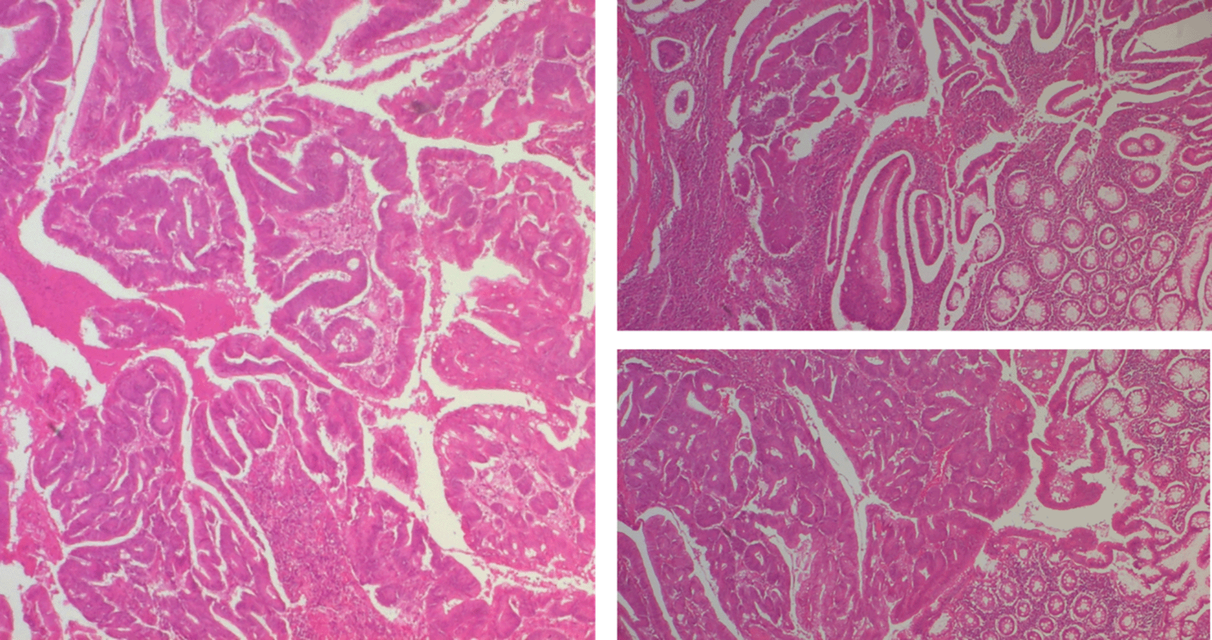
The polypoid lesion was an adenocarcinoma NOS type well differentiated developed on degenerated adenoma stadified pT1N0 (Figure 6).

The patient medical file was discussed with the multidisciplinary consensus staff indicated treatment with imatinib-based targeted therapies. Follow-up at one year postoperatively did not detect tumor recurrence.
Stromal tumors are the most common mesenchymal tumors of the digestive tract. Yearly incidence rates range between 4.3 to 22 per million in the world, which is due to variability, the improving diagnostic criteria and a lack of GIST registries.4 Simultaneous presence of colonic adenocarcinoma and stromal tumor is an uncommon occurrence. Because of the high incidence of adenocarcinoma histological type and the frequency of gastrointestinal stromal tumors (GIST), a fortuitous relationship based on the available data cannot be ruled out. The Genetic pathways of tumorigenesis are different for the two histologic types; c-Kit appears to be occasionally expressed in adenocarcinoma, and there is no evidence if the protein is indeed in the carcinogenetic process; this report is not available for stromal tumors. A review concluded that STI571 blocks the growth of colonic carcinoma cell lines.5 These results justified by preclinical investigations of c-Kit expression in colonic cancers had as objective to evaluate the use of tyrosine kinase inhibitors in the treatment of colorectal carcinomas.
We have presented a case of synchronous invasive colonic GIST with adenocarcinoma. Despite the relative common occurrence of GISTs, reports of synchronous adenocarcinoma and GISTs are quite rare. According to Kover et al., 7 of 43 patients with histologically evidence of GISTs were found with second histological type; three of these GISTs were colorectal adenocarcinomas.5 A second study realized by Au et al., found that nearly 41% of the stromal tumors were synchronous association with second malignant tumor, and 38% of these second malignancies were intestinal.6
Colonic adenocarcinoma and GIST present evidence of familial predisposition, except hereditary cancers. In another case, the patient did not have a family history of gastrointestinal or other malignancies.7 The genetic polymorphism of these two histologic types has been particularly investigated. Through progression from normal colonic epithelium to adenoma and adenocarcinoma, various genetics cancers can occur.8
Mechanisms have been clarified in sporadic colorectal cancer: chromosomal instability is responsible for 85% of the whole cases, and microsatellite instability, in the rest 15%.8 Unusually, none of the most commonly involved genes in colorectal carcinogenesis (APC, DCC, p53, K-ras, DNA mismatch repair genes) have been identified to be associated in the pathway of stromal tumors. Nevertheless, the GISTs seem to be related with the proto-oncogene mutation c-Kit, a tyrosine kinase receptor during embryonic growth and on postnatal. Activation of c-Kit by its ligand, SCF, may generates a cascade of cellular process involving transformation, differentiation, cell proliferation, adhesion, and chemotaxis.9
While the primary oncogenic drivers are structurally distinct (RTK activation via KIT/PDGFRA mutations in GIST versus CIN/MSI via APC/KRAS mutations in CRC), a crucial convergence point exists in the downstream signaling cascades, specifically the RAS/MAPK (Mitogen-Activated Protein Kinase) pathway. Syndromic cases, such as Neurofibromatosis Type 1 (NF1)-associated dual malignancies, demonstrate that the inactivation of NF1 (a negative regulator of RAS) can simultaneously activate the MAPK pathway in both mesenchymal and epithelial tumor types. In sporadic cases, the simultaneous presence of distinct upstream drivers targeting this ubiquitous proliferative axis suggests a shared functional pathway dependency that may facilitate co-tumorigenesis, moving the discussion beyond purely incidental co-occurrence. Therefore, molecular profiling must exhaustively assess the status of the MAPK pathway components (KIT/PDGFRA in GIST; KRAS/BRAF in CRC) in both lesions to fully elucidate potential shared therapeutic vulnerabilities.
When it is possible, surgery is the ideal therapeutic alternative with curative intention for non-metastatic stromal and adenocarcinoma at the same time. The operative strategies are in most situations wide and extensive.10 The integrated surgical approach should be a one-stage radical resection to ensure simultaneous R0 margins and accurate staging. Since nodal involvement is rare, lymph-node clearance is not recommended.11 The prognosis of stromal tumors depends on tumor localization, its size, and the mitotic activity.12,13 The stage of synchronous malignancies is crucial because the dominant one is responsible of the outcome and survival.13 Imatinib provide special focus in the treatment of stromal tumors; particularly, for neoadjuvant process. The benefits of this target therapy are well established to downstage inoperable cases especially by decreasing size. As a result, safe resection margins and therefore an R0 resection are recommended.14
GIST presents a high rate of recurrence (40% within 2 years).15 Just such colonic carcinoma, GISTs usually metastasize to the liver.16 Overall survival after complete resection of stromal tumors ranges from 47% to 66% at 5 years, and seems to be longer in patients with low-grade tumors: 100% at 10 years for tumors with 0–1 mitosis/30 hpf. High-grade lesions:>10 mitosis/10 hpf, have the worst outcome: 0% survival at 10 years. Nevertheless, the absence of a high mitotic index does not guarantee a better outcome.17 Overall, a 5-year survival for colonic adenocarcinoma correlates with the preoperative staging, and ranges from 3–8% for stage IV to 90% for stage I.18
For our case the predominant histologic type was the stromal one (GIST), with a high risk of recurrence due to the innumerable mitoses, which exceeded 100 mitoses per 50 fields at high magnification, and the capsular rupture, according to the Miettinen and Joensuu classification and was classified pT4 according to TNM 2017.
The adenocarcinoma type was well differentiated developed on degenerated adenoma stadified pT1N0. The patient was treated with imatinib-based targeted therapies.
Based on the specific pathological findings in this case (GIST: Very High Risk, pT4, >100 mitoses/50 HPF, capsular rupture; CRC: Stage I, pT1N0), we offer the following evidence-based clinical recommendations for synchronous disease management:
1. Surgical Strategy: A single-stage radical oncologic resection is mandated. The operative strategy must balance the R0 margin requirement for the GIST, which is mandatory, with the definitive staging requirement for the CRC, demanding a complete regional lymphadenectomy (minimum 12 nodes).
Adjuvant Therapy Systemic treatment must be dictated by the malignancy carrying the highest prognostic threat. Given the GIST’s classification as very high-risk, adjuvant Imatinib Mesylate therapy is unequivocally indicated.
Eticulous immunohistochemical and molecular biology study of all resection specimens are highly recommended whenever the combination of two histological types is found in the primary anatomopathological study.
These in-depth and ideally exhaustive studies guarantee the development of new targeted therapies and immunotherapies that would provide these patients with the opportunity of complete remission.
Nonetheless, all such cases must be discussed at a multidisciplinary concertation involving all the medical staff.
Finally, this case certainly illustrates a rare association of two histological entities. There are few cases described in the literature, which limits the possibility of reaching well-codified conclusions regarding management.
However, we believe that this case highlights the necessity for more thorough immunohistochemical and molecular biology studies.
The aim is to draw up recommendations with a high level of scientific evidence.
Synchronous tumors rare cancer of the colonic with the co-existence of two histologically different neoplasms occurring in the same site. This condition is rarely proven in preoperative investigations. Lymph node dissection is essential and must be performed according to the relevant guidelines for oncology. Perfect histopathologic examination with multiple biopsies and pathologic examination of resection specimens is required to detect synchronous tumors. Those with advanced or aggressive behavior has pejorative prognostic significance and should receive adjuvant therapy.
Pathogeneses of like association are still not yet well identified. More studies are required to understand this incident to provide optimal curative management for patients.
However, the biological observation that GIST and CRC pathologies frequently converge on, and share a functional reliance on, the RAS/MAPK signaling cascade provides a compelling molecular mechanism warranting further targeted investigation More studies are required to understand this incident to provide optimal curative management for patients.
We believe that sophisticated molecular biology studies are the bridge to innovative, more effective and targeted therapies.
Written informed consent was obtained from the patient for publication of this case report and accompanying images.
All data underlying the results are available as part of the article and no additional source data are required.
| Views | Downloads | |
|---|---|---|
| F1000Research | - | - |
|
PubMed Central
Data from PMC are received and updated monthly.
|
- | - |
Competing Interests: No competing interests were disclosed.
Reviewer Expertise: General medicine and molecular biology of cancers
Is the background of the case’s history and progression described in sufficient detail?
Yes
Are enough details provided of any physical examination and diagnostic tests, treatment given and outcomes?
Yes
Is sufficient discussion included of the importance of the findings and their relevance to future understanding of disease processes, diagnosis or treatment?
Yes
Is the case presented with sufficient detail to be useful for other practitioners?
Yes
Competing Interests: No competing interests were disclosed.
Reviewer Expertise: General medicine and molecular biology of cancers
Competing Interests: No competing interests were disclosed.
Competing Interests: No competing interests were disclosed.
Is the background of the case’s history and progression described in sufficient detail?
Yes
Are enough details provided of any physical examination and diagnostic tests, treatment given and outcomes?
Partly
Is sufficient discussion included of the importance of the findings and their relevance to future understanding of disease processes, diagnosis or treatment?
Partly
Is the case presented with sufficient detail to be useful for other practitioners?
Partly
Competing Interests: No competing interests were disclosed.
Alongside their report, reviewers assign a status to the article:
| Invited Reviewers | ||
|---|---|---|
| 1 | 2 | |
|
Version 4 (revision) 06 Oct 25 |
read | |
|
Version 3 (revision) 07 Apr 25 |
read | read |
|
Version 2 (revision) 26 Feb 25 |
read | |
|
Version 1 30 Aug 23 |
read | |
Provide sufficient details of any financial or non-financial competing interests to enable users to assess whether your comments might lead a reasonable person to question your impartiality. Consider the following examples, but note that this is not an exhaustive list:
Sign up for content alerts and receive a weekly or monthly email with all newly published articles
Already registered? Sign in
The email address should be the one you originally registered with F1000.
You registered with F1000 via Google, so we cannot reset your password.
To sign in, please click here.
If you still need help with your Google account password, please click here.
You registered with F1000 via Facebook, so we cannot reset your password.
To sign in, please click here.
If you still need help with your Facebook account password, please click here.
If your email address is registered with us, we will email you instructions to reset your password.
If you think you should have received this email but it has not arrived, please check your spam filters and/or contact for further assistance.
Comments on this article Comments (0)