Keywords
Lung Cancer, Radiomics, Computed Tomography, Slice Thickness, CT Parameters
This article is included in the Manipal Academy of Higher Education gateway.
Lung Cancer, Radiomics, Computed Tomography, Slice Thickness, CT Parameters
Radiomics is a new field that seeks to improve the physician’s visual perception of medical images with addition of more quantitative objectivity. The quantitative attributes from radiographic images are utilized to characterize spatial and textural patterns of lesions which can provide information about the heterogeneity associated with biological processes. Radiomics is a rapidly evolving field particularly in oncology to improve patient care, aid in treatment decision making, characterization, response to therapy and prognosis.1–5
Lung cancer/carcinoma (LC) remains one among the most prevalent and familiar types of cancer that results in mortality notwithstanding recent improvements in healthcare. As, most detected LC are in the middle to late phase of the disease progression and have few management options left, hence, people with lung cancer have a 10-20% survival rate at 5 years following the diagnosis in most of the developed nations.6–7 Radiomics and Machine learning methods have been used for classification of histological subtypes of LC, prediction of LC staging and outcome, response to treatment, prognosis of lung cancer.8–11
Radiomics, a rapidly evolving field, employs quantitative attributes from medical images to enhance physician’s interpretation, particularly in oncology. Radiomics and machine learning models developed based on radiomic features play crucial roles in classifying histological subtypes lung cancer. Evaluating the variability of radiomic features (RF) is important as diagnosis and treatment decision made using these quantitative should be precise and reproducible. Recent studies have shown that the CT technical parameters such as exposure factors, slice thickness (ST) and image reconstruction algorithms (IRA) can significantly affect the values of RF. Experts have recommended that for training predictive models using radiomics based machine learning models, only reproducible RF should be considered.12–14 The reproducibility of texture analysis of lung tumors is unclear and there is scarcity of research that has delved into how ST affects variability of RF in lung tumors. Hence, aim of the study is to evaluate the influence of ST on reproducibility of CT -RF for lung tumors.
This is a prospective study. The study was commenced upon approval from the Institutional Ethical committee of Kasturba Medical College and Hospital, Manipal, India on 12th August 2022 (IEC:193/2022) followed by the enrolment of the first subject after registration in the Clinical Trial Registry – India (CTRI) registration (CTRI/2022/09/045554) on 15th September 2022, and continued till 30th April 2023.
Patients with histopathological diagnosis of lung cancer types such as Non-Small Cell Lung Carcinoma (NSCLC) and Small Cell Lung Carcinoma (SCLC) were included. We excluded patients with ground glass nodules (GGN), lesions measuring less than 4mm, scans with motion artifacts and patients that did not consent to take part in the study. Written informed consent to participate was obtained from each patient.
The study was conducted at the Department of Radiodiagnosis, Kasturba Medical College and Hospital, Manipal, India. Both Kasturba Medical College and Hospital (KMC) and Manipal College of Health Professions (MCHP) are constituent colleges of Manipal Academy of Higher Education (MAHE). A total of thirty-two (32) patients with confirmed histopathological diagnosis of lung cancer (NSCLC- 71.8% & SCLC-28.1%) between September 2022 to April 2023 were included and all patients consented. The study population's demographic characteristics are outlined in (Table 1).
All patients underwent Contrast Enhanced CT (CECT) Thorax examination using 128 Slice Incisive CT (Philips Medical Systems). The protocol used for the CECT Thorax examination of the study population is detailed in Table 2. Retrospective reconstruction of the CT images was carried out utilizing CECT images from a standard protocol of 5mm to produce a ST of 2 mm. Contrast scans were performed using Iohexol 300 mgI/ml (General Electric Health care, Wisconsin, USA) as the contrast agent. The contrast media was administered using Dual Head CT Pressure injector, OptiVantage (Guerbet, France, UK).
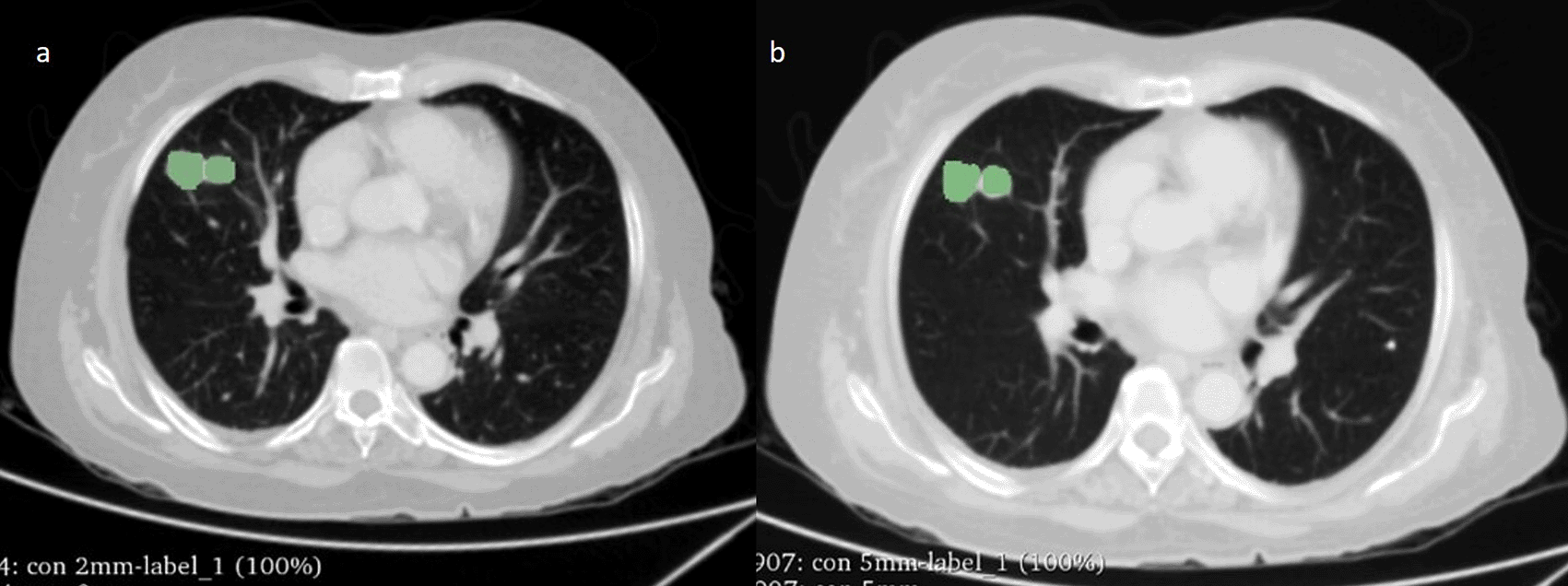
The Digital Imaging and Communication in Medicine (DICOM) CECT sections of two different slice thickness (2 mm and 5 mm) of the same patient were loaded into 3D slicer (version 4.10.2) and a radiologist (Bharath J L) with over 10 years of experience manually delineated the tumours (see Figure 1 for an example). The segmentation was performed using lung window (Window Width (WW): 1500 HU and Window Level (WL): -600 HU). All pulmonary nodules/lesions present in the right, left, and bilateral lungs were segmented rather than solely selecting the largest or most prominent nodule/lesion. The segmentation of the nodule was performed while excluding the airways, blood vessels, or bronchi. We extracted RF from the segmented regions of the lung nodules using both 2-mm and 5-mm ST.
Statistical analysis was done using SPSS version 20.0. A Paired t-test was performed to identify the significant difference in RF between the two slice thickness (2 mm and 5 mm) groups. Lin’s Concordance Correlation Coefficient (CCC) was calculated to assess the reproducibility of RF between two groups (2 and 5 mm). Concordance Correlation Coefficient of > 0.99 suggests excellent reproducibility, > 0.95 to 0.99 suggests good reproducibility, >0.90 to 0.95 suggests moderate reproducibility, ≤ 0.90 suggests weak reproducibility. p-value (<0.05) was considered.
A total of 32 cases (18 males and 14 females) with LC [Non-Small Cell Lung Cancer (NSCLC) – 71.8%, Small Cell Lung Cancer (SCLC) – 28.1%)] with mean age of were included 53.16 ± 10.25.
A total of 3424 RF measurements (107 RF per study) were extracted. Among them 66 (61.6%) RF exhibited significant difference between two the slice thickness measurements, while 41 (38.3%) RF did not show significant difference between the two slice thickness measurements. (Figure 2; Table 3).
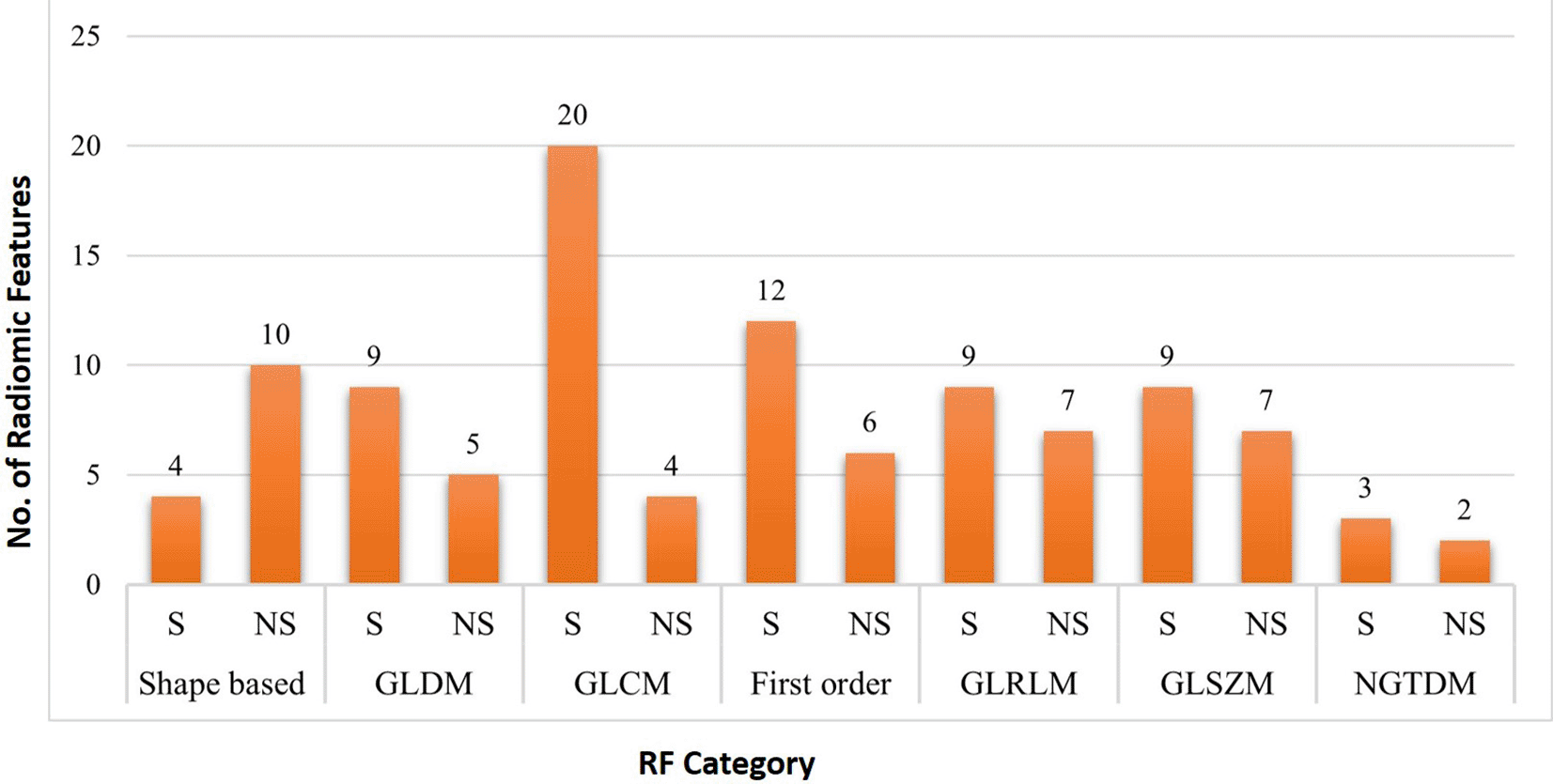
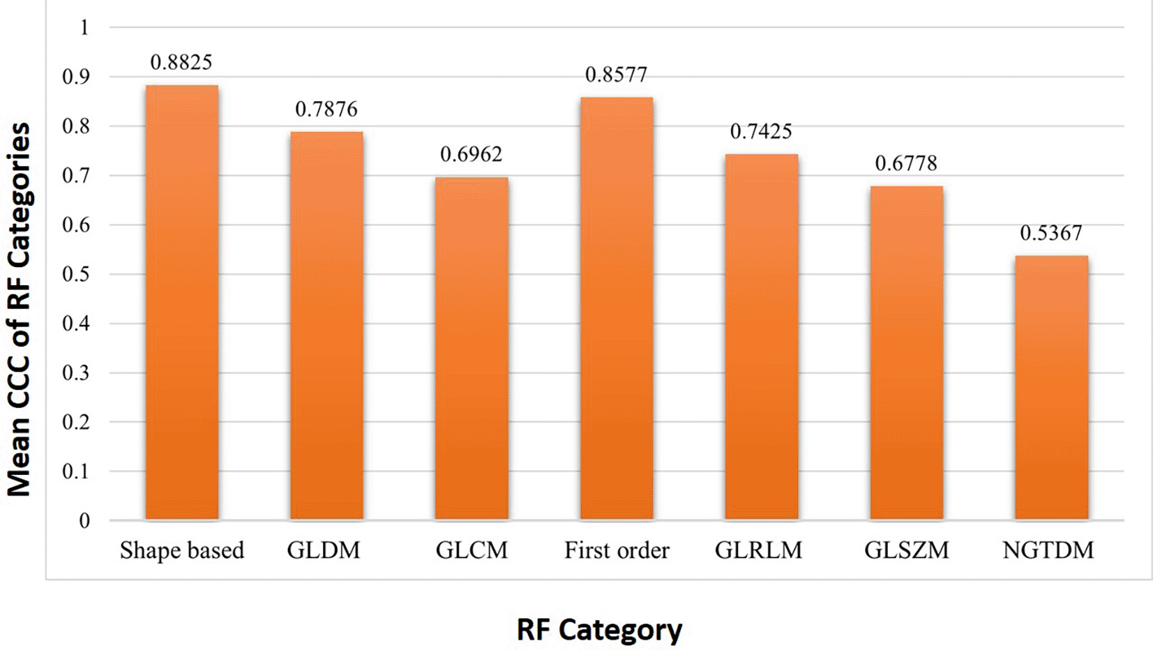
It was found that out of 14 shape-based features 8 (57.1%), out of 14 Gray Level Dependence Matrix (GLDM) RF 5 (35.71%), out of 24 for Gray Level Co-occurrence Matrix (GLCM) RF 3 (12.5%), out of 18 first order RF 8 (44.4%), out of 16 Gray level run length matrix (GLRLM) RF 4 (25%), out of 16 Gray level size zone matrix (GLSZM) RF 1 (6.25%) were found to be reproducible. All 5 neighboring gray tone difference matrix (NGTDM) RF were found to be not reproducible. Among the seven features categories, the shape-based features (57.1%) showed the maximum reproducibility whereas NGTDM based features showed negligible reproducibility (Table 4). The mean CCC of RF categories were shown in (Figure 3).
In shape-based category, features such as Voxel volume (0.997) and Mesh volume (0.997) showed excellent reproducibility. Major (0.973) and minor axis length (0.959), maximum 2D-diameter (0.976) had good reproducibility. Maximum 3D-diameter (0.944), maximum 2D-diameter slice (0.926) and maximum 2D-diameter row (0.903) had moderate reproducibility and rest of the six features showed poor reproducibility between 2- and 5-mm slice thickness.
In GLDM category, features such as high gray level emphasis [HGLE] (0.918), dependence entropy [DE] (0.929), small dependence emphasis [SDE] (0.935), dependence non uniformity normalized [DNU] (0.935) and large dependence high gray level emphasis [LDHGLE] (0.903) showed moderate reproducibility and rest of the nine features showed poor reproducibility between 2- and 5-mm slice thickness.
In GLCM category, features such as Idm (0.930), Id (0.922) and Sum squares (0.908) showed moderate reproducibility and rest of the twenty-one features showed poor reproducibility between 2- and 5-mm slice thickness.
In first order category, features such as 10th percentile (0.961) showed good reproducibility, Skewness (0.948), Uniformity (0.947), Median (0.921), Total energy (0.920), Root mean squared (0.945), Entropy (0.943) and Mean (0.943) showed moderate reproducibility and rest of the ten features showed poor reproducibility between 2- and 5-mm slice thickness.
In GLRLM category, features such as Gray level non uniformity normalized (0.952) showed good reproducibility, Short run emphasis (0.949), Run percentage (0.936) and Run length non uniformity normalized (0.943) showed moderate reproducibility and rest of the twelve features showed poor reproducibility between 2- and 5-mm slice thickness.
In the present study, we assessed the impact of slice thickness on the reproducibility of CT radiomic features (RF) for lung tumors. Few previous studies had addressed the influence of exposure parameters such as tube voltage (kVP), tube current (mA), image reconstruction algorithms (IRA), CT Scanner vendors on RF in CT for conditions like liver fibrosis, metastatic liver lesions, pancreatic neuroendocrine neoplasm.15–18 Variability of acquisition parameters could affect the diagnostic performance of radiomic signatures in oncologic patients.18–19 Limited studies had investigated the impact of ST on reproducibility of CT-RF in lung tumors.
In this study, the category of shape-based RF (57.1%) exhibited the highest reproducibility compared to other RF categories. These shape based features demonstrated robustness due to presence of low-frequency components and the reliance on segmented boundaries resulting in consistent reproducibility across changes in ST. Findings by Erdal et al.20 & Lu et al.21 supported this, revealing that RF describing tumor dimension, shape of boundaries, low-order density frequencies, and rough features were less sensitive to image setting parameters, in contrast to features characterizing sharpness of boundaries, high-order density frequencies and smooth features. Both studies analyzed the combination of ST with IRA (lung and standard) and noted that shape-based features were less effected by change in slice thickness and reconstruction algorithm. They also observed that the thinner slices with sharper reconstructions had fewer reproducible features compared to thicker slices with smoother reconstructions.
The GLDM category features in our study, such as HGLE, DE, SDE, DNU, LDHGLE demonstrated moderate reproducibility. A study by Emaminejad et al.22 in non-contrast chest CT (NCCT) identified that GLDM DE, DNU, GLNU were reproducible against the dose and kernel variations with varying slice thickness. Unlike our study, none of the previous research mentioned the reproducibility of GLDM features concerning slice thickness alone.
Within the GLCM category in our study, only two features showed reproducibility with variations in slice thickness. Similar results were documented by Erdal et al.20 & Kim et al.23 indicating that GLCM category (19.4 % & 25 %) had lower reproducibility compared to other RF categories. We observed that first-order features (44.4%) had the second highest reproducibility. Studies by Erdal et al.,20 Park s et al.,24 Choe J et al.25 reported that first-order features exhibited the most reproducibility across various imaging parameters. Park s et al.24 and Choe J et al.25 reported that convolution network-based super resolution (SR) algorithms and kernel-converted images had reduced effects on the reproducibility of RF with variations in slice thickness and reconstruction kernels. Yang et al.26 employed a resampling technique to standardize the voxel measurement of both thick and thin section CT images to 1x1x1 mm3 using linear interpolation and observed that, following resampling of thicker images, 202 RF (66.2%, 202/305) exhibited a noteworthy reduction in variability of RF compared to the original non-resampled data (Table 5).
| Author name (year) | Our study (2023) | Lu et al.21 (2016) | Erdal et al.20 (2020) | Yang et al.26 (2020) | Emaminejad et al.22 (2021) |
|---|---|---|---|---|---|
| Pathology studied | Lung tumors (SCLC, NSCLCL) | Lung cancer | Lung nodules | Solid pulmonary nodules | Lung cancer |
| Study Procedure | CECT | NCCT | NCCT | CECT | NCCT |
| Technical parameters | ST (2 and 5- mm) | IRA (Lung and standard) ST | Dose levels (4) Kernels (10) Thicknesses (8) | ST (1.25 mm and 5 mm) | Dose levels (100 %, 50%, 25% and 10 %) ST (0.6, 1 and 2 mm) Reconstruction kernel (smooth, medium, sharp) |
| RF extracted | 107 | 89 | 28 | 396 | 226 |
| Features extracted | Shape, GLDM, GLCM First order, GLRLM GLSZM, NGTDM | Tumor size, Shape, Boundary shape, Sharpness, Density distributions with and without spatial information | Histogram, GLCM, RLM, NGDLM,NGTDM | Histogram, Geometry Texture features | First-order, Wavelet Features, GLDM, GLRLM, GLCM, GLSZM, NGTDM |
| Reproducibility of RF | Shape based features (57.1%), First order (44.4%) features showed highest reproducibility compared to other RF categories | Eight of the feature groups associated with dimensions, form, and rough texture exhibited consistent reproducibility across all combinations | Density feature was robust against dose changes, Skewness was robust for kernel and ST, Deviation was weakest feature for all cases. GLCM category was least reproducible | In non-resampled data, 239 features were shown significant differences between thin and thick slice. 66 RF were reproducible. In resampled data, 202 features exhibited significant differences between two thicknesses. 103 features were reproducible. | Seventeen and Eighteen features were reproducible with respect to dose and kernel changes. Only one to five features were reproducible with changes in slice thickness |
For the GLRLM and GLSZM categories, reproducibility rates were 25% and 6.25 %, respectively, in the current study. A Study by Emaminejad et al.22 similarly found that GLRLM Run length non uniformity (1 of 9 features) and GLSZM (1 of 10 features) displayed very limited reproducibility against the dose and kernel variations with varying slice thickness. Contrary to the study reported by Liu J et al.27 which demonstrated that NGTDM exhibited good reproducibility, we did not observe any reproducible features in NGTDM. The reason for this disparity is attributed to differences in technical parameters, specifically in terms of dose variation, rather than slice thickness.
The study has few limitations. Firstly, the sample size was relatively small, as it is time bound study with prospective data collection of patients who underwent CT scan with histopathological proven cases of lung cancer. A larger sample size is required to confirm the reproducibility of RF with slice thickness. Secondly, we did not analyze whether a thinner slice thickness would result in better performance of radiomic models for predicting lung cancer. Thirdly, a single image acquisition variable such as slice thickness was examined to determine how it affects the reproducibility of radiomic features.
Radiomics has the potential to transform lung cancer diagnosis, follow-up, and therapy planning by enabling individualised management in a non-invasive and an economical manner. Our study found that ST is the main parameter impacting the reproducibility of CT-RF for lung tumours. The study also increases awareness regarding the significance of accurately configuring imaging acquisition parameters in the context of radiomic/radio genomic approaches. Standardization of technical parameters and protocols is necessary when conducting multicentre studies, as these factors can impact the diagnostic performance of Machine Learning (ML) models developed using radiomic features.
| Views | Downloads | |
|---|---|---|
| F1000Research | - | - |
|
PubMed Central
Data from PMC are received and updated monthly.
|
- | - |
Provide sufficient details of any financial or non-financial competing interests to enable users to assess whether your comments might lead a reasonable person to question your impartiality. Consider the following examples, but note that this is not an exhaustive list:
Sign up for content alerts and receive a weekly or monthly email with all newly published articles
Already registered? Sign in
The email address should be the one you originally registered with F1000.
You registered with F1000 via Google, so we cannot reset your password.
To sign in, please click here.
If you still need help with your Google account password, please click here.
You registered with F1000 via Facebook, so we cannot reset your password.
To sign in, please click here.
If you still need help with your Facebook account password, please click here.
If your email address is registered with us, we will email you instructions to reset your password.
If you think you should have received this email but it has not arrived, please check your spam filters and/or contact for further assistance.
Comments on this article Comments (0)