Keywords
Arthroscopic Surgery; Popliteus; Plastic Surgery
This article is included in the Manipal Academy of Higher Education gateway.
Arthroscopic Surgery; Popliteus; Plastic Surgery
As per the reviewer’s opinion, the potential limitations of this anatomical research are added in this revised version. These included no description of the sex, age, or statures of the cadavers that were used in this experiment. There was no statistical difference or correlation between these parameters and stature and muscle length. The position of the cadaveric knee joint is also not considered. It is difficult to fixate the cadaver in a certain position because sometimes cadavers are already fixed its position by rigor mortis.
See the authors' detailed response to the review by Graham Louw
See the authors' detailed response to the review by Kyutaro Kawagishi
The popliteus is a muscle in the posterior compartment of the lower extremity, which is located at the leg and innervated by the tibial nerve. This is the only muscle in the back of leg, which acts on the knee joint and not over the ankle joint. This is considered as the unlocking muscle of the knee joint.1 It laterally rotates the femur over the tibia, while walking when one foot is on the ground. It helps in the knee stabilization along with the fibular collateral and popliteo-fibular ligaments.1 The popliteus has dual origin, one from the lateral femoral condyle and the other from the lateral meniscus. Its origin is tendinous and it is interesting to know that there exists variability in its origin like from the styloid process of the fibula.2 On few occasions, an accessory head of popliteus may originate from the sesamoid bone at the gastrocnemius lateral head.3 On rare occasions, there may be a popliteus minor muscle, which originates from the femur over the deep part of the plantaris muscle and its distal attachment is at the posterior aspect of knee joint.2 The popliteal tendon occupies a part of the knee joint capsule; however, it does not enter the synovial cavity. Hence it is intra-capsular, however extra-articular and extra-synovial.4 It runs underneath the fibular collateral ligament and biceps femoris tendon. The popliteus separates the lateral collateral ligament from the lateral meniscus and prevents its injury.5 The popliteus inserts at the dorsal aspect of the tibial upper end just over the soleal line. More clinical and basic anatomical studies are needed to understand the injuries and pathological involvement of popliteus in order to accomplish the better diagnosis and management.6 In this situation, the primary goal was to determine the dimensions of different parameters of popliteal muscle tendon complex at its various parts. The objectives were to measure the dimensions of popliteo-fibular ligament and to study the topographic anatomy of the neurovascular structures supplying the popliteus.
This is a descriptive cross sectional institutional based study, which involved 25 formalin embalmed adult cadavers from Indian population. The sample size is similar to the earlier study performed by Olewnik et al.1 The protocol of this anatomical research is available at dx.doi.org/10.17504/protocols.io.3byl4qqk8vo5/v1. Meticulous dissection was performed to expose the popliteus muscle, tendon and its neurovascular structures. In total, 50 popliteus muscles were analyzed based on the side. The sex differences were not explored in this study. The inclusion criteria were adult embalmed cadavers, which were available at the department of anatomy. Cadavers showing pathological changes and congenital anomalies at the knee joint were excluded from this study. The exclusion criteria also included the previously dissected cadavers. The measurements were performed by using the digital Vernier caliper (Mitutoyo Digital Vernier Caliper 0-150 mm 500-196 made in Japan) and the analysis of the data was done by using the recent version of SPSS (version 27) software after applying the paired t-test. Single person, who is a coauthor in this study, performed all the measurements. This was followed to prevent the inter-observer bias and the measurements were taken on three consecutive times. The average of which was considered to prevent the intra-observer bias. The measurements of the popliteus muscle tendon complex are schematically represented in Figure 1 and tabulated in Table 1.
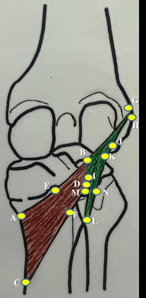
| No. | Parameter | Representation in Figure 1 |
|---|---|---|
| 1 | mediolateral length along the upper border of popliteus | AB |
| 2 | mediolateral length along the lower border of popliteus | CD |
| 3 | width at the muculotendinous junction of popliteus | BD |
| 4 | width at the midpoint of popliteus | EF |
| 5 | width at the insertion of popliteus | AC |
| 6 | thickness at the midpoint of popliteus along the lower border of popliteus | F |
| 7 | length of popliteal tendon | BG |
| 8 | width of popliteal tendon at origin | GH |
| 9 | width of politeal tendon at muculotendinous junction | BI |
| 10 | thickness of popliteal tendon at mid point | J |
| 11 | distance of origin of medial geniculate artery from the intercondylar line | - |
| 12 | distance of origin of lateral geniculate artery from the intercondylar line | - |
| 13 | distance of division of popliteal artery from the intercondylar line | - |
| 14 | distance of origin of nerve to popliteus from the intercondylar line | - |
| 15 | length of nerve to popliteus | - |
| 16 | length of popliteo-fibular ligament | KL |
| 17 | width of popliteo-fibular ligament | MN |
This anatomical research has received the approval from the ethics committee of our institution (Approval Committee Name: Institutional Ethics Committee, Kasturba Medical College, Mangalore, Approval Number: IEC KMC MLR: 09/2022/400, dated 21.09.2022). Since this is a study from the human cadavers, the consent from the participants is not applicable. This was waived by our institutional ethics committee. The consent was already given by the participant to perform the medical teaching and research, while donating his or her body. This present research is following the guidelines of the international ethical standards.
The length of the popliteus muscle belly along the upper and lower border were 44.2±6.63 mm and 89.26±14.41 mm, width of the muscle belly at midpoint, musculotendinous junction and insertion were 28.45±6.85 mm, 11.7±3.5 mm and 75.95±10.7 mm.18 The thickness of muscle belly at the midpoint was 2.55±0.55 mm. The morphometric data of the popliteus muscle belly are given in Table 2. The length of popliteal tendon, width at origin and at musculotendinous junction were 24.85±2.15 mm, 7.55±1.55 mm and 8.5±1.15 mm. The thickness of tendon of popliteus was 2.6±0.75 mm. Table 3 represents the dimensions of the tendon of popliteus of this study.
| Dimension of the tendon | mean±SD |
|---|---|
| length | 24.85±2.15 |
| width at origin | 7.55±1.55 |
| width at musculotendinous junction | 8.5±1.15 |
| thickness | 2.6±0.75 |
The length of nerve to popliteus was 50.44±8.66 mm and its origin was located 27.54±6.18 mm from the intercondylar line. The distance of origin of medial and lateral geniculate arteries from the intercondylar line were 26.26±10.47 mm and 20.76±5.19 mm. The distance of division of popliteal artery was 49.44±16.26 mm from the intercondylar line. Table 4 offers the topographic anatomy of the neurovascular structures of popliteus. The length and width of the popliteo-fibular ligament was 17.84±3.43 mm and 7.36±1.9 mm individually. They are summarized in Table 5 and the sidewise comparison of all the parameters, which are measured in this study are given in Table 6, Table 7, Table 8 and Table 9. The statistical significance was not there, when the right and left side comparison was considered (p>0.05). The only significant difference was observed for the width of the popliteal muscle at the insertion, which was higher for the left side (p<0.05).
| Popliteo-fibular ligament | mean±SD |
|---|---|
| length | 17.84±3.43 |
| width | 7.36±1.9 |
| Dimension of the muscle | Right side (n=25) | Left side (n=25) |
|---|---|---|
| mediolateral length along upper border | 44.88±7.44 | 43.52±5.82 |
| mediolateral length along lower border | 88.12±17.06 | 90.4±11.77 |
| width at the musculotendinous junction | 12.4±4.3 | 11±2.7 |
| width at the midpoint | 28.8±6.5 | 28.1±7.2 |
| width at the insertion* | 72.5±9.9 | 79.4±11.5 |
| thickness at the midpoint | 2.6±0.7 | 2.5±0.4 |
| Dimension of the tendon | Right side (n=25) | Left side (n=25) |
|---|---|---|
| length | 25±2.5 | 24.7±1.8 |
| width at origin | 7.1±1.8 | 8±1.3 |
| width at musculotendinous junction | 8.6±1.2 | 8.4±1.1 |
| thickness | 2.4±0.7 | 2.8±0.8 |
| Popliteo-fibular ligament | Right side (n=25) | Left side (n=25) |
|---|---|---|
| length | 17.84±3.23 | 17.84±3.63 |
| width | 6.92±1.57 | 7.8±2.23 |
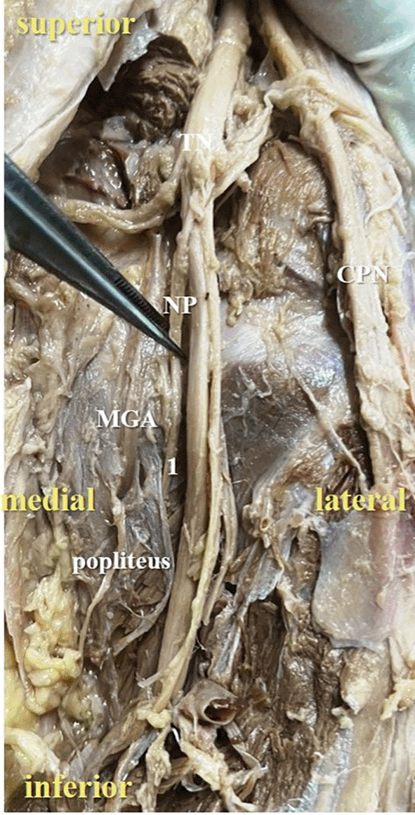
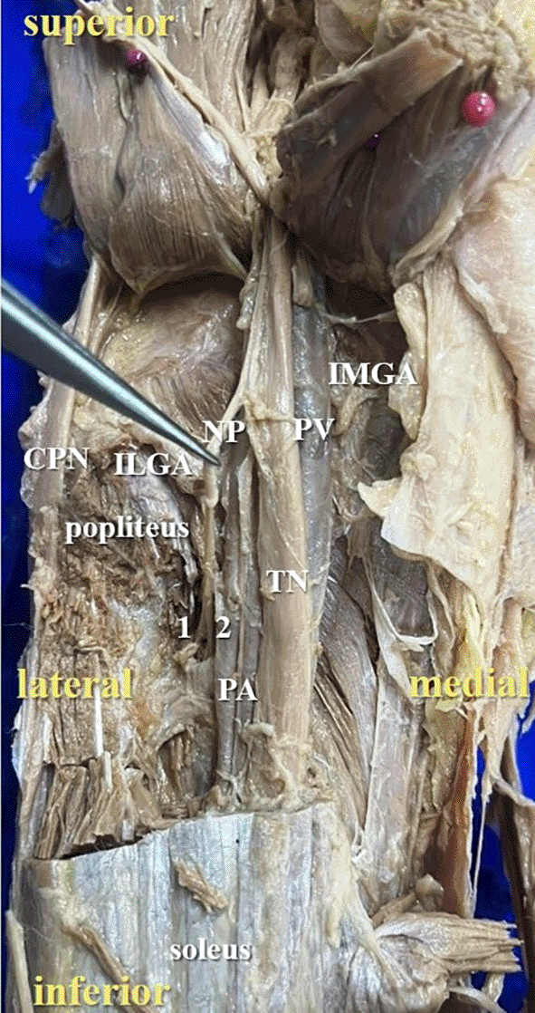
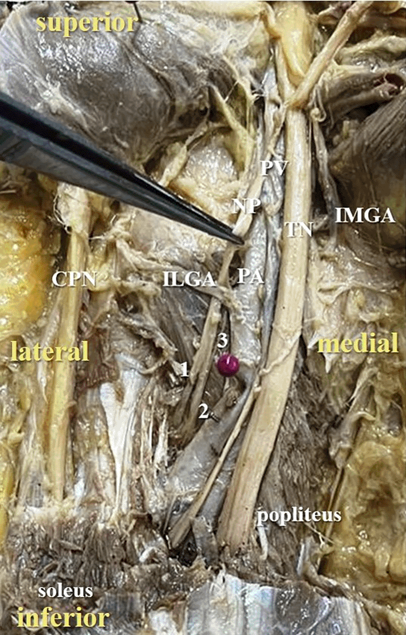
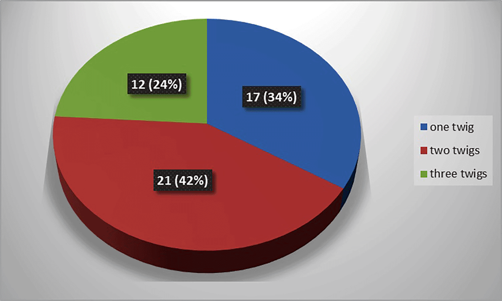
The present study observed that, there was single twig (Figure 2) of nerve to popliteus in 17 lower limbs (34% cases), there were two twigs (Figure 3) in 42% cases (in 21 lower limbs) and the nerve to popliteus was giving 3 twigs (Figure 4) in 12 lower extremities (24%). The frequency of distribution of nerve to popliteus is represented in Figure 5.
The popliteus muscle is the unlocking muscle of the knee and avoids the medial rotation of femur over tibia.7 It is known for variations and this is explained on the basis of phylogeny.8 In reptiles, fibula directly articulates with the lateral femoral condyle, so popliteus is more occupied between the proximal parts of tibia and fibula. In mammals, femur articulates with tibia, leading to the migration of popliteus muscle proximally until the lateral femoral condyle. In humans, attachment of popliteus to fibula is represented by the popliteo-fibular ligament. This is very important as it stabilizes the posterolateral aspect of the femoro-tibial articulation. According to Vani and Raveendranath,9 the length and width of tendon of popliteus was 35.12 mm and 9.52 mm, which was comparable to the dimensions by Jung et al.10 and Osti et al.11 LaPrade et al.12 reported that the length of popliteus tendon measures 54.5 mm. In our research, the same parameters were 24.85 ± 2.15 mm and the width at the musculotendinous junction was 8.5±1.15 mm. These dimensions are slightly lower in comparison to the data by Vani and Raveendranath.9 In their study, the distance of distal attachment of popliteus from its musculotendinous junction was 107.14±13.45 mm and widest part of popliteus measured 32.38±4.33 mm. Kurtoglu et al.13 reported in their study that popliteus muscle belly length and width were 107.14mm and 32.38mm. However, the mediolateral length of popliteus along the lower border was 89.26±14.41 mm in our study and the width of popliteus at the midpoint was 28.45±6.85 mm. These dimensions are small in comparison to Kurtoglu et al.,13 may be because of ancestral variations. Hwang et al.14 reported the popliteal length at its lateral border, which was 119±15 mm. This was almost parallel to the findings of Vani and Raveendranath.9 In the present study, this dimension was not performed and the morphometric data of the length and width of popliteus is different in our study in comparison to previous studies, as the different points were used for the measurements. The positive outcome of this anatomical research was we measured the length of the popliteus at both the upper and lower borders.
Vani and Raveendranath9 reported that, the distance of origin of nerve to popliteus from the intercondylar line ranged between 12.10±10.54 mm above the intercondylar line to 18.74±11.51 mm below the intercondylar line. In the present study, this distance was measuring 27.54±6.18 mm below the intercondylar line. In most of our specimens, it was observed that, nerve to popliteus was arising separately and was not giving the nerve to soleus or nerve to tibialis posterior. We could observe that; these nerves were separate branches coming from the tibial nerve. However, previous authors mentioned that, nerve to tibialis posterior originates from the nerve to popliteus.14
In the present study, it was observed that the nerve to popliteus along with the blood vessels, descend anterior to the popliteus muscle and enter at its anterior surface, which is obvious in Figure 2. The basic anatomical knowledge of these structures can enlighten the plastic surgeons during the reconstruction surgeries of popliteus. The anatomy and biomechanics of popliteus makes it an important structure, which keeps the knee stable. But its involvement is ignored in the complex injury of the knee joint.6 The isolated involvement of popliteus is seen in sports injuries and it may be misinterpreted as a tear of lateral meniscus. The sports like tennis, basketball and downhill running may put additional stress on the tendon of popliteus.6 The present study provided the data about the neurovascular structures in relation to the popliteus and it is believed that these details are clinically important for the effective treatment of the popliteus muscle spasticity.15 Popliteus muscle tendon complex is a landmark to the operating surgeon during the sling reconstruction of popliteus tendon.16 Popliteus is commonly injured in the posterolateral impact at the femoro-tibial articulation and gets torn. The muscular strains are also common in the sports injuries, which commonly affect the popliteus at its tendino-muscular junction.17 Due to all these implications, the present study was undertaken. The literature search did not reveal much studies about the morphometry of the popliteus and particularly, the dataset is not available from the Indian population. In this context, the data of the present study is enlightening to the orthopedic surgeons, particularly for the posterior knee approach procedures like baker’s cyst excision, fixation of tibial plateu fractures and meniscal tears. However, the present study has limitations like the smaller sample size, and sex differences, which were not explored. In this study, repeatability of the measurements by asking a secondary observer was not performed. It would have been better if a subset of the sample was measured by a secondary observer and intraclass correlation was applied statistically. If the measurements are consistently taken by one observer incorrectly and then averaged to one value it still might not be representative of the 'true' dimensions of the vertebrae. Statistically confirming the agreement between these measurements would strengthen the quality of the research. If the intra-observer error is high, it might suggest the requirement for better measurement definitions or suggest using different tools for them in the field of vertebral morphometry in general. The correction factor for overall height of the body is not given. This is also another limitation of this study. The anatomical points for the morphometry were approximately considered, which may not be the perfect method.
There was no description of the sex, age, or statures of the cadavers that were used in this experiment. This study has failed to provide the statistical difference or correlation between these parameters. In anatomical measurements, sex differences which are related to the difference in the average stature are often observed. Especially in the lower extremities, length dramatically changes with growth. There is no correlation between stature and muscle length or any other parameters in this study. The comparison of the muscle belly thickness in the formaldehyde-fixed cadavers is one of the most challenging methods, because we might not know whether the muscle is in the contracting or relaxing phase. Moreover, the muscle belly easily changes its shape while dissecting the cadaver, even in formaldehyde-fixed cadavers.
We also did not describe the position of the knee joint. It is difficult to fixate the cadaver in a certain position because sometimes cadavers are already fixed its position by rigor mortis. These are the potential limitations of this anatomical research.
The present study offered the detailed morphometric data about the dimensions of the popliteus muscle belly and its tendon along with the popliteo-fibular ligament. It is believed that, the data of popliteal muscle tendon complex of this study will be enlightening to the orthopedic surgeons particularly in the field of arthroscopic and plastic surgery like the reconstruction. The data can be considered as the database for our population.
| Views | Downloads | |
|---|---|---|
| F1000Research | - | - |
|
PubMed Central
Data from PMC are received and updated monthly.
|
- | - |
Provide sufficient details of any financial or non-financial competing interests to enable users to assess whether your comments might lead a reasonable person to question your impartiality. Consider the following examples, but note that this is not an exhaustive list:
Sign up for content alerts and receive a weekly or monthly email with all newly published articles
Already registered? Sign in
The email address should be the one you originally registered with F1000.
You registered with F1000 via Google, so we cannot reset your password.
To sign in, please click here.
If you still need help with your Google account password, please click here.
You registered with F1000 via Facebook, so we cannot reset your password.
To sign in, please click here.
If you still need help with your Facebook account password, please click here.
If your email address is registered with us, we will email you instructions to reset your password.
If you think you should have received this email but it has not arrived, please check your spam filters and/or contact for further assistance.
Comments on this article Comments (0)