Keywords
Aggregatibacter actinomycetemcomitans, Enterococcus faecalis, photodynamic inactivation, diode laser, diseases, periodontitis, curcumin, chlorophyll
This article is included in the Plant Science gateway.
Aggregatibacter actinomycetemcomitans, Enterococcus faecalis, photodynamic inactivation, diode laser, diseases, periodontitis, curcumin, chlorophyll
The oral cavity is one of the most important parts of the body that must be maintained. Infectious diseases of the teeth and mouth that are often found are periodontitis and endodontics. Periodontitis is a bacterial infection of the teeth that causes inflammation of the supporting tissues of the teeth, which include the gingiva, ligaments, cement, and alveolar bone.1 Periodontitis is caused by pathogenic bacteria, predominantly gram-negative, anaerobic, or microaerophilic in the subgingival area.2 Aggregatibacter actinomycetemcomitans bacteria are found in dental plaque, periodontal pockets, and buccal mucosa in up to 36% of the normal population.3 Aggregatibacter actinomycetemcomitans bacteria can infect patients when the human immune system decreases and inhibits other organisms' growth in the oral mucosa, teeth, and nasopharynx.
In general, gram-positive bacteria, Enterococcus faecalis (E. faecalis), are found in the root canals of teeth. The bacterium Enterococcus faecalis is ovoid, with a diameter between 0.5 and 1 μm.4 These bacteria are facultative anaerobes and can survive in extreme environments such as highly alkaline pH and high salt concentration conditions. The number of these bacteria in the human body can be minimized by paying attention to the food consumed and environmental conditions such as humidity. Furthermore, E. faecalis bacteria resist calcium hydroxide and antibiotics such as tetracycline.5 Systemic treatment in the form of antibiotics has been widely used to treat periodontitis. However, several studies have reported cases of antimicrobial resistance to certain types of antibiotics. So alternative therapy is needed that is effective and does not cause antibiotic resistance.6 Therefore, the recommended alternative therapy in this study is photodynamic inactivation (PDI).
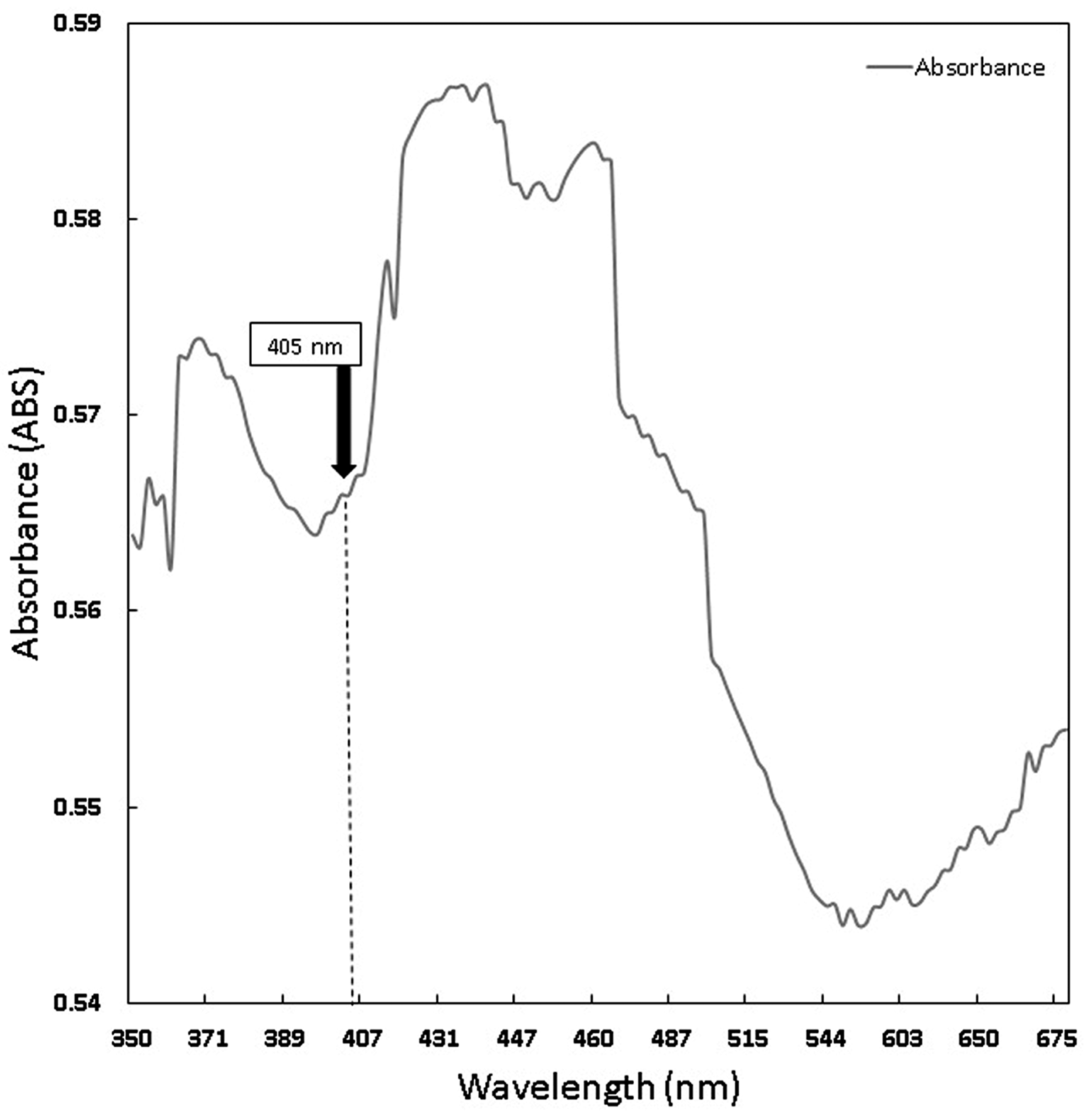
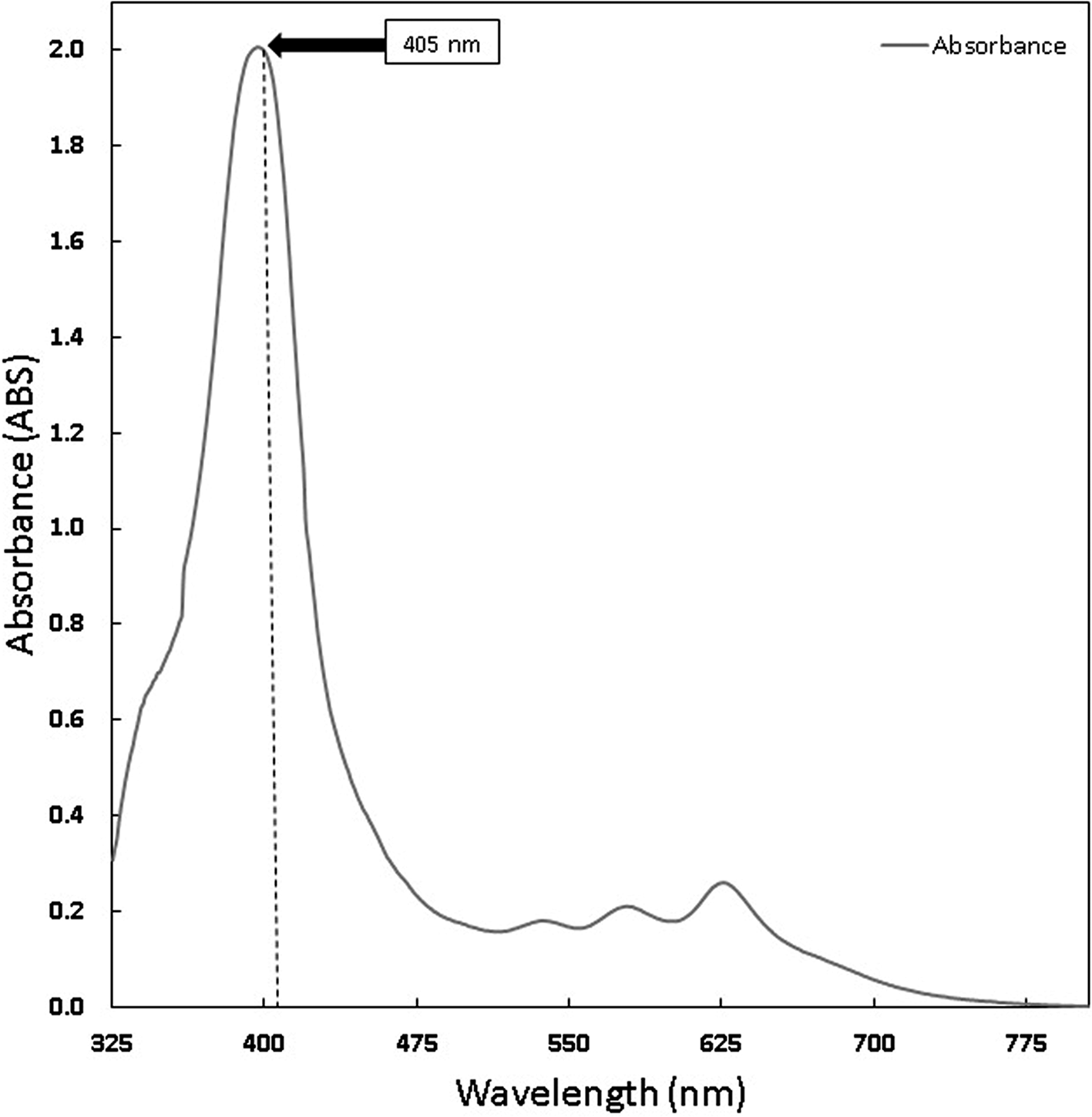
Photodynamic inactivation (PDI) is a method of inactivating microorganisms by utilizing light to activate a photosensitizer (PS) agent that produces reactive oxygen species (ROS), causing cell lysis.7,8 The suitability of the light spectrum with the PS absorption spectrum is the key to photophysical reactions, namely the absorption of light energy by PS agents, which will trigger photochemical and photobiological reactions to produce antimicrobial effects9,10 and biomodulation.11 PS is a light-sensitive molecule that plays a role in absorbing light energy. PS is divided into two types, namely endogenous and exogenous photosensitizers. The addition of exogenous PS aims to increase the effectiveness of light energy absorption.12 Some natural ingredients that are exogenous PS include chlorophyll and curcumin. Chlorophyll is a green substance found in green plants that photosynthesizes.13 In photosynthesis, chlorophyll acts as a light catcher, energy transfer, and light conversion and can absorb a maximum wavelength between 400-700 nm.14 Curcumin is a curcuminoid compound with yellow pigment in turmeric rhizome, which is antitumor, antioxidant, anticarcinogenic, anti-inflammatory, antiviral, antifungal, antispasmodic, and hepatoprotective.15 The absorption spectrum of curcumin is in the wavelength range of 375-475 nm.16
Previous studies have reported the effectiveness of using PS chlorophyll in alfalfa leaves with a blue LED activator of 20.48 J/cm2 for the inactivation of A. actinomycetemcomitans bacteria by 81%.17 The results of another study with the addition of PS curcumin and diode laser activator 403 nm 15.83 J/cm2 in Staphylococcus aureus resulted in a mortality rate of 85.48%.16 Then, another study using curcumin and blue LEDs on S. aureus bacteria resulted in a mortality rate of 91.49%.18 Continuing previous studies,15,16,18 this study aims to compare the effectiveness of antimicrobial reduction from photodynamic inactivation (PDI) on bacteria A. actinomycetemcomitans and E. faecalis with PS curcumin and chlorophyll Medicago sativa L. using a 405 nm diode laser. Diode laser irradiation was carried out at various lengths of irradiation time, namely (30, 60, 90, 120, 150, and 180) seconds.
Bacteria A. actinomycetemcomitans ATCC 43718 and E. faecalis ATCC 29212 were cultured in Tryptone Soy Broth (TSB). Then, they were incubated for 24 hours at 37oC until the colonies reached ~108 CFU/mL or 1.0 McFarland standard. The culture was placed 100 L on 96-well microplates and incubated for 48 hours.
Chlorophyll was extracted from Medicago sativa L (K-Link liquid, Indonesia) and Curcuma standard (Sigma Aldrich) with a concentration of 1.6 mg/ml diluted with sterile normal saline. The absorption spectrum of chlorophyll was measured using Shimadzu UV-VIS 1800 spectrometer.
The light source of a laser diode is 405 nm, and characterization was carried out using Jasco CT–10 monochromators to determine the peak wavelength. The power output was 2.49 mW, measured with power meter OMM-6810B-220V. The spot beam area size was 0.28 cm2. Diode laser irradiation was carried out with variations in the length of the irradiation time of 30, 60, 90, 120, 150, and 180 seconds. The energy density value can be calculated using equation 19:
The treatment samples consisted of two types of bacteria, A. actinomycetemcomitans and E. faecalis. The bacterial PDI treatment consisted of a negative control group without treatment (T0), a positive control group with the addition of chlorophyll and curcumin (T1), a 405 nm diode laser treatment group at various energy densities of 0.26; 0.53; 0.79; 1.06; 1.32; 1.59 J/cm2 (S1), a 405 nm diode laser treatment group with the addition of chlorophyll Medicago sativa L 1.6 mg/ml (S2), and a 405 nm diode laser treatment group with the addition of curcumin PS 1.6 mg/ml (S3). In groups S2 and S3, samples were given chlorophyll or curcumin and they were incubated for 10 minutes, then irradiated with 405 nm diode laser with an exposure time (30, 60, 90, 120, 150, and 180 seconds). The treated samples were grown on TSA media, incubated for 24 hours at 37C, and the number of bacterial colonies grown was counted by the Total Plate Count method.
Each treatment was calculated as CFU/ml using equation 2. Next, the percentage of bacterial reduction was calculated using equation 3 based on the control group. The results of bacterial reduction were statistically analyzed by ANOVA test using IBM SPSS Statistics Version 21.
The chlorophyll and curcumin extracts were tested using UV-Vis at a wavelength range of 325 nm to 705 nm to determine the absorption spectrum of light. Then, the results of the characterization of the absorption spectrum of chlorophyll and curcumin to light are obtained, as shown in Figures 1 and 2. Based on Table 1, the characterization results show the peak wavelength of the laser diode at 405 nm with the stability of the output power at a distance of 1 cm (2.49 ±0.07) mW. The temperature characterization showed the optimum temperature stability (26.60±0.01)oC for bacterial growth. Thus, the irradiation energy density of the laser diode is 405 nm with an output power of 2.49 mW and a beam area of 0.28 at various exposure times (30, 60, 90, 120, 150, 180) seconds are 0.26; 0.53; 0.79; 1.06; 1.32 and 1.59 J/cm2.
After that, antibacterial tests were carried out on Aggregatibacter actinomycetemcomitans and Enterococcus faecalis bacteria, which were exposed to a diode laser with and without a photosensitizer. The viability of the bacteria Aggregatibacter actinomycetemcomitans and Enterococcus faecalis are shown in Figures 3 and 4. Based on bacterial viability, the percentages of death of A. actinomycetemcomitans and E. faecalis bacteria by diode laser irradiation with the addition of curcumin photosensitizer and Medicago sativa L chlorophyll treatment are shown in Figures 5 and 6.
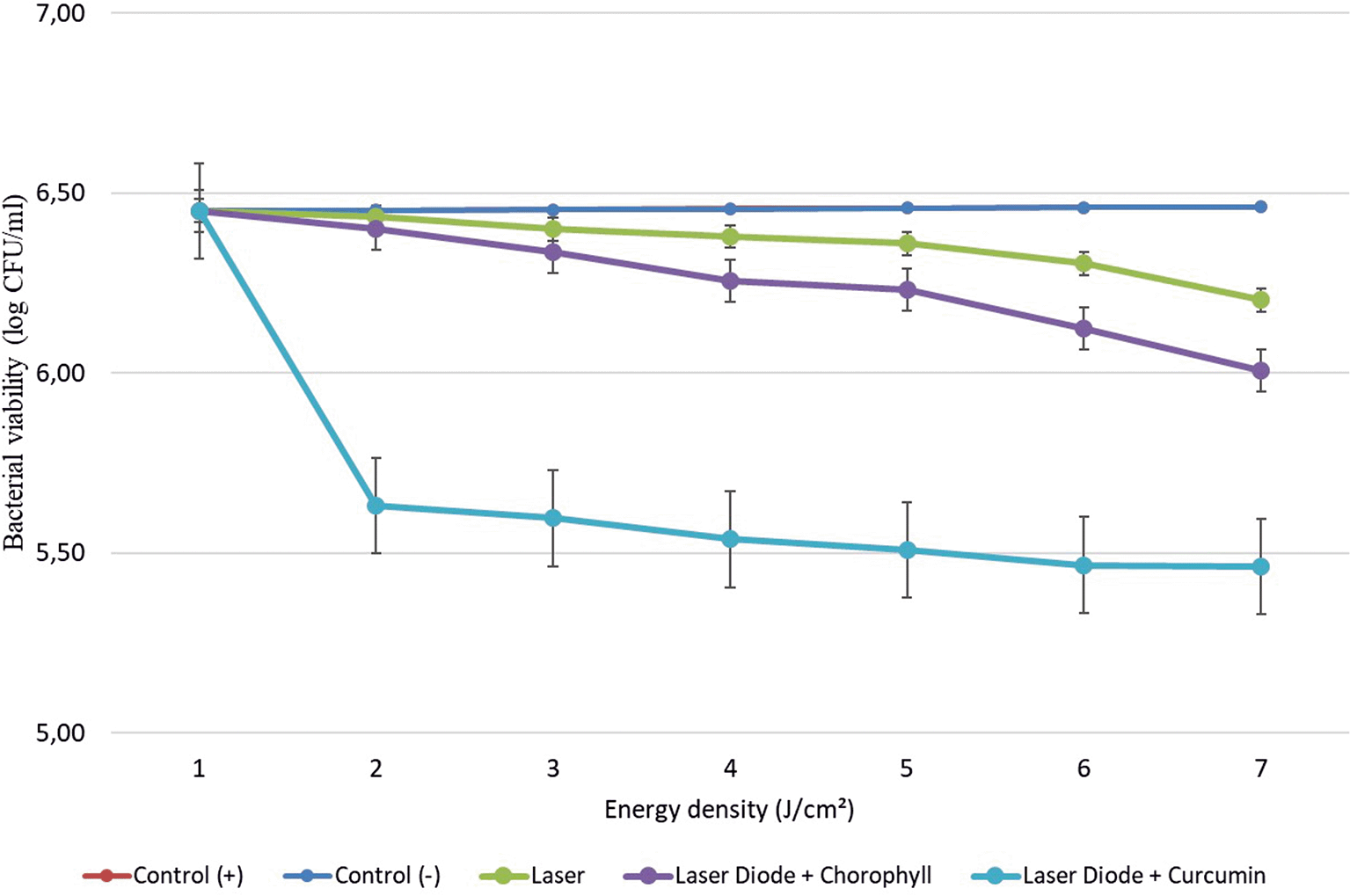
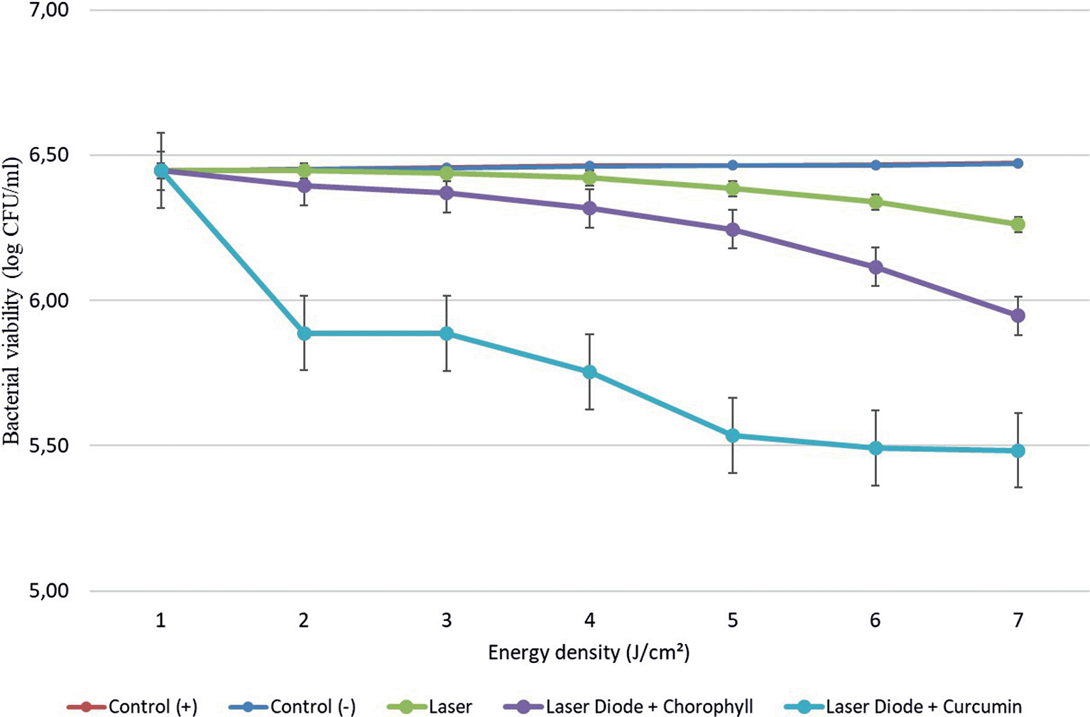
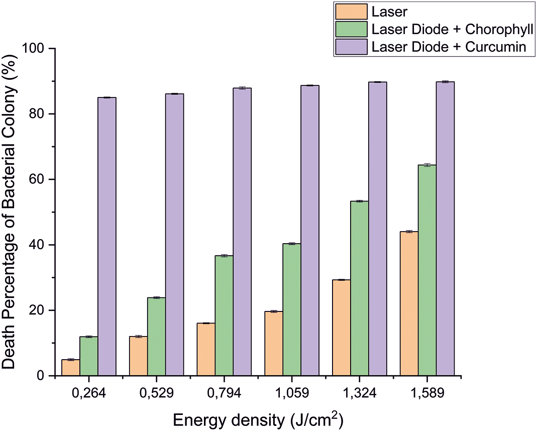
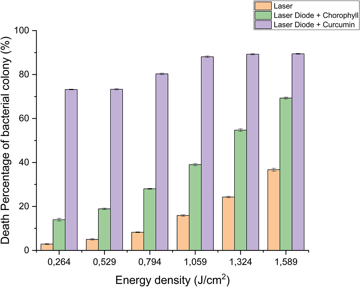
Based on the results of statistical tests, it was shown that diode laser irradiation with an energy density of 1.59 J/cm2 gave a percentage of E. faecalis bacteria death of 36.7% without adding a photosensitizer. Then, the percentage of death of E. faecalis bacteria was 69.30% with the addition of PS chlorophyll Medicago sativa L. and 89.42% with the addition of PS curcumin. Meanwhile, the results of statistical tests on bacteria A. actinomycetemcomitans with diode laser irradiation at an energy density of 1.59 J/cm2 gave the percentage of bacterial death of 35.81% without the addition of PS. Then, the death of A. actinomycetemcomitans was 64.39% with the addition of PS chlorophyll Medicago sativa L and 89.82% with the addition of PS curcumin.was 64.39% with the addition of PS chlorophyll Medicago sativa L and 89.82% with the addition of PS curcumin.
This research was conducted using the PDI technique using a blue diode laser as a light source, chlorophyll Medicago sativa L and curcumin as PS to reduce bacteria E. faecalis and A. actinomycetemcomitans. The wavelength of light is an important factor in the photoinactivation process. The laser diode used in this study has a wavelength of 405 nm and output power of 2.49 mW. The results of the characterization of power against time and temperature show the stability of power and temperature so that the temperature factor does not cause the death of bacteria.
PS is a light-sensitive molecule. Exogenous PS is PS that is added to assist the photoinactivation process. This study used exogenous PS chlorophyll Medicago sativa L and curcumin. Medicago sativa L chlorophyll absorbance used for a laser wavelength of 405 nm was 85.1% and for curcumin was 80.64%.18 The photoinactivation process occurs due to a photophysical mechanism initiated by the absorption of light by PS. The energy of the absorbed photon will cause the excitation of the electron to increase to a higher energy level. If the energy excitation state overlaps with the triplet excitation state, an intersystem crossing occurs, a spin reversal that places the electron in a triplet excited state and triggers a photochemical reaction.
Photochemical reactions are divided into two types. The first type is the transfer of electrons to a biological substrate in the form of a redox reaction and produces singlet oxygen. The second type is the transfer of energy to the triplet electrons to produce singlet oxygen. Singlet oxygen is radical. If attached to lipids and membrane proteins, it will cause peroxidation and damage cell membranes, causing leakage and cell lysis.18 The photochemical reactions in PDI are generally of the second type.
The study's results on E. faecalis bacteria showed a significant difference between treatments. Diode laser irradiation with an energy density of 1.59 J/cm2 gave the percentage of bacterial death of E. faecalis 36.7% without the addition of PS, 69.30% with the addition of PS chlorophyll Medicago sativa L and 89.42% with the addition of PS curcumin. Meanwhile, in A. actinomycetemcomitans bacteria with energy density diode laser irradiation 1.59 J/cm2, the percentage of bacterial death was 35.81% with the addition of PS, 64.39% with the addition of PS chlorophyll Medicago sativa L and 89.82% with the addition of PS curcumin.
The results showed that adding PS curcumin increased the effectiveness of reducing E. faecalis and A. actinomycetemcomitans bacteria. PDI with PS curcumin was effectively used to reduce bacteria because its absorption followed endogenous porphyrins.19 The addition of energy density will increase the reduction effect without and with the addition of PS Medicago sativa L17 and curcumin.18
Based on statistical tests, the results of research on E. faecalis bacteria showed that laser irradiation with an energy density of 1.59 J/cm2 gave a percentage of E. faecalis bacteria mortality of 36.7% without the addition of PS, 69.30% with the addition of PS chlorophyll Medicago sativa L and 89.42% with the addition of PS curcumin. Meanwhile, A. actinomycetemcomitans showed that the energy density diode laser irradiation of 1.59 J/cm2 gave the percentage of bacterial death 35.81% without the addition of PS, 64.39% with the addition of PS chlorophyll Medicago sativa L and 89.82% with the addition of PS curcumin. So it can be concluded that the role of PS is significant for the success of PDI. The addition of PS curcumin increased the effectiveness of reducing bacteria E. faecalis and A. actinomycetemcomitans compared to chlorophyll Medicago sativa L.
DA contributes to the data curation, methodology, validation, original draft preparation of the work and editing of the work. SDA contributes to the conception, methodology, analysis, funding acquisitions, project administration, supervision, validation, review, original draft preparation of the work and editing of the work. SRM, SBL, DZIN, and DAH contribute to the conception, data curation, methodology, investigation, validation, original draft preparation of the work and editing of the work. S contributes to the conception, methodology, investigation, analysis, supervision, validation, review, original draft preparation of the work and editing of the work. YS contributes to the conception, data curation, methodology, investigation, validation, original draft preparation of the work and editing of the work. AS contributes to the conception, methodology, analysis, supervision, validation, review, original draft preparation of the work and editing of the work.
| Views | Downloads | |
|---|---|---|
| F1000Research | - | - |
|
PubMed Central
Data from PMC are received and updated monthly.
|
- | - |
Provide sufficient details of any financial or non-financial competing interests to enable users to assess whether your comments might lead a reasonable person to question your impartiality. Consider the following examples, but note that this is not an exhaustive list:
Sign up for content alerts and receive a weekly or monthly email with all newly published articles
Already registered? Sign in
The email address should be the one you originally registered with F1000.
You registered with F1000 via Google, so we cannot reset your password.
To sign in, please click here.
If you still need help with your Google account password, please click here.
You registered with F1000 via Facebook, so we cannot reset your password.
To sign in, please click here.
If you still need help with your Facebook account password, please click here.
If your email address is registered with us, we will email you instructions to reset your password.
If you think you should have received this email but it has not arrived, please check your spam filters and/or contact for further assistance.
Comments on this article Comments (0)