Keywords
Central Serous Chorioretinopathy, Choroidal Thickness, Swept Source Optical Coherence Tomography, Retina, Ophthalmology
This article is included in the Eye Health gateway.
Central Serous Chorioretinopathy, Choroidal Thickness, Swept Source Optical Coherence Tomography, Retina, Ophthalmology
Central serous chorioretinopathy (CSCR) typically affects men of working age, according to Kitzmann et al. (2008).1 The estimated incidence rate for males is 10 cases per 100,000, while for women it is 2 cases per 100,000.2 The presence of neuroretinal separation, produced by the buildup of serous subretinal fluid (SRF), distinguishes clinical central serous chorioretinopathy.3 This buildup is caused by a malfunctioning retinal pigment epithelium (RPE), as well as increased permeability and thickness of the underlying choroid. The condition is characterized by the degradation of central vision, the existence of a central scotoma, and the presence of micropsia or metamorphopsia.4 CSCR is a retinal disorder that can be identifiable by the separation of the neurosensory layer of the retina.5 The disease is commonly categorized into two types: acute central serous chorioretinopathy, which usually heals on its own around a period of 4 months, and chronic CSCR.6 The categorization of these types is determined by the duration it takes for the subretinal fluid to subside.7 At present, a definitive consensus regarding the precise period differentiating the acute and chronic forms is lacking.8
In a previous study, the occurrence of both side’s appearance was estimated to be 14%,9 while a different study disclosed it to be 34%.10 This variation can be attributed, in particular, because of the lack of a standardized clinical categorization system for central serous chorioretinopathy among experienced retina professionals.11
The etiology of central serous chorioretinopathy remains incompletely elucidated. Over the past several decades, there has been a transition concerning previous theories that primarily emphasized the involvement of retinal pigment epithelium (RPE),12 either by the occurrence of localized RPE leaks or a reverse of RPE cell polarity leading to the release of fluid to the subretinal space.13 More recent hypotheses have emerged, highlighting the choroid’s significance in the etiology of CSCR.14
Indocyanine green angiography (ICGA) research studies have demonstrated the presence of extensive and multifocal regions of choroidal hyperpermeability surrounding active retinal pigment epithelium leaking.15
Liu et al. conducted a comprehensive review and meta-analysis, which yielded findings suggesting that several factors, including elevated blood pressure, infection with Helicobacter pylori, steroid medication consumption, sleeping disorder, immune-mediated illness, psychopharmacologic intake, and Type-A personality, may be associated with an increased risk of developing central serous chorioretinopathy.16 A recent study conducted by Jia Yu et al. has established a correlation between various factors, including being pregnant, obstructive sleep apnea, transplantation of organs, kidney failure, and a history of methylenedioxymethamphetamine (MDMA), commonly referred to as ecstasy addiction, and the development of the illness as mentioned earlier.17
It is hypothesized that an elevation in choroidal vascular permeability may result from arterial vascular spasm induced by adrenaline, which is further intensified by the presence of steroid hormones.18 This process eventually results in choroidal ischemia and a rise in vascular hyper-permeability. Consequently, the level of oncotic pressure within the choroidal space rises, and when paired with the malfunction of the retinal pigment epithelium, the fluid accumulates in the sub-retinal space.19
The objective of this study is to assess the choroidal thickness in eyes exhibiting active central serous chorioretinopathy along with comparing it with the uninfluenced opposite eyes of the exact same participants, as well as with the regards of a control group consisting of healthy individuals.
On May 1, 2019, the research proposal, assigned to Research No. 24 and Approval No. 119, was granted approval by the research ethics committee of the Iraqi Board of Ophthalmology. Following a thorough discussion of the study objectives, every participant officially provided written informed consent. The personal information of individuals involved in the study was maintained anonymously.
The present research was an observational case-control study carried out on a group of Iraqi individuals who were looking for medical care at the retina clinic, which is a part of the ophthalmology division at Ghazi Al-Hariri Hospital for Surgical Specialties within the Medical City Complex in Baghdad, Iraq. The data collection period for this study ran from October 1, 2019, to March 31, 2020. At the completion of the data acquisition process, 49 individuals were successfully included, resulting in a sample size of 65 eyes.
The recruited cases consisted of confirmed cases of active central serous chorioretinopathy, which is defined as the presence of subretinal fluid on swept-source OCT imaging. A time frame of six months from the onset of signs was determined as the threshold for distinguishing between acute and chronic central serous chorioretinopathy cases.
Any patient presenting with one or more of the following conditions was excluded from the study, refractive errors characterized by a spherical equivalent exceeding 1 diopter, hypertension that is not under control characterized by elevated systolic blood pressure exceeding 139 mmHg and/or alternatively, diastolic blood pressure exceeding 89 mmHg, the patient has a history of receiving intravitreal injections of anti-vascular endothelial growth factor agents, the patient has a history of prior intraocular surgery, the presence of cataracts and media opacities can lead to a decline in image quality, a preceding photodynamic therapy, glaucoma, diabetes type 1 or 2, the history of uveitis or trauma.
The determination and computation of the sample size were conducted using the software application G*Power 3.1.9.7 (RRID: SCR 013726). The minimum sample size observed in this study was 42 individuals. The effect size was determined to be 0.5, with a power of 95% at a two-tailed alpha level of 0.05. Additionally, a 95% confidence interval was calculated for the observed effect size. A total of 49 participants were chosen for the study, while the remaining individuals met the criteria for exclusion as presented in Figure 1.
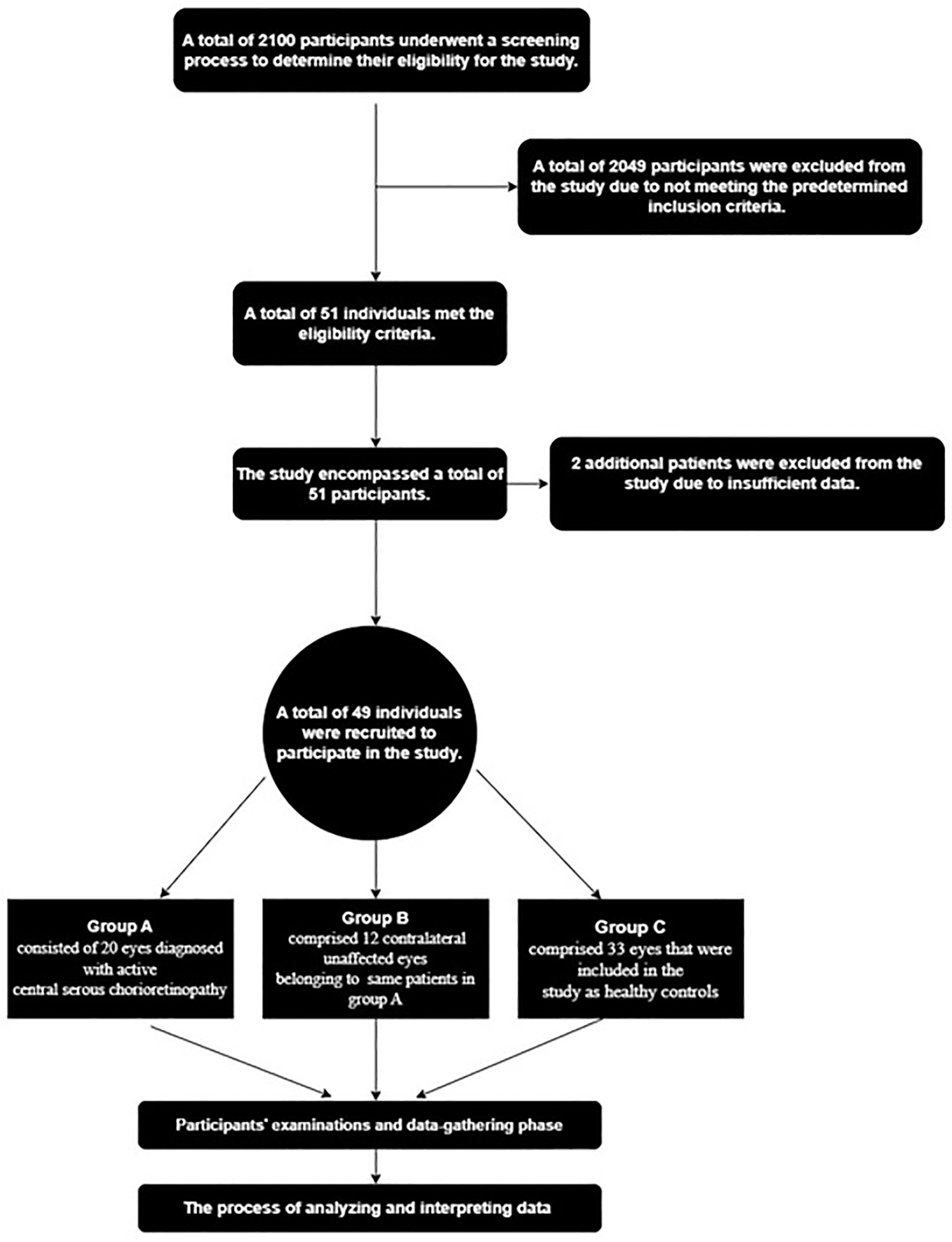
Group A consisted of 20 eyes diagnosed with active central serous chorioretinopathy. It is worth noting that four individuals in this set had both sides of active disease, resulting in the inclusion of each of their retinas in the analysis.
Group B comprised 12 contralateral unaffected eyes belonging to patients with active central serous chorioretinopathy.
Group C comprised 33 eyes that were included in the study as healthy controls.
One of the most successful techniques used by researchers to mitigate sampling bias is simple random sampling, when samples are selected purely based on chance. This research ensured that each member of the population had an equal probability of being selected as a participant.
During the study period, individuals who had been diagnosed with central serous chorioretinopathy were recruited from a specialized medical retina clinic. These patients had already undergone fundus fluorescein angiography and provided informed consent. Subsequently, they were referred to the assessment unit, where they underwent a thorough evaluation conducted by a specialized ophthalmologist. The participants were paired according to their ages and sex. The control group was comprised of people who were chosen from the population of family members accompanying patients to the ophthalmology department for problems that were not connected to central serous chorioretinopathy. The procedure included the collection of patient medical records by direct interaction and the performance of a comprehensive examination while the patient was sitting. In order to minimize any diurnal variations in choroidal thickness, all measurements were performed only in the morning, precisely between the hours of 9:00 AM and 12:00 PM. The procedures conducted in this study encompassed various components, including interviews pertaining to the aforementioned aspects. Additionally, a sequential approach was followed for the measurements, commencing with the assessment of visual acuity using a Snellen chart. Subsequently, refraction was determined using an autorefractor, followed by the measurement of intraocular pressure (IOPA) utilizing an air puff tonometer. Furthermore, an evaluation of the anterior segment was performed, and subsequently, one drop of 1% Tropicamide was administered to induce pupil dilation. This facilitated the examination of the posterior segments of the eye using a slit lamp and a +78 diopter condensing lens. Moreover, images of the fundus were captured employing a fundus camera. Additionally, the participant’s height and weight were recorded to facilitate the calculation of their body mass index (BMI). Lastly, arterial pressure was measured in a seated position utilizing a mercury sphygmomanometer. The assessment of choroidal thickness was conducted using the deep-range imaging swept-source optical coherence tomography (SS-OCT) equipment developed by Topcon Corp. in Tokyo, Japan utilizing the default configurations. Each assessment was done by a single trained person using the provided software (version 10.0x). The results were shown using an Early Treatment Diabetic Retinopathy Study (ETDRS) graphic. The measurement of choroidal thickness was conducted in nine distinct sectors located inside the posterior pole, as seen in Figure 2. A summary of the equipment used during the study has been included in Table 1.
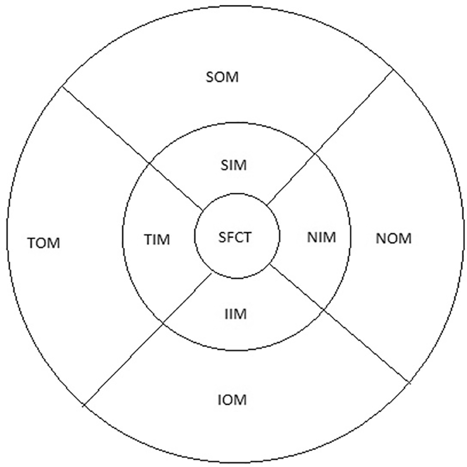
The inner circle has a diameter of 1 mm, the middle circle has a diameter of 3 mm, and the outer circle has a diameter of 6 mm (TOM: temporal outer macula, SOM: superior outer macula, NOM: nasal outer macula, IOM: inferior outer macula, TIM: temporal inner macula, SIM: superior inner macula, NIM: nasal inner macula, IIM: inferior inner macula, SFCT: sub foveal choroidal thickness).
The statistical process was conducted with SPSS version 23 (RRID:SCR_002865). All data were presented as mean±standard deviation. We employed the independent samples t-test and Fisher’s exact test for analyzing demographic information among our various study groups. To assess the differences in choroidal thickness throughout the study groups, we utilized the Kruskal-Wallis test with Dunn-Bonferroni pairwise comparisons. Significance levels were set at p<0.05 and p<0.001, denoting statistical significance and high statistical significance, respectively.
The current research study included a total of 49 individuals, corresponding to 65 eyes.20 Among these participants, there were 16 individuals diagnosed with central serous chorioretinopathy, accounting for 32 eyes. More precisely, eight participants had acute type, while the remaining eight were chronic cases. The average age of the patients was 38.38 years, with 14 males and two females included in this group. Additionally, there were 33 healthy subjects included in the study, with only one eye per subject being analyzed, resulting in a total of 33 eyes serving as a control group. The average age of the healthy participants was 36.36 years, with 28 males and five females included in this group. The best-corrected visual acuity (BCVA) observed among individuals with central serous chorioretinopathy varied from 6/6 to 6/24, whereas the whole group of control subjects exhibited a BCVA of 6/6.
Table 2 demonstrates that there were no statistically significant differences observed among patients with central serous chorioretinopathy and the control group in terms of age, sex, body mass index, or spherical equivalent.
| Parameters | Central serous chorioretinopathy group | Control group | P-Value (Statistical significance) |
|---|---|---|---|
| Mean±SD | Mean±SD | ||
| Age | 38.38±8.7 | 36.36±8.3 | p>0.05* |
| Gender (M/F) | 14/2 | 28/5 | p>0.05** |
| Body mass index | 25.88±3.2 | 27.12±4.3 | p>0.05* |
| Spherical equivalent | 0.83±0.2 | 0.92±0.2 | p>0.05* |
In statistical terms, significant disparities in choroidal thicknesses were observed across every sector whenever comparing group A to group C, and also when comparing group B to group C, despite controlling for variables such as age, sex, body mass index, and spherical equivalent. Furthermore, the choroid exhibited greater thickness in all sectors within group A compared to group B, although this discrepancy did not reach statistical significance, as can be seen in Table 3.
| Parameters | Group A | Group B | Group C | P-Value (Statistical significance) |
|---|---|---|---|---|
| Mean±SD | Mean±SD | Mean±SD | ||
| SFCT | 474.5±89.6 | 437.5±90.8 | 292.0±59.5 | <0.001/<0.001* |
| TIM | 460.3±94.3 | 418.7±95.5 | 285.6±56.6 | 0.001/<0.001* |
| SIM | 467.7±82.5 | 427.5±75.8 | 291.5±58.1 | <0.001/<0.001* |
| NIM | 462.3±89.7 | 425.7±77.3 | 295.4±63.2 | <0.001/<0.001* |
| IIM | 469.5±105.2 | 438.9±84.2 | 303.2±60.6 | <0.001/<0.001* |
| TOM | 403.9±78.3 | 364.3±92.4 | 269.2±53.2 | 0.005/<0.001* |
| SOM | 437.9±80.5 | 395.8±73.1 | 283.8±50.7 | <0.001/<0.001* |
| NOM | 401.8±83.5 | 373±65.3 | 247.5±58.3 | <0.001/<0.001* |
| IOM | 443.5±89.5 | 417.1±84 | 291.3±62.8 | <0.001/<0.001* |
The evaluation of the choroid has historically posed difficulties when using conventional imaging methods, such as indocyanine green angiography and B-scan ultrasound imaging. Nevertheless, the advent of optical coherence tomography has enabled the in vivo visualization of the choroid in a manner comparable to retinal imaging, thus providing a more comprehensive inspection.21 Estimating choroidal thickness has presented novel opportunities for investigating various pathologies affecting the choroid.22 One limitation of SD-OCT is its inability to effectively image the sclera-choroidal junction within some eyes, particularly in individuals with media opacities. In contrast, SS-OCT enables the clear imaging of both the retina and the sclera-choroidal junction in such cases.23
The current research revealed a substantial rise in choroidal thickness throughout all evaluated eye regions exhibiting active central serous chorioretinopathy. Specifically, the subfoveal choroidal thickness was determined to be 474.55 μm in CSCR eyes, whereas it was 292.03 μm in the control group. These findings align with previous investigations utilizing indocyanine green angiography, demonstrating choroidal vessel congestion and heightened permeability of vascular walls (as evidenced by raised choroidal thickness) in CSCR cases.15,24
Jirarattanasopa et al. disclosed comparable findings, indicating that the subfoveal choroidal thickness measured 374.3 μm, whereas healthy eyes exhibited an average value of 248.4 μm. It is worth noting that their study employed a prototype swept-source optical coherence tomography device and using traditional measurement techniques, which may account for the observed disparities in comparison to our own measures.25
In a research project conducted by Cano-Hidalgo et al. in Mexico, it was noted the fact that the average choroidal thickness in the influenced eyes was measured to be 435 μm, similar to the aforementioned study, measurements performed by hand were employed, and the study participants consisted of an older patient group with an average age of 44.23 years±11.57 years. Furthermore, the researchers observed a negative correlation between choroidal thickness and age among patients with central serous chorioretinopathy.26
The findings of this study indicate that the choroidal thickness in the unaffected eyes of individuals with central serous chorioretinopathy was noticeably greater compared to that observed in control eyes. Specifically, the subfoveal choroidal thickness measured 437.55 μm in CSCR patients’ unaffected eyes, while it measured 292.03 μm in control eyes. This finding was also corroborated by Cano-Hidalgo et al., as well as by other studies utilizing enhanced depth imaging optical coherence tomography.27,28
Numerous studies have demonstrated that eyes afflicted with active central serous chorioretinopathy exhibit a greater thickness of the choroid in comparison to their unaffected counterpart eyes.26,28 The present study yielded comparable findings; however, it did not attain statistical significance, potentially attributable to the limited size of the sample.
The observed elevation in choroidal thickness in the unaffected fellow eyes implies that the disorder under investigation may have systemic implications, particularly regarding the malfunction of the choroidal vasculature due to abnormalities in the mineralocorticoid receptor pathway among affected individuals.14 This hypothesis is further reinforced by the identified risk factors for central serous chorioretinopathy, including sex, anxieties, steroid consumption, and pregnancy, as well as the susceptibility of choroidal vessels to systemic factors due to their lack of autoregulation.
A number of the limitations that accompany this study pertain to the relatively small sample size employed, which hindered our ability to conduct a comparative analysis of choroidal thickness between acute and chronic central serous chorioretinopathy.
The findings of the present research indicate that the choroid exhibits thickening not solely in eyes displaying active central serous chorioretinopathy, but also in the unaffected contra lateral eyes of the same individuals. It is recommended that future research endeavors employ a greater number of participants in order to conduct further investigations utilizing fluorescein angiography as a means to establish associations between choroidal thickness and angiographic findings.
| Views | Downloads | |
|---|---|---|
| F1000Research | - | - |
|
PubMed Central
Data from PMC are received and updated monthly.
|
- | - |
Provide sufficient details of any financial or non-financial competing interests to enable users to assess whether your comments might lead a reasonable person to question your impartiality. Consider the following examples, but note that this is not an exhaustive list:
Sign up for content alerts and receive a weekly or monthly email with all newly published articles
Already registered? Sign in
The email address should be the one you originally registered with F1000.
You registered with F1000 via Google, so we cannot reset your password.
To sign in, please click here.
If you still need help with your Google account password, please click here.
You registered with F1000 via Facebook, so we cannot reset your password.
To sign in, please click here.
If you still need help with your Facebook account password, please click here.
If your email address is registered with us, we will email you instructions to reset your password.
If you think you should have received this email but it has not arrived, please check your spam filters and/or contact for further assistance.
Comments on this article Comments (0)