Keywords
Sebaceous carcinoma, Muir- Torre syndrome, eyelid cancer
This article is included in the Oncology gateway.
This article is included in the Datta Meghe Institute of Higher Education and Research collection.
Sebaceous carcinoma, Muir- Torre syndrome, eyelid cancer
We have added certain details suggested by the reviewer for better understanding of the disease.
See the authors' detailed response to the review by Jianmin Ma
Sebaceous carcinoma originates from cutaneous sebaceous glands, gland of Zeis or the Meibomian glands.1 It is more commonly found in the Asian population.2 It has a syndromic association with Muir-Torre syndrome, with hereditary nonpolyposis colorectal cancer syndrome.3 It can occur secondary to radiation therapy for retinoblastoma.4 It clinically mimics some benign conditions such as blepharitis, chalazion and sometimes basal cell carcinoma or squamous cell carcinoma which causes difficulty in diagnosing the disease.5 It is very essential to diagnose this fatal disease due to its potential of metastasis and local invasion. Early diagnosis can prevent patient’s vision and may increase survival. Here, we report a case of sebaceous carcinoma of the upper eyelid on histopathology of orbital exenteration specimen.
A 50-year-old male, farmer by occupation, presented with an 8-month history of an ulcerated growth over the left eye. The patient stated that the lesion started as a pea sized cystic swelling which gradually increased in size. He also gave a history of pain and maggot infestation in the same eye for which he took symptomatic treatment. Condition of patient deteriorated further, so he came to the tertiary care centre for further management.
Routine investigations were performed, on ocular examination no light perception was present. Slit lamp examination of the left showed a mass of 6.3 × 3 × 1.5 cm with ulcerated appearance. Eyelashes were spared. The eyeball appeared as degenerated and distorted. On general examination no lymphadenopathy and organomegaly was noted.
MRI (Magnetic resonance imaging) reports showed an ulcerated mass in the left periorbital and orbital region appearing heterogeneously hyperintense on T2 and hypointense on T1 which was involving the left orbital group, adjacent intraorbital optic nerve and extraocular muscles.
Fine needle aspiration cytology (FNAC) was performed which showed polymorphs, lymphocytes and islands of tumor cells. These tumor cells appeared large in size, oval in shape and were arranged in groups. These cells had hyperchromatic nuclei. Cells also showed malignant epithelial differentiation.
One day prior to surgery general physical examination and orbital examination was done. Patient was considered fit for the intervention. A surgical intervention named intraoperative frozen section was performed from the lesion and diagnosis suggestive of carcinoma either from sebaceous or squamous origin was made. After that, left eye exenteration was done.
The resected orbital exenteration specimen [Figure 1] was sent to histopathology. Specimen measured 6.2 × 6 × 3.5 cm. A tumor mass involving the upper eyelid extending from the medial margin measuring 5 × 2 × 1.5 cm was identified.
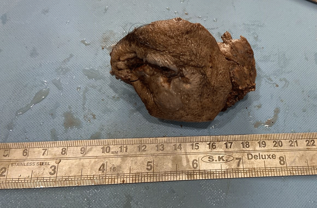
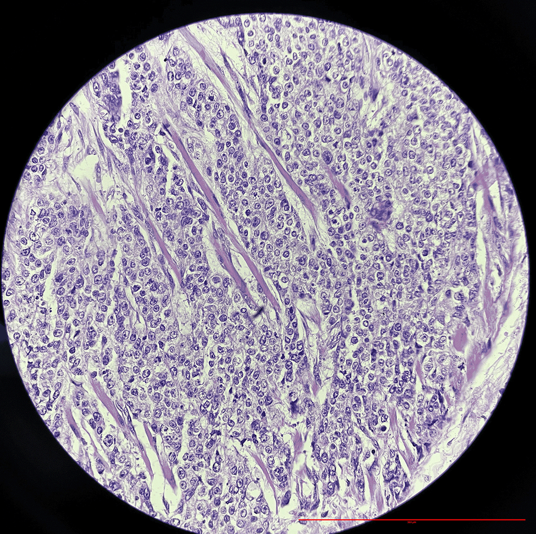
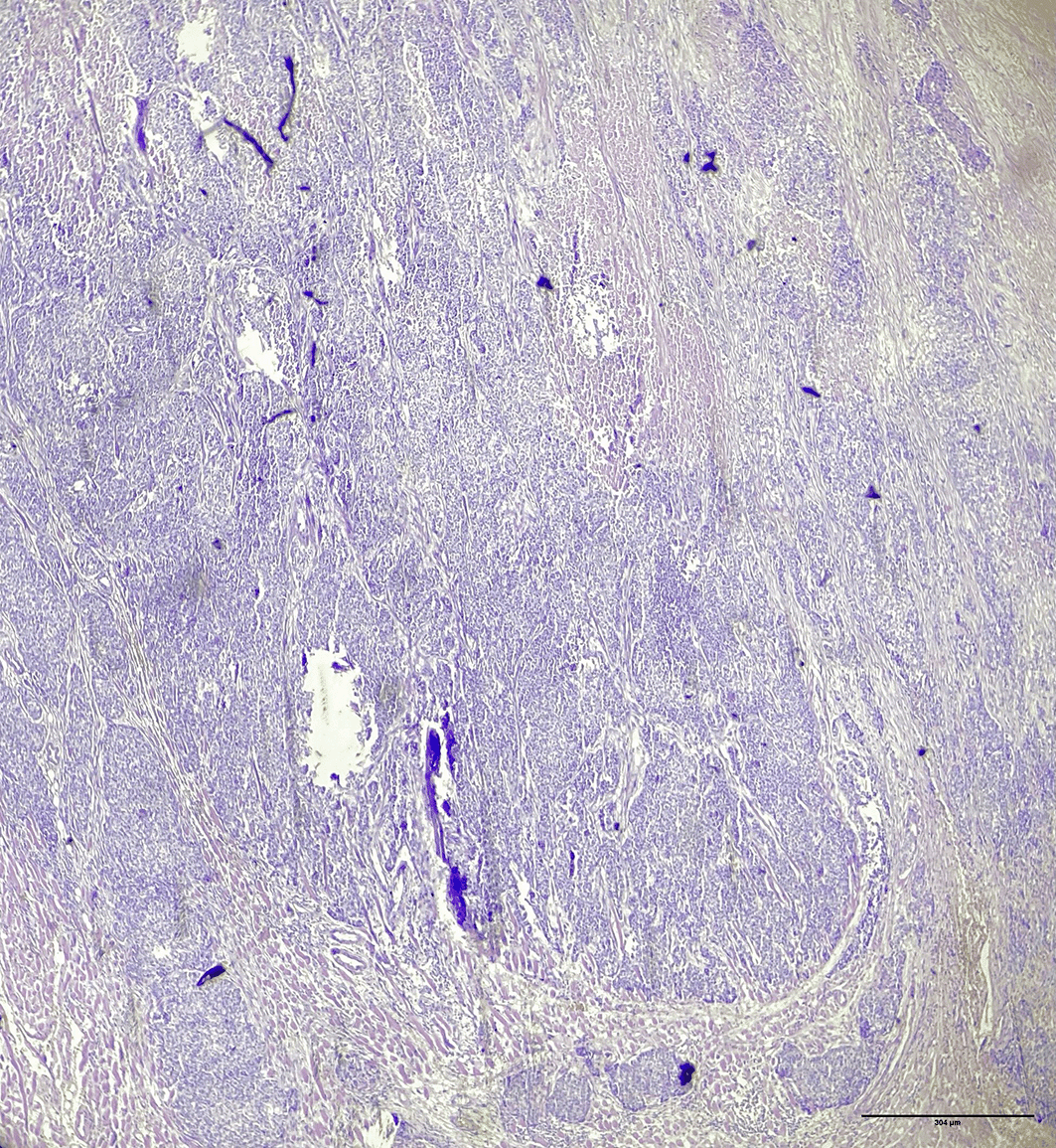
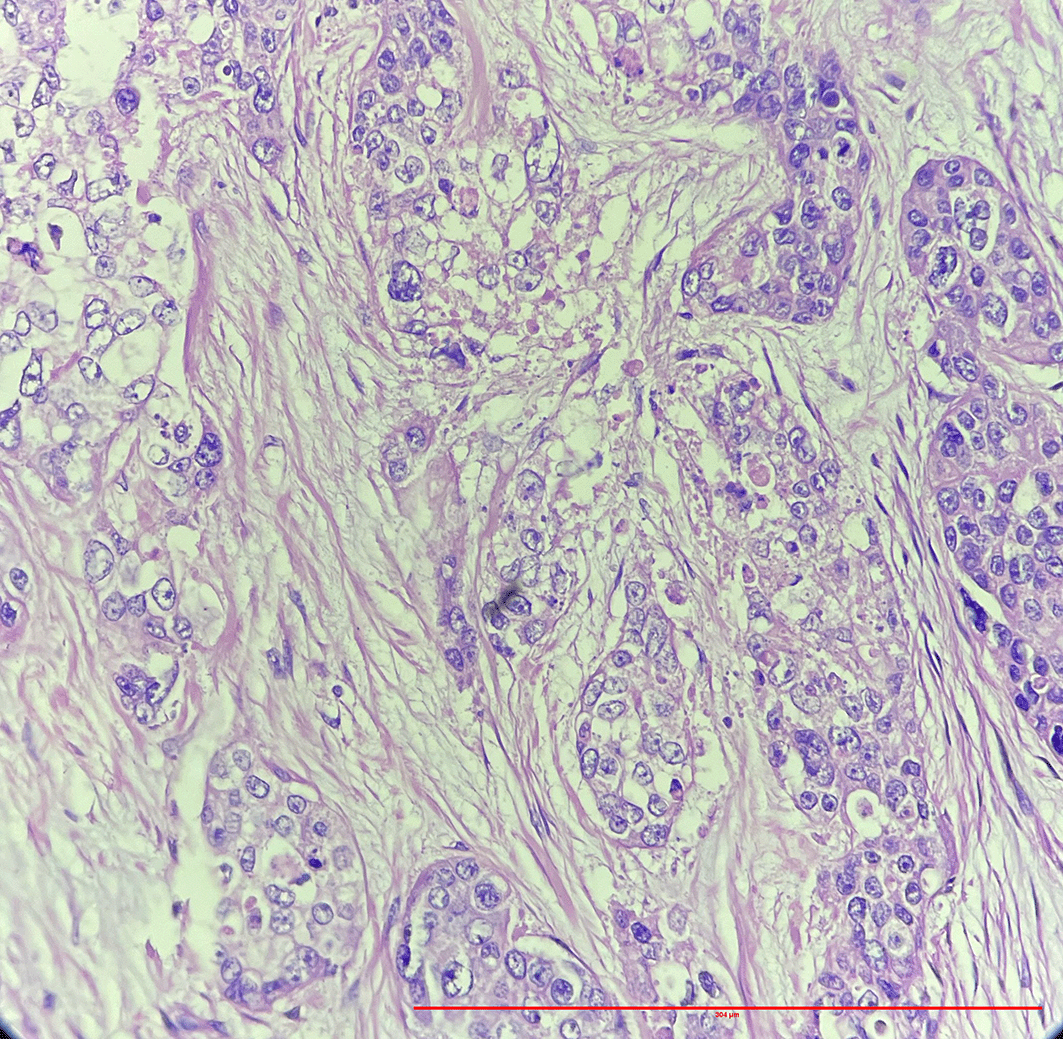
Microscopy showed pigmented lining epithelium. A scanner view of the section [Figure 2] also showed tumor cells arranged in lobules and sheets at places. High power view i.e. 40× showed sebaceous differentiation [Figure 3], individual tumor cells, which were polygonal in shape with scant, multivacuolated cytoplasm, it also showed round to oval, enlarged pleomorphic nuclei with 1–2 prominent nucleoli [Figure 4]. Deeper tissues showed fibro collagenous tissue, necrotic hemorrhagic tissue and infiltration by malignant cells.
Sebaceous carcinoma is a great masquerader.6 Although sebaceous carcinoma has great tendency to arise in the ocular region, especially in the eyelids, it does occur in extraocular regions such as parotid. It accounts for 1 to 3% of malignant orbital tumors.7 Among the reported malignancies of eyelid tumors it is the third most common malignancy.8 It has a female predominance.9 The upper eyelid is affected more than the lower lid due to the abundance of meibomian glands.10 Periocular sebaceous carcinoma can clinically mimic a range of conditions and are misdiagnosed as basal cell carcinoma or squamous cell carcinoma.11
The most common presentation of sebaceous carcinoma in the eyelid is as round highly cellular nests of poorly differentiated tumor cells. Sometimes better differentiated cells with vacuolated cytoplasm are identified.12
Many studies have shown that sebaceous carcinoma of the eyelid has a poor prognosis and if there is an orbital or vascular invasion present prognosis further worsens.13
The treatment of choice for sebaceous carcinoma is surgery, with complete excision verified by negative margins.14
Sebaceous carcinoma of the eyelid is a rare entity, but might be difficult to diagnose because of its ability to masquerade as the periocular lesions. However, accurate and prompt diagnosis is crucial for planning further management of the disease and prevention of any complications such as loss of vision, metastasis to other organs etc, therefore tissue diagnosis is the gold standard method and it can be aided by a panel of immunohistochemistry stains.
Written informed consent for publication of their clinical details and clinical images was obtained from the patients.
Jayashree Bhawani: drafting the case report and overview of patient management. Dr. Samarth Shukla: Reporting of the excised specimen sent for histopathological investigation and giving the suitable diagnosis. All the authors read and approved the final version of this manuscript.
| Views | Downloads | |
|---|---|---|
| F1000Research | - | - |
|
PubMed Central
Data from PMC are received and updated monthly.
|
- | - |
Provide sufficient details of any financial or non-financial competing interests to enable users to assess whether your comments might lead a reasonable person to question your impartiality. Consider the following examples, but note that this is not an exhaustive list:
Sign up for content alerts and receive a weekly or monthly email with all newly published articles
Already registered? Sign in
The email address should be the one you originally registered with F1000.
You registered with F1000 via Google, so we cannot reset your password.
To sign in, please click here.
If you still need help with your Google account password, please click here.
You registered with F1000 via Facebook, so we cannot reset your password.
To sign in, please click here.
If you still need help with your Facebook account password, please click here.
If your email address is registered with us, we will email you instructions to reset your password.
If you think you should have received this email but it has not arrived, please check your spam filters and/or contact for further assistance.
Comments on this article Comments (0)