Keywords
Hydtidosis, multi visceral, muscles
Hydtidosis, multi visceral, muscles
Hydatidosis is a human disease caused by the larval form of Echinococcus spp., which live in the gut of dogs, wild canines and other carnivorous animals. Humans become the accidental intermediate hosts by ingesting Taenia spp. eggs. Echinococcus spp. are endemic in many countries where sheep, dogs and man live in close contact.
All organs in the human body may be affected by hydatid disease. The hydatid cyst from Echinococcus granulosus commonly involves the liver and lung but may also be found in other unusual organs, including the brain, heart and bones. Hydatid cysts rupture into left-sided cardiac chambers may cause systemic emboli, and if ruptured into right-sided cardiac chambers may cause pulmonary emboli.
Cases of multi visceral echinococcosis with atypical localization are rare. We report here the case of a male with 13 hydatid cysts including the lung, the heart, the muscles, the liver, the kidney and abdomen cavity.
This is the case of a 53-year-old maghribian male farmer. This patient, with no personal or family history, was referred to the pulmonology department, for the management of a pulmonary Aspergilloma on a sequelary lung cavity following lung surgery.
At the time of the encounter the patient had no subjective complaints, expect for right lower limb paresthesia. The physical exam showed no abnormalities.
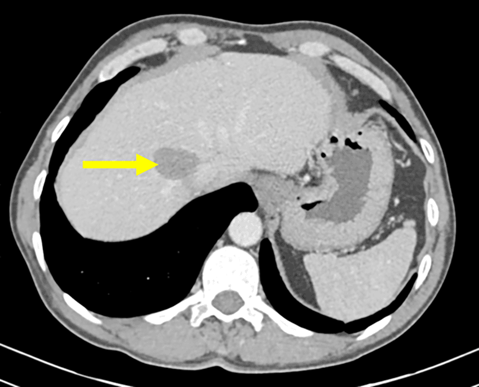
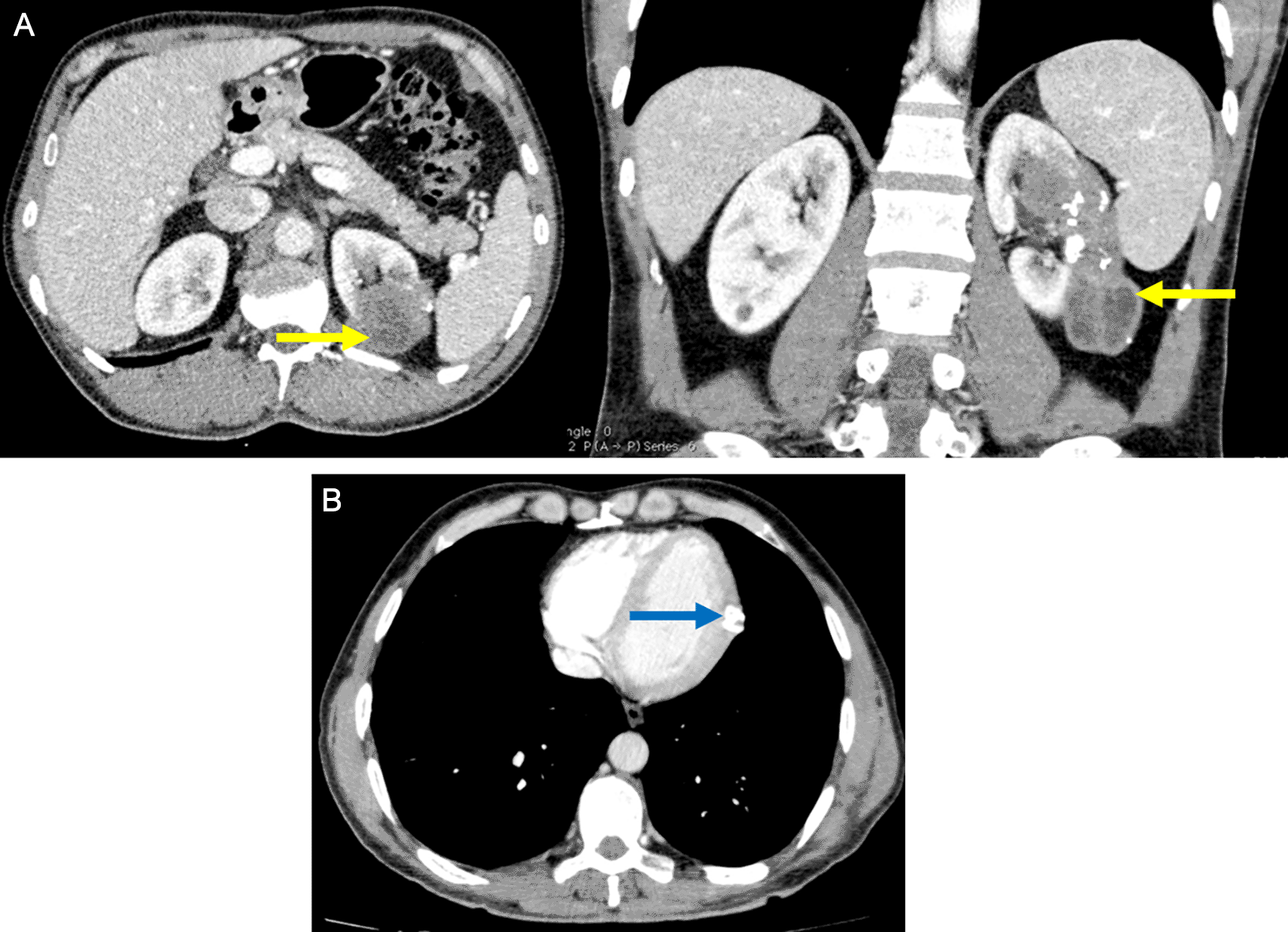
The patient was followed up in the abdominal surgery department for a multiple organ hydatidosis that included 13 hydatid cysts: the lungs, the liver (Figure 1), the left heart ventricle, the left kidney (Figure 2), the abdomen cavity, muscles (psoas, adductors), and subcutaneous gluteal area. The majority of these cysts was already treated surgically and some are still pending to be removed (Table 1). The patient has received oral Albendazole 400 mg twice-daily for 2 years.
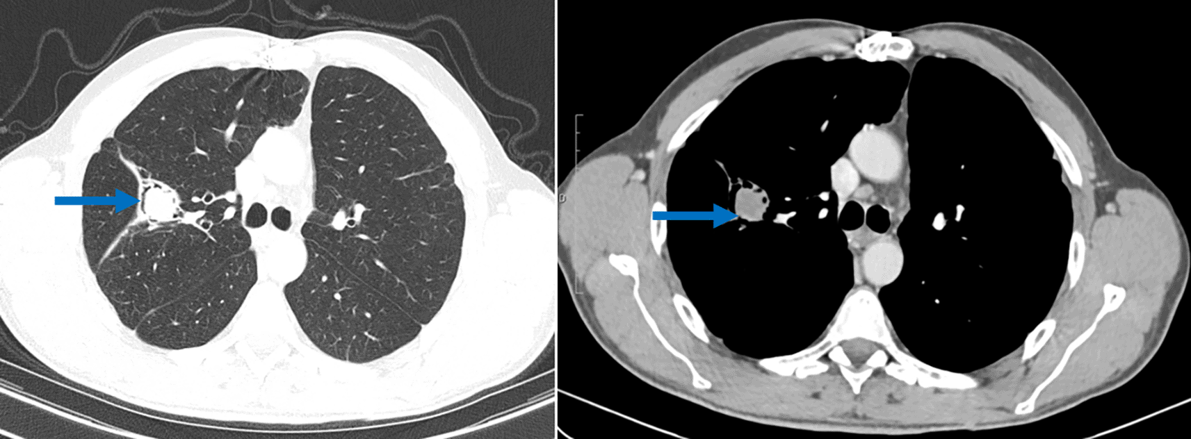
During a regular post-surgical follow up, a scannographic image of a fluid-density endo-bronchial material in the right superior lobe (lung cavity sequelary to the previous cystectomy) of the lung separated from the cavity wall by an airspace (“air crescent” sign) was found, this image is typical of pulmonary aspergilloma (Figure 3). Even though the Aspergillus serology showed doubtful results, the clinical context coupled to the CT scan findings were highly suggestive of a pulmonary aspergilloma. Blood work up showed high levels of IgE. A surgical resection of the cavity is programmed but not yet performed.
Hydatid cyst remains an important issue in Tunisian health that affects both humans and animals, especially in rural areas.
The annual incidence of hydatidosis is 11.3 per 100000 inhabitant.1 Usually, hydatidosis presents as a single cyst usually of the liver or of the lungs, but in certain circumstances multiple organs may be affected. In our case we are reporting multiple atypical locations including heart, kidney, muscles and subcutaneous tissue. The conjunction of cysts in these locations simultaneously has not been described in the literature. We will not focus on the liver and the lung involvement given that they are a classic location of this parasitosis, and they are well commonly cited in the literature, instead we will discuss the other locations separately.
Heart involvement is uncommon and accounts for less than 0.5% of the cases, it is usually part of a disseminated infection.2 This localization is potentially fatal without surgical treatment but fortunately with the improvement of surgical techniques, its morbidity has declined drastically. Our patient underwent open heart surgery to remove a left ventricular wall cyst without local recurrence and a post-surgical echocardiography without abnormalities.
The invasion of the myocardium usually occurs hematogeniously through the coronary arteries and since the majority of the population have a left dominant circulation, the left ventricle is the most commonly involved part of the heart (60%),3 other explanation is the dissemination from the lungs either following a pulmonary vein rupture and migration of the cysts4 or by a direct contact with hydatid cysts originating from the lung.5
Renal involvement is also rare (2–3%) and it is usually associated to a disseminated disease, they are most commonly asymptomatic, like the case of our patient. The diagnosis was made by an abdominal CT which has a sensitivity of 98% to diagnose hydatid disease.6
Psoas cysts is also uncommon, our patient presented with two psoas cysts, a finding never been described before in literature.
The patient has also presented with a 30-mm gluteal subcutaneous cyst, this involvement was described in rare cases in literature and usually the patient will have a painless palpable mass history of at least 3 months, and it is usually larger than 3 cm, which is the case in our patient.7 Subcutaneous cysts tend to involve trunk and limb roots, possibly due to the rich vascularization and relatively less muscle activity in these areas.8
Another intriguing finding in this case report is the discovery of an aspergilloma, on a lung cavity. Pulmonary aspergilloma occurs as a colonizer of pre-existing pulmonary cavity of any etiology such as sequelae tuberculosis, cavitary neoplasia or operated hydatid cyst and it is a saprophytic infection.9 Aspergilloma has rarely been described in operated hydatid cyst cavities in immunocompetent patients.10 For this patient, the aspergilloma was discovered two years after the lung surgery. A very similar case of a 56-year-old patient, who presented with an aspergilloma of the upper right lobe following cystectomy, have been described by M. El Hammoumi et al.10
Despite the existence of multiple cysts, our patient is doing well with good tolerance and he is asymptomatic.
Multiple hydatidosis is a rare condition which can endanger the vital and functional prognosis. Imaging is essential for the diagnosis and finds its place for the assessment of extension and detection of asymptomatic localization to ensure early treatment. Prevention remains the best treatment for hydatid cyst.
| Views | Downloads | |
|---|---|---|
| F1000Research | - | - |
|
PubMed Central
Data from PMC are received and updated monthly.
|
- | - |
Provide sufficient details of any financial or non-financial competing interests to enable users to assess whether your comments might lead a reasonable person to question your impartiality. Consider the following examples, but note that this is not an exhaustive list:
Sign up for content alerts and receive a weekly or monthly email with all newly published articles
Already registered? Sign in
The email address should be the one you originally registered with F1000.
You registered with F1000 via Google, so we cannot reset your password.
To sign in, please click here.
If you still need help with your Google account password, please click here.
You registered with F1000 via Facebook, so we cannot reset your password.
To sign in, please click here.
If you still need help with your Facebook account password, please click here.
If your email address is registered with us, we will email you instructions to reset your password.
If you think you should have received this email but it has not arrived, please check your spam filters and/or contact for further assistance.
Comments on this article Comments (0)