Keywords
Cisplatin, melatonin, doxorubicin, SKOV3
This article is included in the Oncology gateway.
Cisplatin, melatonin, doxorubicin, SKOV3
There are several new addition in the mechanism of melatonin in other cancer that have been added to discussion. Also this study was performed only in one cell line, to show several pathway of melatonin, there should be other study that performed on several cell line, so we have been added the limitation section.
See the authors' detailed response to the review by Tharcisio C. Tortelli
See the authors' detailed response to the review by Pavel Souček
See the authors' detailed response to the review by Seyed Mostafa Mir
To date, the management of ovarian cancer, especially epithelial ovarian cancer, has not yet given satisfactory results (Razi et al., 2016). Ovarian cancer is known to have high morbidity and mortality rates as well as poor prognosis (Coburn et al., 2017). It ranks third in the world after cervical and uterine cancer (Bray et al., 2018). According to the Global Burden Cancer 2020 database, the number of ovarian cancer cases worldwide is 313,959, with the number of deaths being 207,252 (Sung et al., 2021). In Indonesia, the rate of ovarian cancer is still high at around 3,398 from 2016 to 2020 (INASGO, 2021).
At present, ovarian cancer recurrence rate is still high and influences the morbidity and mortality rates. One of the reasons why the recurrence rate remains high is chemotherapy resistance, especially by cisplatin at cancer stem cell (CSC), because evasion from apoptosis caused the cell to redevelop after therapy has been performed (Woo et al., 2012). There are multiple factors in cisplatin resistance, and the mechanism is still unclear from the perspective of cellular mechanism (Sousa et al., 2014).
There are many intracellular mechanisms that affect the evasion of a cell from the cytotoxic effect of chemotherapy, such as the process that regulates drug bioavailability, mesenchymal epithelial transition, and oncogenic signals producing a phenotype that causes resistance to chemotherapy (Yeldag et al., 2018).
Melatonin has been considered an alternative for managing chemotherapy resistance, especially in ovarian cancer (Chuffa et al., 2017). It is synthesized at the pineal gland. Melatonin exerts antioxidant and antiapoptotic effects on normal cells but exerts pro-oxidative, antiproliferative, antiangiogenic, and immunomodulatory effects on cancer cells, especially hormone-dependent cancer (Su et al., 2017). Melatonin has been known for it’s oncostatic acitivity in several malignancies. Melatonin has anticancer and oncostatic effects, e.g., it potentiates antimetastasis, induces anticancer immunity, modulation of cell cycle, induces autophagy, modulate oncogene expression, enhances drug sensitivity, induces apoptosis, inhibits cancer growth, and exhibits antiangiogenic and antiinvasive activities (Su et al., 2017; Targhazeh et al., 2022). In addition, melatonin has a good effect on clinical outcomes in several cancers such as colon, breast, lung, and ovarian cancers (Li et al., 2017). However, to date, the study of melatonin in cisplatin-resistant cancer is scarce. Therefore, this study was conducted to find new alternatives for ovarian resistant cancer cell by using a combination of cisplatin and melatonin.
In this study, a laboratory experiment on the combination of cisplatin and melatonin in cisplatin-resistant ovarian cancer cell was conducted using SKOV3 obtained from the American Type Culture Collection (no. HTB-77). This research was conducted at the SCTE (Stem Cell and Tissues Engineering Research Cluster) at the Medical Faculty of Universitas Indonesia. Furthermore, flow cytometry (BD FACS ARIA III) was performed at the Integrated Laboratory of the Faculty of Medicine, Universitas Indonesia, from September 2020 to November 2021. This research has been approved by the ethics committee of Universitas Indonesia with 419/UN2.F1/ETIK/PPM.00.02/2021 on May, 3rd 2021.
The materials used were IC50 melatonin [Liftmode], IC50 doxorubicin [MBS], IC50 cisplatin [Mybiosource], sodium bicarbonate [Sigma-aldrich], aquabidest, microcarrier beads [Sigma-aldrich], trypsin-EDTA [Gibco], phospate buffer saline (PBS) [Gibco], dimethyl sulfoxide (DMSO) [Gibco], Roswel Park Memorial Institute [RPMI] medium, antibiotic [Penstrep], antimycotic [Gibco], tryphan blue [Gibco], heparin [Gibco], fetal bovine serum (FBS) [Amresco], ethanol 70%, aquadest, whitening [Bayclin], Tris-Cl 0,5 M pH 6,8 [Invitrogen], and 3-(4,5-dimethylthiazol-2-yl)-5-(3-carboxymethoxyphenyl)-2-(4-sulfophenyl)-2H-tetrazolium (MTS) [Promega]. This research used 8 groups of samples. Mead’s formula was used to calculate sample size. Minimum samples were 16 with three times repetition in each group, so the total was 24 samples. The samples were described in Table 1 and for the protocols can be accessed from dx.doi.org/10.17504/protocols.io.yxmvm2539g3p/v1.
The groupings were as follows: cell control (without any treatment), IC50 doxorubicin, IC50 melatonin, IC50 cisplatin, C1 (combination of 1× IC50 cisplatin and 1x melatonin), C2 (combination of ¾× IC50 cisplatin and ¾× melatonin), C3 (combination of ½× IC50 cisplatin and ½× melatonin), and C4 (combination of ¼× IC50 cisplatin and ¼× melatonin). The dependent variables were the morphology of SKOV3 cells visualized using a microscope with 100x magnification, percentage of cancer cell viability determined via MTS assay, and cell percentage that expressed CTR-1 (as an influx marker) [MBS], p-glycoprotein (Pgp) (as an efflux marker) [FITC], gamma-glutamylcysteinylglycine (GSH) (as a drug inactivation marker) [GSH Kit], excision repair cross-complementation 1 (ERCC 1) (as a damage repair marker) [PE 647], e-cadherin (as a mesenchymal epithelial transition marker) [FITC], and apoptosis (as an annexin V marker) [Mybiosource] using flow cytometry. Positive control was performed with doxorubicin.
Treated cells were harvested by adding 1 mL tripsin-EDTA, centrifuged at 2000 rpm for 5 minutes. The fifth passage of the SKOV3 cell was obtained from a cryopreservation tank and thawed at 37°C for 2 min. Then, the cell was centrifuged for 10 min, and the supernatant was discharged; 1–2 mL of the complete medium was added for cell counting. The cells were cultured and harvested, 2–3 mL of trypsin was added and incubated for 5 min, then the suspension was centrifuged for 10 min and then re-harvested from the 96-well plate for the MTS assay [Promega].
IC50 was used to express the 50% decrease in cell viability. To determine the IC50, each concentration of solutions was tested in each cell. The IC50 values of melatonin, cisplatin, and doxorubicin were assessed. Different concentrations were tested to find the IC50 of each variable as follows: melatonin (0.1, 0.5, 1, 2, 5 mM), doxorubicin (25, 50, 100, 150, 200, 300 μM), and cisplatin (10, 20, 40, 50, 80, 100, 200 μM). Spectrophotometry [Shimadzu] was employed with λ = 490 nm for the absorbency. Graphpad software was used to analyze the IC50 values. The IC50 values of the materials were 1.841 mM for melatonin, 117.5 μM for cisplatin, and 14.72 μM for doxorubicin.
Cells were treated using Roswell Park Memorial Institute (RPMI) medium consisting of 1% penicillin-streptomycine [Penstrep], 1% fungizone [Gibsco], 10% fetal bovine serum [Amresco], and ethanol 70% in a flask incubated at 37°C with CO2 5%. SKOV3 cells were seeded onto the 96-well plate with 25x104 cell/well density, then incubated at 37°C for 24 h, then each cell was exposed to each material and incubated for 48 h. To measure the cell viability percentage and cytotoxic activity, MTS assay was employed. The principle is formazan crystal will be produced by the viable cell much more than the nonviable cell. Each of the materials were exposed to the cells and MTS solution was added at each of the wells, then read in λ = 490 nm.
Flow cytometry was performed by adding EDTA into the plate and then centrifuging several times, and cell pellets were washed once with PBS. The cell was put into flow cytometry tube and added 1 mL stain buffer solution, then centrifuged at 2100 rpm for 5 minutes. The supernatant was then separated and the sediment was collected.
P-glycoprotein antibody was added into the sediment and incubated for 15 minutes at room temperature, washed with buffer stain, ad centrifuged at 2100 rpm for 5 minutes. 1 mL cytofix was then added and incubated at room temperature for 10 minutes. The cell was centrifuged at 2100 rpm for 5 minutes, the supernatant was discharged and washed with 1 mL of permwash buffer, then recentrifuged at 2100 rpm for 5 minutes. The supernatant was separated, CTR-1 antibody was added, and it was incubated for 20 minutes at room temperature, then washed with permwash buffer and recentrifuged at 2100 rpm for 5 minutes. Samples were analyzed with flow cytometry (BD FACS ARIA III).
5 μL of GSH antibody was added into the sample, which was then incubated at 37°C for 30 minutes, washed with 1 mL buffer, and centrifuged at 2100 rpm for 5 minutes. The sediment was dissolved with stain buffer. Samples were analyzed using flow cytometry (BD FACS ARIA III).
Antibodies of ERCC and E-Cadherin were added into the sample, incubated at room temperature for 20 minutes, then washed with 1 mL permwash buffer for 5 minutes and centrifuged at 2100 rpm for 5 minutes. The sediment was collected and dissolved with stain buffer. Samples were analyzed using flow cytometry (BD FACS ARIA III).
0.1 mL annexin V reagent was added into the sediment, incubated at room temperature for 20 minutes, washed with 1 mL stain buffer and recentrifuged at 2100 rpm for 5 minutes. The sediment was collected and stain buffer was added. Samples were analyzed using flowcytometry (BD FACS ARIA III).
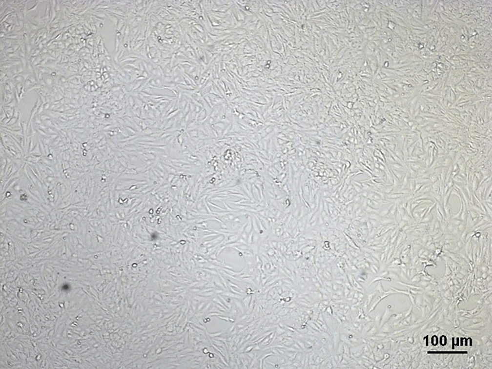
In this study, SKOV3 cells were used. SKOV3 cell culture was observed under a microscope, as presented in Figure 1. After calculating the IC50 values of the materials (melatonin, 1.841 mM; cisplatin, 117.5 μM; and doxorubicin, 14.72 μM), cell viability was calculated via MTS assay and observed under microscope as in Figure 2. The highest rate of cell viability decrease was in group C1 (IC50 melatonin and cisplatin) at 37.57%, as shown in Table 2 (Adella, 2023a, 2023b, 2023c).

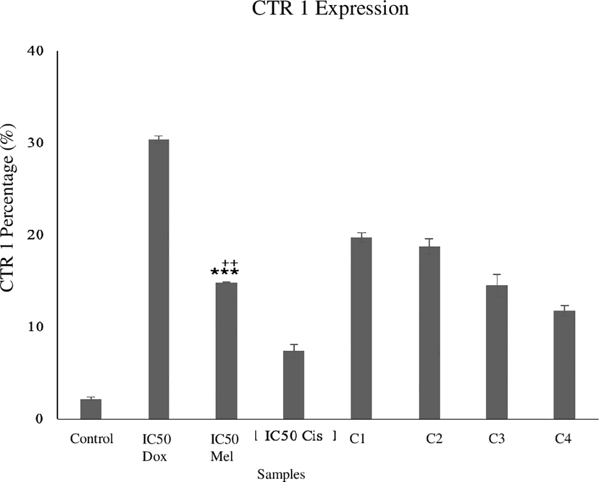
CTR1 is a marker of drug influx. An increase in CTR1 expression indicates an increase of drug influx into the cell, which suggests decreased cancer cell resistance to chemotherapy. As can be seen from Table 3 and Figure 3, the control group has the lowest mean expression among the groups. On the contrary, the doxorubicin group has the highest CTR1 expression, indicating that doxorubicin has a higher influx rate than other groups. However, among the combination groups, C1 and C2 had the highest CTR1 expression (P < 0,001), suggesting that these groups have the highest influx rate among the experimental groups. The IC50 melatonin group had lower CTR1 expression than groups C1 and C2, which shows that chemotherapy influx was better in C1 and C2 than the melatonin-only group. If compared with the IC50 of cisplatin, all the combination groups show that the CTR1 expression was higher than the cisplatin alone, which indicated that the combination of cisplatin and melatonin groups were better to increase the influx of the drug than the cisplatin-only group.
| Groups | Mean (SD) | pa | Posthocb | ||||||
|---|---|---|---|---|---|---|---|---|---|
| Dox | Mel | Cis | C1 | C2 | C3 | C4 | |||
| Control | 2.17 (0.21) | <0.001 | <0.001 | <0.001 | <0.001 | <0.001 | <0.001 | <0.001 | <0.001 |
| IC50 Doxorubicin | 30.33 (0.4) | <0.001 | <0.001 | <0.001 | <0.001 | <0.001 | <0.001 | ||
| IC50 Melatonin | 14.8 (0.1) | <0.001 | <0.001 | <0.001 | 1.000 | 0.001 | |||
| IC50 Cisplatin | 7.37 (0.7) | <0.001 | <0.001 | <0.001 | <0.001 | ||||
| C1 | 19.73 (0.49) | 1.000 | <0.001 | <0.001 | |||||
| C2 | 18.73 (0.84) | <0.001 | <0.001 | ||||||
| C3 | 14.53 (1.14) | 0.002 | |||||||
| C4 | 11.77 (0.55) | ||||||||
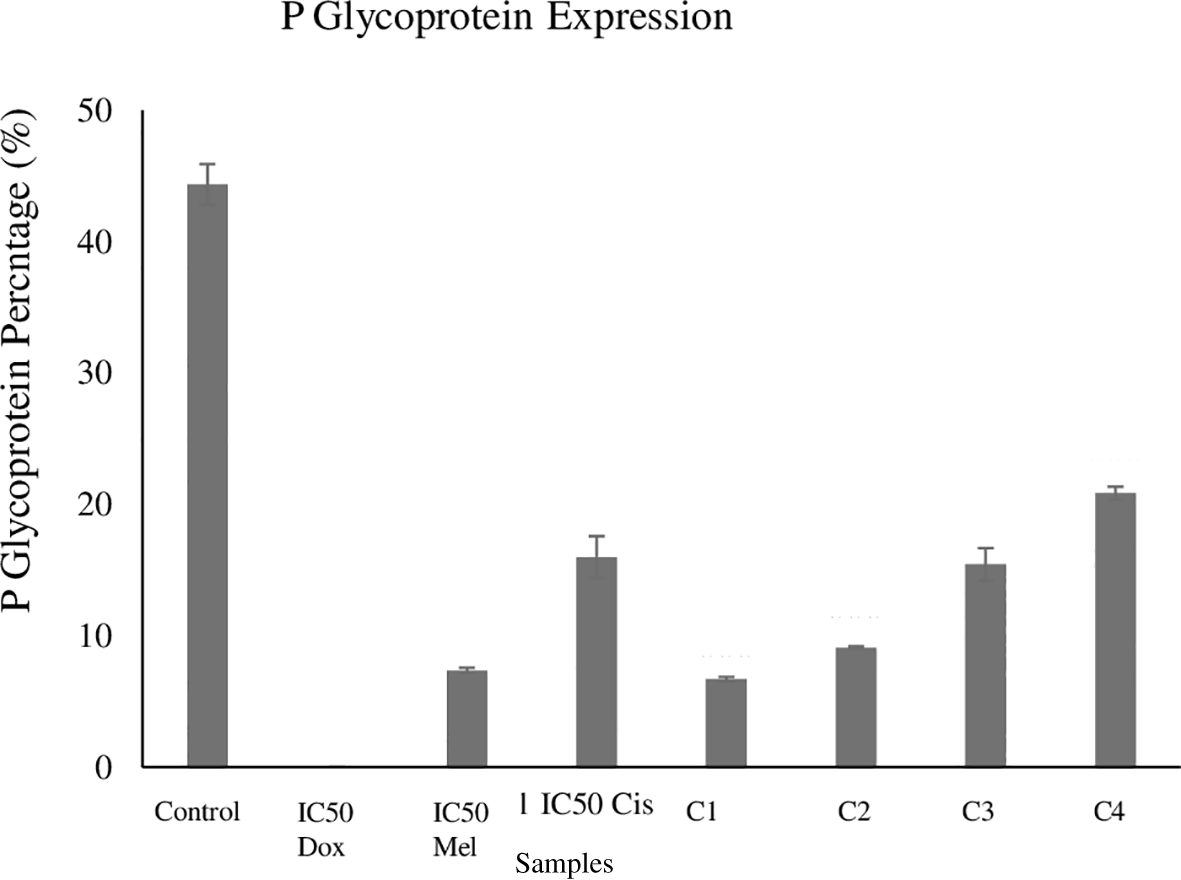
Pgp is an efflux membrane transporter of toxins, cell endogen metabolites, and chemotherapy especially in resistance cells. A high percentage of Pgp indicates a high ability of the cell to inhibit a cytotoxic response, suggesting high chemotherapy resistance of cancer cells. As can be seen from Table 4 and Figure 4, the control group has the highest Pgp expression (44.37%). Among the combination groups, from the mean expression, C1 group has the lowest Pgp expression (6.7%) of all combination groups and C4 has the highest Pgp expression (20.87%). This result indicates that group C1 had the ability to decrease Pgp expression more than the cisplatin-only group (16%). From pair of IC50 melatonin and C1 group, it was shown no difference statistically, but from other pairs shown significantly difference in posthoc analysis. A low rate of drug efflux activity indicates decreased chemotherapy resistance and better outcome. The combination groups had better ability to decrease the efflux mechanism than the cisplatin-only group.
| Groups | Mean (SD) | pa | Posthocb | ||||||
|---|---|---|---|---|---|---|---|---|---|
| Dox | Mel | Cis | C1 | C2 | C3 | C4 | |||
| Control | 44.37 (1.55) | <0.001 | <0.001 | <0.001 | <0.001 | <0.001 | <0.001 | <0.001 | <0.001 |
| IC50 Doxorubicin | 0 | <0.001 | <0.001 | <0.001 | <0.001 | <0.001 | <0.001 | ||
| IC50 Melatonin | 7.37 (0.21) | <0.001 | 1.000 | 0.986 | <0.001 | <0.001 | |||
| IC50 Cisplatin | 16 (1.59) | <0.001 | <0.001 | 1.000 | <0.001 | ||||
| C1 | 6.7 (0.17) | 0.161 | <0.001 | <0.001 | |||||
| C2 | 9.1 (0.1) | <0.001 | <0.001 | ||||||
| C3 | 15.43 (1.25) | <0.001 | |||||||
| C4 | 20.87 (0.49) | ||||||||
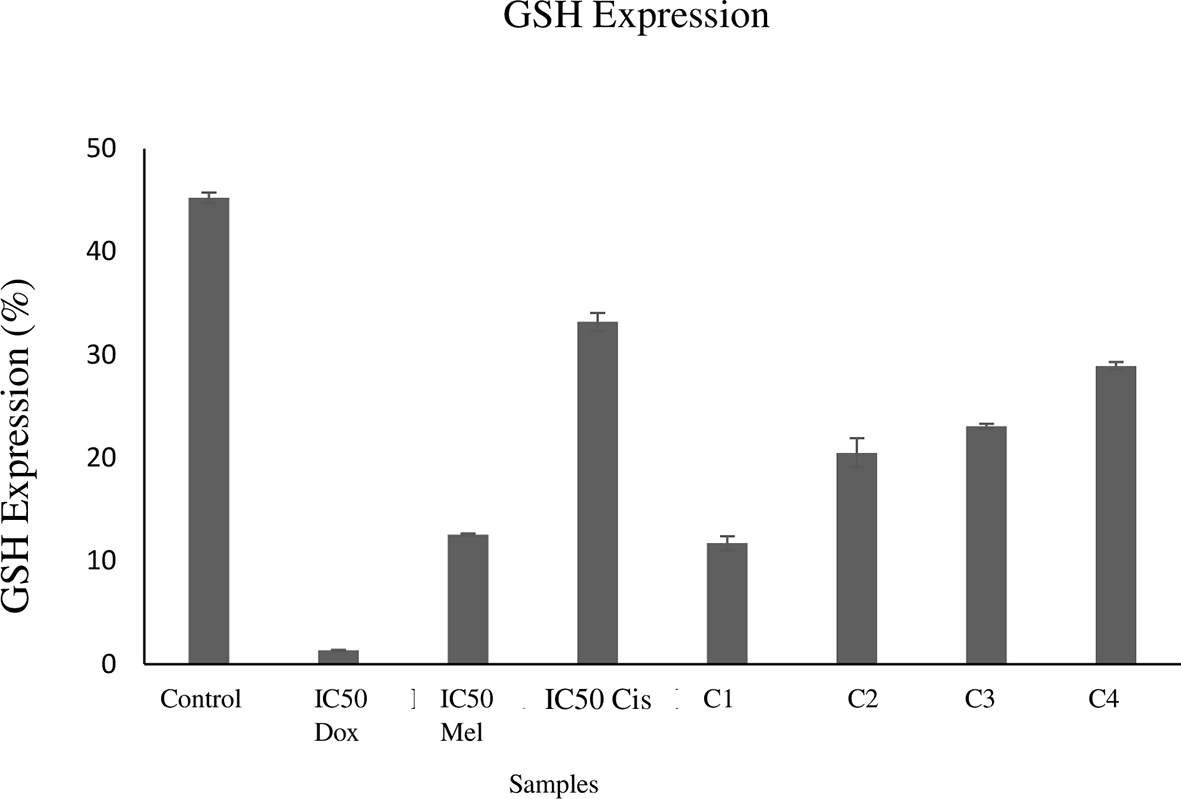
GSH is a peptide, which involved in drug inactivation protein marker. As can be seen from Table 5 and Figure 5, the control group has the highest GSH peptide level (45.23%), whereas the positive control group has the lowest (1.33%), indicating that without giving the materials, the GSH level would high. But if doxorubicin is given, the GSH level would be lowered. Among the combination groups, group C1 has the lowest GSH level (11.73%). When compared with the melatonin-only group (12.57%), group C1 had a lower GSH level. On the other hand, when compared with the cisplatin-only group, all of the combination groups had a higher ability to decrease the GSH level than the cisplatin-only group. But, from pair of IC50 melatonin and C1 in the post hoc analysis, shown no difference statistically. This suggested that the combination groups had decreased drug inactivation activity and chemotherapy resistance as well as a better outcome than the cisplatin-only group. This is presented in Table 5.
| Groups | Mean (SD) | pa | Posthocb | ||||||
|---|---|---|---|---|---|---|---|---|---|
| Dox | Mel | Cis | C1 | C2 | C3 | C4 | |||
| Control | 45.23 (0.5) | <0.001 | <0.001 | <0.001 | <0.001 | <0.001 | <0.001 | <0.001 | <0.001 |
| IC50 Doxorubicin | 1.33 (0.06) | <0.001 | <0.001 | <0.001 | <0.001 | <0.001 | <0.001 | ||
| IC50 Melatonin | 12.57 (0.12) | <0.001 | 1.000 | <0.001 | <0.001 | <0.001 | |||
| IC50 Cisplatin | 33.2 (0.87) | <0.001 | <0.001 | <0.001 | <0.001 | ||||
| C1 | 11.73 (0.67) | <0.001 | <0.001 | <0.001 | |||||
| C2 | 20.5 (1.42) | 0.008 | <0.001 | ||||||
| C3 | 23.07 (0.23) | <0.001 | |||||||
| C4 | 28.93 (0.38) | ||||||||
ERCC1 is a transcription factor used as a DNA repair marker. A high ERCC1 expression indicates high DNA repair mechanism in cancer cells. As can be seen from Table 6 and Figure 6, the control group has the highest ERCC1 expression (48.07%), whereas the positive control group has the lowest (1.57%). Among the combination groups, groups C1, C2, and C3 have the ability to decrease the ERCC1 expression more than the melatonin-only and cisplatin-only groups, but C1 group had the highest ability. The lower the DNA repair activity, the more the chemotherapy resistance decreases, which is shown in combination of melatonin and cisplatin groups.
| Groups | Mean (SD) | pa | Posthocb | ||||||
|---|---|---|---|---|---|---|---|---|---|
| Dox | Mel | Cis | C1 | C2 | C3 | C4 | |||
| Control | 48.07 (0.45) | <0.001 | <0.001 | <0.001 | <0.001 | <0.001 | <0.001 | <0.001 | <0.001 |
| IC50 Doxorubicin | 1.57 (0.06) | <0.001 | <0.001 | <0.001 | <0.001 | <0.001 | <0.001 | ||
| IC50 Melatonin | 14.63 (0.49) | <0.001 | <0.001 | <0.001 | <0.001 | <0.001 | |||
| IC50 Cisplatin | 20.2 (0.17) | <0.001 | <0.001 | <0.001 | 0.001 | ||||
| C1 | 4.27 (0.21) | <0.001 | <0.001 | <0.001 | |||||
| C2 | 9.53 (0.6) | 0.001 | <0.001 | ||||||
| C3 | 11.63 (0.6) | <0.001 | |||||||
| C4 | 22.8 (0.52) | ||||||||

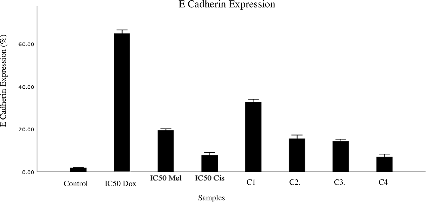
E-cadherin is a marker of epithelial mesenchymal transition. A high e-cadherin expression indicates low epithelial mesenchymal transition, which has a low impact on tumor invasion and drug resistance. As can be seen from Table 7 and Figure 7, the positive control group has the highest e-cadherin expression (64.2%), whereas the control group has the lowest (1.7%). When compared with the cisplatin-only group, the combination groups C1-C3 had higher e-cadherin expression than the cisplatin-only group. This suggested that the combination of melatonin and cisplatin might decrease epithelial mesenchymal transition (increased expression of e-cadherin) more than the cisplatin-only group; a decrease in drug resistance could also be observed.
| Groups | E_Chaderin | p-valuea | P-valueb | |||||||
|---|---|---|---|---|---|---|---|---|---|---|
| Mean ± SD | Control | IC50 Doxorubicin | IC50 Melatonin | IC50 Cisplatin | C1 | C2 | C3 | C4 | ||
| Control | 1.70 ± 0.10 | <0.001 | <0.001 | <0.001 | <0.001 | <0.001 | <0.001 | <0.001 | <0.001 | |
| IC50 Doxorubicin | 64.2 ± 1.76 | <0.001 | <0.001 | <0.001 | <0.001 | <0.001 | <0.001 | |||
| IC50 Melatonin | 19.2 ± 0.36 | <0.001 | <0.001 | <0.001 | <0.001 | <0.001 | ||||
| IC50 Cisplatin | 7.20 ± 0.59 | <0.001 | <0.001 | <0.001 | 1.00 | |||||
| C1 | 32.2 ± 1.60 | <0.001 | <0.001 | <0.001 | ||||||
| C2 | 15.53 ± 0.85 | 0.588 | <0.001 | |||||||
| C3 | 14.30 ± 0.46 | <0.001 | ||||||||
| C4 | 6.93 ± 0.65 | |||||||||
Annexin V was used as an apoptosis marker in this research. A high annexin V activity indicates high apoptosis activity. As can be seen from Table 8 and Figure 8, the positive control group has the highest annexin V activity (70.9%), whereas the control group has the lowest (1.2%). Among the combination groups, group 1 has the highest annexin V activity (53.57%). Compared with the melatonin-only group, all combination groups had a higher ability to decrease annexin V expression. Additionally, compared with the cisplatin-only group, the combination groups all had a better ability to increase apoptosis than the cisplatin-only group. This suggested that the combination of melatonin and cisplatin increases apoptosis to reduce the drug resistance mechanism.
| Groups | Mean (SD) | pa | Posthocb | ||||||
|---|---|---|---|---|---|---|---|---|---|
| Dox | Mel | Cis | C1 | C2 | C3 | C4 | |||
| Control | 1.2 (0) | <0.001 | <0.001 | <0.001 | <0.001 | <0.001 | <0.001 | <0.001 | <0.001 |
| IC50 Doxorubicin | 70.9 (0.1) | <0.001 | <0.001 | <0.001 | <0.001 | <0.001 | <0.001 | ||
| IC50 Melatonin | 15.77 (0.21) | 0.005 | <0.001 | <0.001 | <0.001 | <0.001 | |||
| IC50 Cisplatin | 10.87 (0.91) | <0.001 | <0.001 | <0.001 | <0.001 | ||||
| C1 | 53.57 (1.33) | <0.001 | <0.001 | <0.001 | |||||
| C2 | 46.77 (0.06) | <0.001 | <0.001 | ||||||
| C3 | 32.43 (2.83) | <0.001 | |||||||
| C4 | 23.2 (1.31) | ||||||||

The IC50 of doxorubicin was used as a positive control in this study as doxorubicin has much evidence as an anticancer agent and has been demonstrated to be effective when cells are resistant to cisplatin (Yi S et al., 2020). Doxorubicin has a DNA intercalation chain mechanism, inhibition of topoisomerase II to destroy DNA and increase apoptosis (Johnson and Dubey, 2021). The combination groups had higher cell viability decrease than the cisplatin group. These groups may have the capability to increase the cytotoxic effect of chemotherapy more than that of the single-use therapy. Melatonin exhibits anti-inflammatory, anti-tumor, and anti-proliferative activities, with minimal side effects. In addition as oncostatic agent, it was found to reduce cell proliferation, metastasis, angiogenesis and act as a pro-apoptosis and immunomodulator agent in ovarian cancer. Melatonin exhibits anti-inflammatory, anti-tumor, and anti-proliferative activities, with minimal side effects. In addition as oncostatic agent, it was found to reduce cell proliferation, metastasis, angiogenesis and act as a pro-apoptosis and immunomodulator agent in ovarian cancer. Melatonin reduces p52 and p65 binding activity by downregulate MMP-9, regulates cell motility and activation of MMP-9 by Akt-mediated JNK1/2 and ERK1/2 pathways, this is shown in metastasis activity. Melatonin acts as pro-apoptosis and tumor growth inhibitor by downregulation of MDM2 (E3 ubiquitin ligase), that promotes acetylation of p53 acetylation which lead to cell cycle arrest by elevating p21 levels. Melatonin also inactivates Akt, reduces apoptosis resistance of tumor cells, lowers Snail and vimentin levels and enhances e-cadherin activity. Melatonin decreases ROS and inactivates HIF-α and VEGF (Vascular Endothelial Growth Factor) in malignancy (Chuffa et al., 2017; Heydari et al., 2023). Melatonin has no issues regarding bioavailability compared with other drugs because it has faster solubility through the cell and nuclear membranes (Tamura et al., 2020).
CTR1 and Pgp were found to be associated with cell resistance. Low influx and high efflux may contribute to drug resistance (Zhang et al., 2019). CTR1 plays a role in the absorption of platinum drugs, such as cisplatin and oxaliplatin, and decreases transporter expression, thus influencing tumor resistance (Chen and Chang, 2019). Pgp may pump the drug outside of the cell to reduce cytotoxic activity. A high Pgp indicates a high ability of the cell to inhibit the cytotoxic agent (Amiri-Kordestani et al., 2012). Melatonin is capable of reducing Pgp in diffuse large B-cell lymphoma and activating the NF-kB pathway. In a study using the combination of epirubicin and melatonin, melatonin made the lymphoma cell sensitive to epirubicin from the inhibition of Pgp expression through the pathway of NF-kB. Melatonin decreases P65 in the nucleus and inhibits Pgp expression (Liu et al., 2021). In colorectal cancer that resist to oxaliplatin (using LS174T/DR cell line), combination of melatonin and oxaliplatin reduce oxaliplatin resistance by lowering Pgp activities in LS174T/DR and also suppress colorectal proliferation (Dehghanzad et al., 2024).
GSH has the ability to detoxify intracellular toxins, but the GSH in cell cancer is capable of inhibiting and inactivating chemotherapy (Tapia et al., 2013). GSH binds to cisplatin to inhibit cisplatin binding with DNA, reduce reactive oxygen species, and decrease cell sensitivity to apoptosis (Galluzzi et al., 2012). Melatonin can decrease the GSH level and increase the GSH peroxidase activity (Medina-Leendertz et al., 2018). Furthermore, it induces antioxidant synthesis by inducing gamma glutamylcysteine synthetase (Meng et al., 2017). In addition, melatonin increases antioxidant activity that increases glutathione level and induces glutathione peroxidase (Harderland, 2017). Melatonin increases the apoptosis activity of cisplatin, reduces oxidative stress by decreasing the GSH level, and increases glutathione synthesis (Fernandez et al., 2019).
Melatonin has the ability to inhibit mTOR and ERCC1 expressions and increase the activity of intracellular autophagosomes. It is used as a cancer therapy adjuvant to repair the sensitivity of chemotherapy and managing the side effects of cisplatin (Bennukul et al., 2014). Melatonin affects the DNA repair mechanism by increasing ERCC-1 XPF activity, a NER (Nucleotide Excision Repair) that functions as 5’ endonuclease in the repair mechanism pathway (Mir et al., 2021). Melatonin reduce DNA damage and apoptosis, increase ERCC1 gene which is involved in DNA damage repair in hepatocellular carcinoma (HepG2) cells (Bennukul et al., 2014). ERCC1 overexpression has poor prognosis in patients with osteosarcoma or lung cancer who received cisplatin. Li et al. (2017) demonstrated that low ERCC1 expression increases the sensitivity of platinum chemotherapy in ovarian cancer. The low repair activity of the DNA decreases chemotherapy resistance (Li et al., 2017).
E-cadherin has an impact in cell adhesion and influences cell growth. A decrease in e-cadherin expression increases activity of epithelial mesenchymal transition, thus reducing the cell adhesion strength (Rosso et al., 2017). Loss of e-cadherin will activates signalling pathways and transcription factors such as β-catenin (Su et al., 2017). High e-cadherin was associated with low activities of invasive cancer and metastasis (Loh et al., 2019). Melatonin administration increased e-cadherin expression, decreased N-cadherin and vimentin expressions in CSC from CMT-U229. Melatonin may decrease the migration and invasion activities of cancer cells (Goncalves et al., 2016). It also decreases proteins associated with inflammation, oxidative stress, cell-cycle, proliferation, and apoptosis (Zare et al., 2019). In gastric cancer, melatonin upregulate e-cadherin by involving in interference of interaction between C/EBPβ and NF-κB through induction of endoplasmic reticulum stress that activates calpain (Su et al., 2017). Melatonin also increase e-cadherin, reduce MMP-9 expression and inhibit NF-κB pathway in esophageal cancer (Gu et al., 2020).
Apoptosis indicates low chemotherapy resistance. In this study, we used annexin V as the marker. High annexin V expression indicates high apoptosis activity in the cell (Wang et al., 2012). Melatonin increases p53 expression and activates it, increasing apoptosis in several cancers such as colon and uterine cancer. Melatonin is involved in the BAX gene expression, decreases the expression of BCL-2 as an antiapoptotic gene, and regulates the Bax/Bcl-2 ratio (Chuffa et al., 2017). In human myeloid leukemia, melatonin also inhibit progression of G1 to S phase by up-regulate Bax and down-regulate Bcl-2 (Shen et al., 2017). Other studies also reported melatonin activity in inducing apoptosis by upregulation of pro-apoptotic (p53, Bax, total and cleaved caspase-3) and anti-apoptotic (Bcl-2 and survivin) also downregulation of cyclin dependent kinase (Chuffa et al., 2017). Melatonin and cisplatin potentiate apoptosis by increasing the depolarization of mitochondrial membrane, activating caspase-3/7, and inducing cell-cycle arrest, if compared with the cisplatin-only group (Plaimee et al., 2015).
This study has shown that cisplatin combine with melatonin may reduce the cisplatin resistance in ovarian cancer, but this study only use one cell line which is SKOV3. In order to see the impact of the combination, study on the several cell lines should be performed. The result of this study may just generally indicative, so another study must be performed and added to see the effect of the melatonin and cisplatin combination in reducing the chemoresistance. The authors also suggest that there should be another research that performed in vivo to see the bioavailability and dose of the combination melatonin and cisplatin, so it can be optimized the cancer treatment.
This study has shown that combination of melatonin and cisplatin may reduce the chemoresistance in SKOV3 cell line with several pathways such as decreasing cancer cell viability, increased influx activity by increasing CTR-1 expression, reduced efflux activity by reducing Pgp expression, decrease drug inactivity by reducing GSH expression, decrease DNA repair mechanism shown by reduced ERCC-1 expression, decreased epithelial mesenchymal transition by increasing e-cadherin expression, and increase apoptosis mechanism by increase annexin V expression. This study may describe the melatonin effect on chemotherapy resistance.
| Views | Downloads | |
|---|---|---|
| F1000Research | - | - |
|
PubMed Central
Data from PMC are received and updated monthly.
|
- | - |
Provide sufficient details of any financial or non-financial competing interests to enable users to assess whether your comments might lead a reasonable person to question your impartiality. Consider the following examples, but note that this is not an exhaustive list:
Sign up for content alerts and receive a weekly or monthly email with all newly published articles
Already registered? Sign in
The email address should be the one you originally registered with F1000.
You registered with F1000 via Google, so we cannot reset your password.
To sign in, please click here.
If you still need help with your Google account password, please click here.
You registered with F1000 via Facebook, so we cannot reset your password.
To sign in, please click here.
If you still need help with your Facebook account password, please click here.
If your email address is registered with us, we will email you instructions to reset your password.
If you think you should have received this email but it has not arrived, please check your spam filters and/or contact for further assistance.
Comments on this article Comments (0)