Keywords
Uniprot ID Q13501, SQSTM1, Sequestosome-1, antibody characterization, antibody validation, Western blot, immunoprecipitation, immunofluorescence
This article is included in the YCharOS (Antibody Characterization through Open Science) gateway.
This article is included in the Cell & Molecular Biology gateway.
Uniprot ID Q13501, SQSTM1, Sequestosome-1, antibody characterization, antibody validation, Western blot, immunoprecipitation, immunofluorescence
Sequestosome-1, alternatively known as p62, is an adaptor protein required for selective autophagy and proteasomal degradation.1–3 Delivering polyubiquitinated proteins to the autophagosome or proteasome, Sequestosome-1/p62 plays a key role in the degradation of aggregate prone proteins.1
SQSTM1 gene mutations may act as a potential threat by causing altered autophagy, resulting in pathogenic protein aggregation and the development of a variety of neurodegenerative diseases, including ALS and FTD.4 Furthermore, SQSTM1 mutations have been identified in patients with ALS and FTD.5 Serving as a signalling hub for neurodegenerative pathways, Sequestosome-1/p62 poses as a prospective therapeutic target in the treatment of neurodegenerative diseases.6 Mechanistic studies would be greatly facilitated with the availability of high-quality antibodies.
Here, we compared the performance of a range of commercially available antibodies for Sequestosome-1 and validated high-performing antibodies for Western blot, immunoprecipitation and immunofluorescence, enabling biochemical and cellular assessment of Sequestosome-1 properties and function.
Our standard protocol involves comparing readouts from wild-type (WT) and knockout (KO) cells.7–9 To identify a cell line that expresses adequate levels of Sequestosome-1 protein to provide sufficient signal to noise we examined public proteomics databases, namely PAXdb (RRID:SCR_018910)10 and DepMap (RRID:SCR_017655).11 U2OS was identified as a suitable cell line and thus U2OS was modified with CRISPR/Cas9 to knockout the corresponding SQSTM1 gene (Table 1).
| Institution | RRID (Cellosaurus) | Cell line | Genotype |
|---|---|---|---|
| Montreal Neurological Institute | CVCL_0042 | U2OS | WT |
| Montreal Neurological Institute | CVCL_A6LP | U2OS | SQSTM1 KO |
For Western blot experiments, we resolved proteins from WT and SQSTM1 KO cell extracts and probed them side-by-side with all antibodies in parallel8,9 (Figure 1).
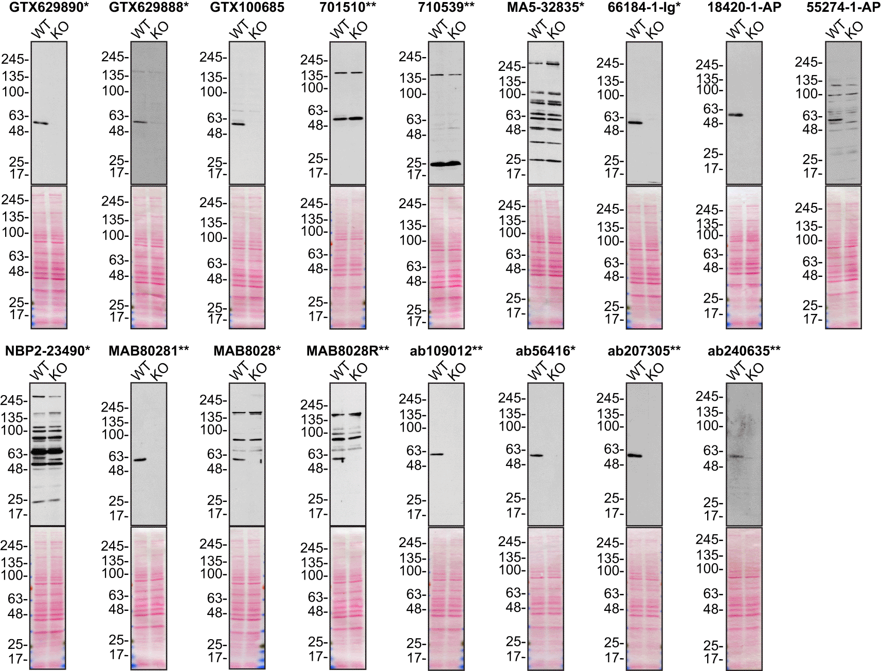
Lysates of U2OS (WT and SQSTM1 KO) were prepared and 25 μg of protein were processed for Western blot with the indicated Sequestosome-1 antibodies. The Ponceau stained transfers of each blot are presented to show equal loading of WT and KO lysates and protein transfer efficiency from the acrylamide gels to the nitrocellulose membrane. Antibody dilutions were chosen according to the recommendations of the antibody supplier. Exceptions were given for antibodies MA5-32835*, 66184-1-Ig* and 18420-1-AP, which were titrated to 1/200, 1/1000 and 1/1000, respectively, as the signals were too weak when following the supplier’s recommendations. Antibody dilution used: GTX629890* at 1/1000; GTX629888* at 1/1000; GTX100685 at 1/1000; 701510** at 1/1000; 710539** at 1/200; MA5-32835* at 1/200; 66184-1-Ig* at 1/1000; 18420-1-AP at 1/1000; 55274-1-AP at 1/1000; NBP2-23490* at 1/1000; MAB80281** at 1/1000; MAB8028* at 1/1000; MAB8028R** at 1/1000; ab109012** at 1/10000; ab56416* at 1/1000; ab207305** at 1/1000; ab240635** at 1/1000. Predicted band size: ~62 kDa. *= monoclonal antibody, **= recombinant antibody.
For immunoprecipitation experiments, we used the antibodies to immunopurify Sequestosome-1 from U2OS cell extracts. The performance of each antibody was evaluated by detecting the Sequestosome-1 protein in extracts, in the immunodepleted extracts and in the immunoprecipitates8,9 (Figure 2).
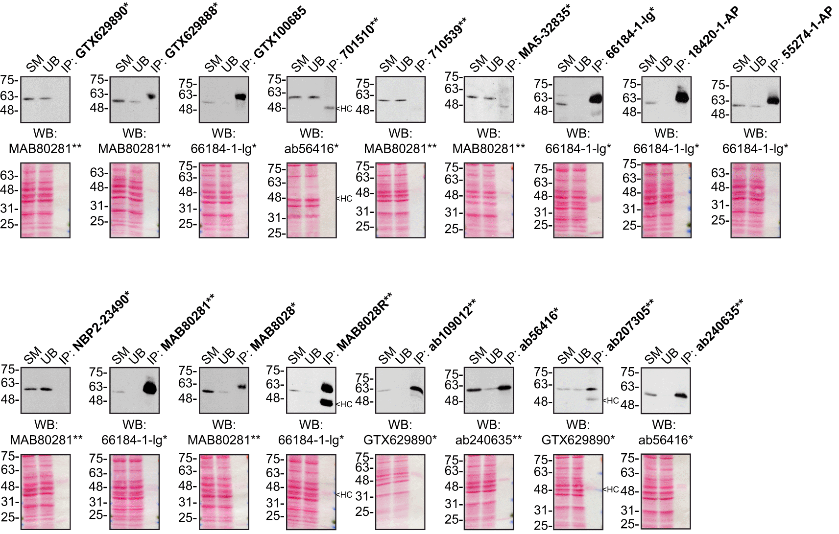
U2OS lysates were prepared, and IP was performed using 1.0 μg of the indicated Sequestosome-1 antibodies pre-coupled to protein G or protein A Sepharose beads. Samples were washed and processed for Western blot with the indicated Sequestosome-1 antibody. For Western blot, MAB80281** was used at 1/3000, 66184-1-lg* at 1/3000, ab56416* at 1/5000, ab207305** at 1/10000 and GTX629890* at 1/5000. The Ponceau stained transfers of each blot are shown for similar reasons as in Figure 1. SM= 10% starting material; UB=10% unbound fraction; IP=immunoprecipitate, HC= antibody heavy chain. *= monoclonal antibody, **= recombinant antibody.
For immunofluorescence, as described previously, antibodies were screened using a mosaic strategy.12 In brief, we plated WT and KO cells together in the same well and imaged both cell types in the same field of view to reduce staining, imaging and image analysis bias (Figure 3).
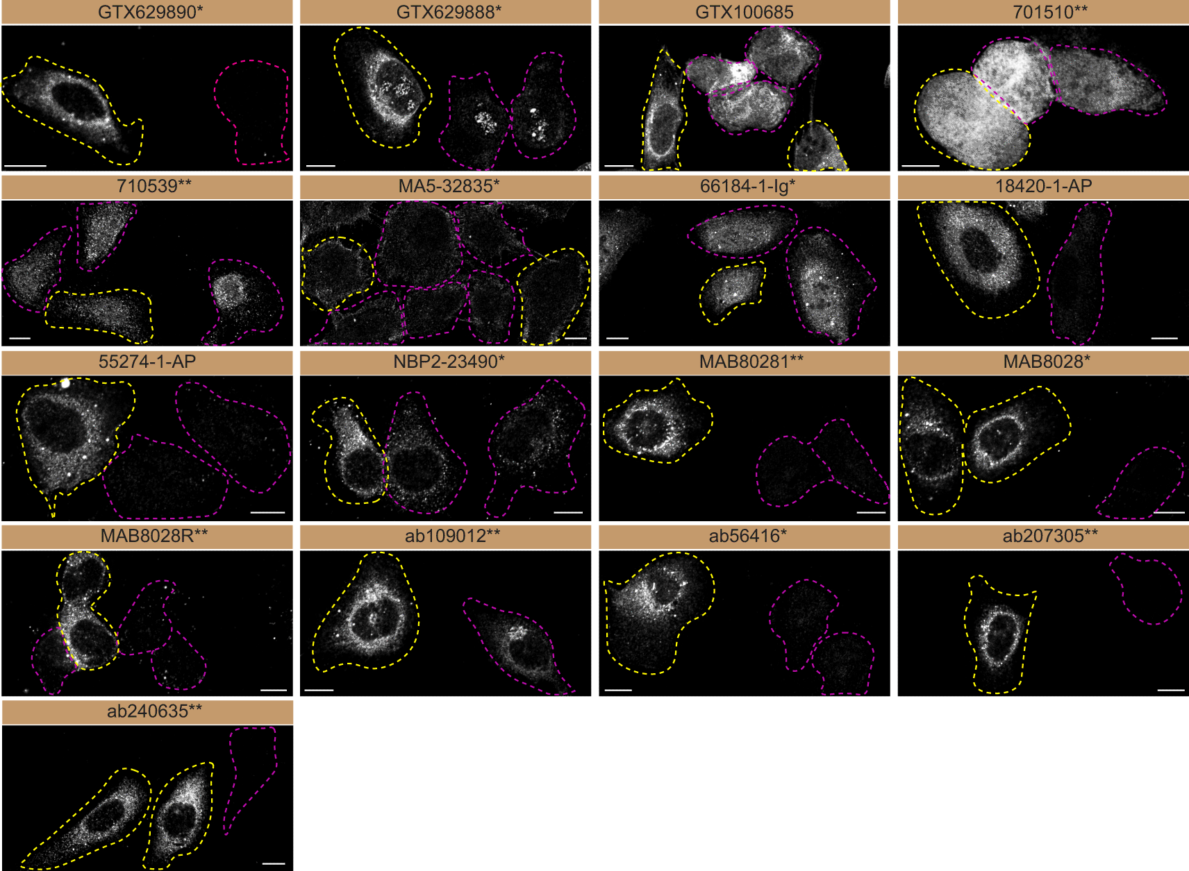
U2OS WT and SQSTM1 KO cells were labelled with a green or a far-red fluorescent dye, respectively. WT and KO cells were mixed and plated to a 1:1 ratio on coverslips. Cells were stained with the indicated Sequestosome-1 antibodies and with the corresponding Alexa-fluor 555 coupled secondary antibody. Acquisition of the green (WT), red (antibody staining) and far-red (KO) channels was performed. Representative images of red (grayscale images) channel are shown. WT and KO cells are outlined with yellow and magenta dashed line, respectively. Antibody dilutions were chosen according to the recommendations of the antibody supplier. Exceptions were given for antibodies MA5-32835*, 18420-1-AP and NBP2-23490*, which were titrated to 1/2000, 1/300 and 1/1000, respectively, as the signals were too strong when following the supplier’s recommendations. When the concentration was not indicated by the supplier, we tested antibodies at 1/500 and 1/1000. At this concentration, the signal from each antibody was in the range of detection of the microscope used. Antibody dilution used: GTX629890* at 1/1000; GTX629888* at 1/1000; GTX100685 at 1/700; 701510** at 1/500; 710539** at 1/500; MA5-32835* at 1/2000; 66184-1-Ig* at 1/300; 18420-1-AP at 1/300; 55274-1-AP at 1/1300; NBP2-23490* at 1/1000; MAB80281** at 1/500; MAB8028* at 1/500; MAB8028R** at 1/500; ab109012** at 1/500; ab56416* at 1/1000; ab207305** at 1/200; ab240635** at 1/500. Bars = 10 μm. *= monoclonal antibody, **= recombinant antibody.
In conclusion, we have screened Sequestosome-1 commercial antibodies by Western blot, immunoprecipitation and immunofluorescence and characterized several high-quality antibodies under our standardized experimental conditions. The underlying data can be found on Zenodo.13,14
All Sequestosome-1 antibodies are listed in Table 2, together with their corresponding Research Resource Identifiers, or RRID, to ensure the antibodies are cited properly.15 Peroxidase-conjugated goat anti-rabbit and anti-mouse antibodies are from Thermo Fisher Scientific (cat. number 65-6120 and 62-6520). Alexa-555-conjugated goat anti-mouse and anti-rabbit secondary antibodies are from Thermo Fisher Scientific (cat. number A21424 and A21429).
| Company | Catalog number | Lot number | RRID (Antibody Registry) | Clonality | Clone ID | Host | Concentration (μg/μl) |
|---|---|---|---|---|---|---|---|
| GeneTex | GTX629890* | 41470 | AB_2885144 | monoclonal | GT1478 | mouse | 1.00 |
| GeneTex | GTX629888* | 41470 | AB_2885143 | monoclonal | GT239 | mouse | 1.00 |
| GeneTex | GTX100685 | 42893 | AB_2038029 | polyclonal | - | rabbit | 0.67 |
| Thermo Fisher Scientific | 701510** | 2315239 | AB_2532489 | recombinant-mono | 11HC14LC25 | rabbit | 0.50 |
| Thermo Fisher Scientific | 710539** | RF229394 | AB_2532735 | recombinant-poly | 11HCLC | rabbit | 0.50 |
| Thermo Fisher Scientific | MA5-32835* | VL3152616 | AB_2802482 | monoclonal | 10-E10 | mouse | 2.00 |
| Proteintech | 66184-1-Ig* | not provided | AB_2881579 | monoclonal | 1H5C1 | mouse | 1.33 |
| Proteintech | 18420-1-AP | not provided | AB_10694431 | polyclonal | - | rabbit | 0.35 |
| Proteintech | 55274-1-AP | not provided | AB_11182278 | polyclonal | - | rabbit | 0.43 |
| Bio-Techne | NBP2-23490* | A-1 | AB_2885153 | monoclonal | 5H7E2 | mouse | 1.00 |
| Bio-Techne | MAB80281** | CMRM0120031 | AB_2888658 | recombinant-mono | 2533b | rabbit | 0.50 |
| Bio-Techne | MAB8028* | CHZL0520071 | AB_2885150 | monoclonal | 864807 | mouse | 0.50 |
| Bio-Techne | MAB8028R** | CLJP0118091 | AB_2885151 | recombinant-mono | 864807R | mouse | 0.50 |
| Abcam | ab109012** | GR3241806-8 | AB_2810880 | recombinant-mono | EPR4844 | rabbit | 0.43 |
| Abcam | ab56416* | GR3374761-2 | AB_945626 | monoclonal | Not provided | mouse | 1.00 |
| Abcam | ab207305** | GR323335-4 | AB_2885112 | recombinant-mono | EPR18351 | rabbit | 0.21 |
| Abcam | ab240635** | GR3314160-2 | AB_2885121 | recombinant-mono | EPR23101-103 | rabbit | 0.49 |
SQSTM1 KO clone was generated in Cas9-expressing U2OS cell line7 with low passage cells using an open-access protocol available on Zenodo.org: https://zenodo.org/record/3875777#.ZA9VdC-96Tf. Two guide RNAs were used to introduce a STOP codon in the SQSTM1 gene (sequence guide 1: CCACCGCCCACCGUGUGCUC, sequence guide 2: AUGCGAGCUUGGUGUGCCCC).
Both U2OS WT and SQSTM1 KO cell lines used are listed in Table 1, together with their corresponding RRID, to ensure the cell lines are cited properly.16 Cells were cultured in DMEM high glucose (GE Healthcare cat. number SH30081.01) containing 10% fetal bovine serum (Wisent, cat. number 080450), 2 mM L-glutamate (Wisent cat. number 609065), 100 IU penicillin and 100 μg/ml streptomycin (Wisent cat. number 450201).
Western blots were performed as described in our standard operating procedure.17 U2OS WT and SQSTM1 KO were collected in RIPA buffer (50 mM Tris pH 8.0, 150 mM NaCl, 1.0 mM EDTA, 1% Triton X-100, 0.5% sodium deoxycholate, 0.1% SDS) supplemented with protease inhibitor (MilliporeSigma, cat. number P8340). Lysates were sonicated briefly and incubated for 30 min on ice. Lysates were spun at ~110,000 x g for 15 min at 4°C and equal protein aliquots of the supernatants were analyzed by SDS-PAGE and Western blot. BLUelf prestained protein ladder (GeneDireX, cat. number PM008-0500) was used.
Western blots were performed with large 4-15% polyacrylamide gels and transferred on nitrocellulose membranes. Proteins on the blots were visualized with Ponceau staining, which is scanned to show them together with individual Western blots. Blots were blocked with 5% milk for 1 hr, and antibodies were incubated overnight at 4°C with 5% bovine serum albumin (BSA) (Wisent, cat. number 800-095) in TBS with 0.1% Tween 20 (TBST) (Cell Signaling Technology, cat. number 9997). Following three washes with TBST, the peroxidase conjugated secondary antibody was incubated at a dilution of ~0.2 μg/ml in TBST with 5% milk for 1 hr at room temperature followed by three washes with TBST. Membranes were incubated with regular ECL (cat. number 32106) or super signal West Femto (cat. number 34096) from Thermo Fisher Scientific prior to detection with the HyBlot CL autoradiography films from Denville (cat. number 1159T41).
Immunoprecipitation was performed as described in our standard operating procedure.18 Antibody-bead conjugates were prepared by adding 1.0 μg of antibody to 500 μl of phosphate buffered saline (PBS) (Wisent, cat. number 311-010-CL) with 0.01% triton X-100 (Thermo Fisher Scientific, cat. number BP151-500) in a 1.5 mL microcentrifuge tube, together with 30 μl of protein A- (for rabbit antibodies) or protein G- (for mouse antibodies) Sepharose beads. Tubes were rocked overnight at 4°C followed by several washes to remove unbound antibodies.
U2OS WT were collected in HEPES lysis buffer (20 mM HEPES, 100 mM sodium chloride, 1 mM EDTA, 1% Triton X-100, pH 7.4) supplemented with protease inhibitor (MilliporeSigma, cat. number P8340). Lysates were rocked 30 min at 4°C and spun at 110,000 x g for 15 min at 4°C. One ml aliquots at 1.0 mg/ml of lysate were incubated with an antibody-bead conjugate for ~2 hours at 4°C. The unbound fractions were collected, and beads were subsequently washed three times with 1.0 ml of HEPES lysis buffer and processed for SDS-PAGE and Western blot on a 4-15% polyacrylamide gels as described above.
Immunofluorescence was performed as described in our standard operating procedure.8,9,12 U2OS WT and SQSTM1 KO were labelled with a green and a deep red fluorescence dye from Abcam (cat. number ab176735 and ab176736), respectively. WT and KO cells were plated on glass coverslips as a mosaic and incubated for 24 hrs in a cell culture incubator at 37°C, 5% CO2. Cells were fixed in 4% paraformaldehyde (PFA) (Beantown chemical, cat. number 140770-10ml) in PBS for 15 min at room temperature and then washed three times with PBS. Cells were permeabilized in PBS with 0.1% triton X-100 for 10 min at room temperature and blocked with PBS with 5% BSA, 5% goat serum (Gibco, cat. number 16210-064) and 0.01% Triton X-100 for 30 min at room temperature. Cells were incubated with IF buffer (PBS, 5% BSA, 0.01% Triton X-100) containing the primary Sequestosome-1 antibodies overnight at 4°C. Cells were then washed 3 x 10 min with IF buffer and incubated with corresponding Alexa Fluor 555-conjugated secondary antibodies in IF buffer at a dilution of 1.0 μg/ml for 1 hr at room temperature. Cells were washed 3 x 10 min with IF buffer and once with PBS. Coverslips were mounted on a microscopic slide using fluorescence mounting media (DAKO).
Imaging was performed using a Zeiss LSM 880 laser scanning confocal microscope equipped with a Plan-Apo 40x oil objective (NA = 1.40). Analysis was done using ImageJ (RRID:SCR_003070). All cell images represent a single focal plane. Figures were assembled with Adobe Photoshop (version 24.2.1) (RRID:SCR_014199) to adjust contrast and apply 1-pixel Gaussian blur, and then they were assembled with Adobe Illustrator (version 27.3.1) (RRID:SCR_010279).
| Views | Downloads | |
|---|---|---|
| F1000Research | - | - |
|
PubMed Central
Data from PMC are received and updated monthly.
|
- | - |
Provide sufficient details of any financial or non-financial competing interests to enable users to assess whether your comments might lead a reasonable person to question your impartiality. Consider the following examples, but note that this is not an exhaustive list:
Sign up for content alerts and receive a weekly or monthly email with all newly published articles
Already registered? Sign in
The email address should be the one you originally registered with F1000.
You registered with F1000 via Google, so we cannot reset your password.
To sign in, please click here.
If you still need help with your Google account password, please click here.
You registered with F1000 via Facebook, so we cannot reset your password.
To sign in, please click here.
If you still need help with your Facebook account password, please click here.
If your email address is registered with us, we will email you instructions to reset your password.
If you think you should have received this email but it has not arrived, please check your spam filters and/or contact for further assistance.
Comments on this article Comments (0)