Keywords
Regenerative Endodontics, Mineral Trioxide Aggregate, Platelet Rich Fibrin
This article is included in the Manipal Academy of Higher Education gateway.
Regenerative Endodontics, Mineral Trioxide Aggregate, Platelet Rich Fibrin
‘Regenerative endodontics’ using biomaterials, stem cells, biomimetic scaffold and bioactive growth factors have contributed immensely to the clinical management of teeth with pulp necrosis and underdeveloped roots. Success in the form of regression of apical lesion, continued root maturation in length and thickness may return the tooth to vitality.1 Pulp revascularization techniques have gathered much consideration due to their feasibility, cost-effectiveness and reasonable success rate.
Current regenerative endodontic procedures utilize various host-derived scaffolds such as intracanal blood clots (BC) or platelet substitutes like platelet-rich fibrin (PRF),2 platelet-rich plasma (PRP), which supply necessary signalling molecules with growth factors for tissue regeneration. The alkaline irrigants (sodium hypochlorite), medicaments (triple antibiotic paste and calcium hydroxide) and biomaterials like mineral trioxide aggregate (MTA) or biodentine with different biological properties can also impact the outcome.
On hydration calcium silicates present in MTA produce calcium silicate hydrate and calcium hydroxide. Some of the hydration products such as calcium hydroxide dissociate into Ca+ and OH ions, increasing the pH, contributing to antibacterial activity, osteogenic differentiation and bone formation.3 It has been observed that acidic environments increase the solubility of materials, inhibiting the setting reaction and the sealing ability.4
Calcium ions released from the MTA have been shown to pass through the cell membrane by depolarization or activation of membrane-bound calcium channels stimulating the expression of bone-associated proteins which may contribute to the repair process.5 It can further activate ATP leading to osteoblast and cementoblast differentiation and hard tissue mineralization.6
There is limited literature on the influence of scaffolds like PRF and BC on the pH and Ca+ ion release of MTA. Interaction of PRF and BC with MTA may positively or negatively affect the pH and Ca+ ion release. This knowledge may contribute to the understanding of the regenerative process and help modify regenerative treatment strategies. Therefore, this study evaluated the influence of PRF and BC on the pH and Ca+ ion release from MTA.
The study protocol was approved by the Human Research Ethics Committee of M S Ramaiah University of Applied Sciences, Bangalore, Karnataka. Whole fresh blood was collected from a healthy volunteer after obtaining written informed consent.
Fifteen previously extracted, single-rooted human teeth were selected. The teeth were sectioned at the level of the cementoenamel junction (CEJ) using a slow-speed diamond disc.7 Only roots with a single canal were selected. The apical end of the sectioned root was further trimmed to obtain a uniform root length of 10mm for all the specimens. The root canal was then prepared using Peeso reamers Number 1–6 (Kerr, Kerr Corporation, Orange, CA) followed by the use of long straight diamond point to achieve a final canal diameter of 4mm. After mechanical instrumentation, the canals were irrigated with 1.5 % sodium hypochlorite (20mL, 5 min) 5mL sterile physiological saline followed by a final rinse with 20 mL of 17% EDTA and dried using sterile paper points.
A 5 mL blood volume was drawn by venipuncture of the antecubital vein from a healthy volunteer and transferred to a 5mL sterile PRF tube (BD vacutainer serum tubes 5mL) without anticoagulant and centrifuged immediately at 3000 revolutions/min (rpm) for 10 min. The resultant PRF clot between the acellular platelet poor plasma and red blood cells was separated out, squeezed on a piece of sterile gauze to obtain PRF membrane.8
Coronal end of the root was sealed with wax. MTA was mixed (one scoop powder was mixed with 0.33mL of liquid) and condensed into the prepared canal to obtain a thickness of 4mm and confirmed by a graduated probe. Fifteen samples thus prepared were divided into three groups.
PRF membrane cut into fragments was placed into the root canal and gently compacted over the MTA using a hand plugger to achieve a thickness of 5mm.
Blood drawn by venipuncture of the antecubital vein of a healthy volunteer was introduced into the root canal and left to form a clot.9
MTA was mixed according to the manufacturer’s instructions (one scoop of powder was mixed with 0.33 mL of liquid). Cement was then placed into the prepared canal to obtain a thickness of 4 mm and confirmed by a graduated probe.
The prepared specimens were transferred to sterile falcon tubes containing 10 ml of distilled water7 and stored at 37oC and 100% relative air humidity throughout the testing period. At the end of every experimental period (three hours, seven days and 14 days), the specimens were transferred to a fresh falcon tube containing 10mL of distilled water and the collected solutions were analysed for pH and Ca+ ion release.
The digital pH meter (HI5221 Hanna Instruments Cal Check, United States) was calibrated to pH 7 with standard buffer solution before use. The refillable calomel electrode was placed into the falcon tube containing 10mL of solution to record the pH. The electrode was washed with distilled water and wiped dry between readings.
To determine the release of Ca+ ions, ICP-OES Inductively Coupled Plasma- Optical Emission Spectrometry (Thermo Fisher ICAP 7400 ICP-OES, Radial N. American, USA). equipment was used, owing to its high sensitivity and stability for inorganic analysis. It provides rapid and multi-element analysis of the solutions.
The pH and Ca+ ion release were presented as mean values. One-way ANOVA followed by post hoc Bonferroni test were done to determine the significant differences between groups using IBM SPSS software version 22.0 (IBM Corp., Armonk, NY, USA). The results were considered statistically significant if the p-value was less than 0.05.
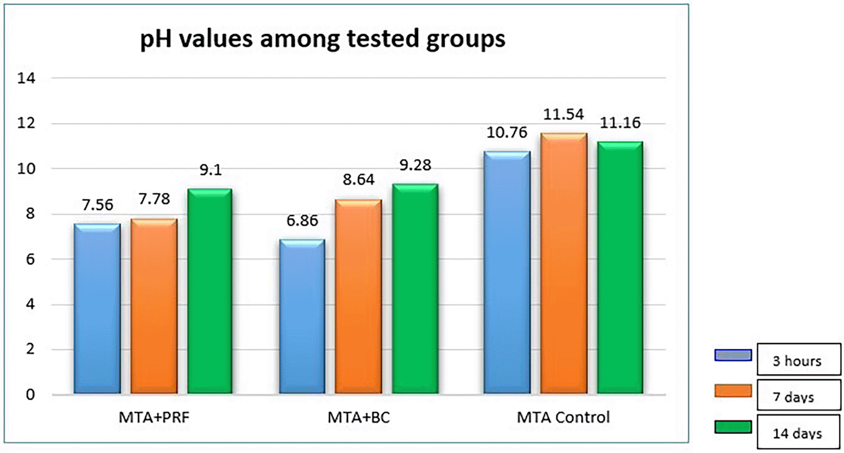
The mean pH values of the individual groups at different time periods are presented in Table 1. The control group (MTA) exhibited a mean value of 10.76 at 3h, 11.54 on the seventh day, and 11.16 on the 14th day, which was significantly higher than the other groups (p=0.000). pH readings for Group 1 (MTA +PRF) were 7.56 at 3h, 7.78 at seven days which increased to 9.10 on the 14th day. Group 2 (BC) presented pH of 6.68 at 3h which increased to 8.64 on the seventh day and 9.28 on the 14th day. There was a significant difference in pH between Groups 1 and 2 at 3h and seven days (p < 0.001) but on the 14th day there was no significant difference between the groups (p > 0.973).
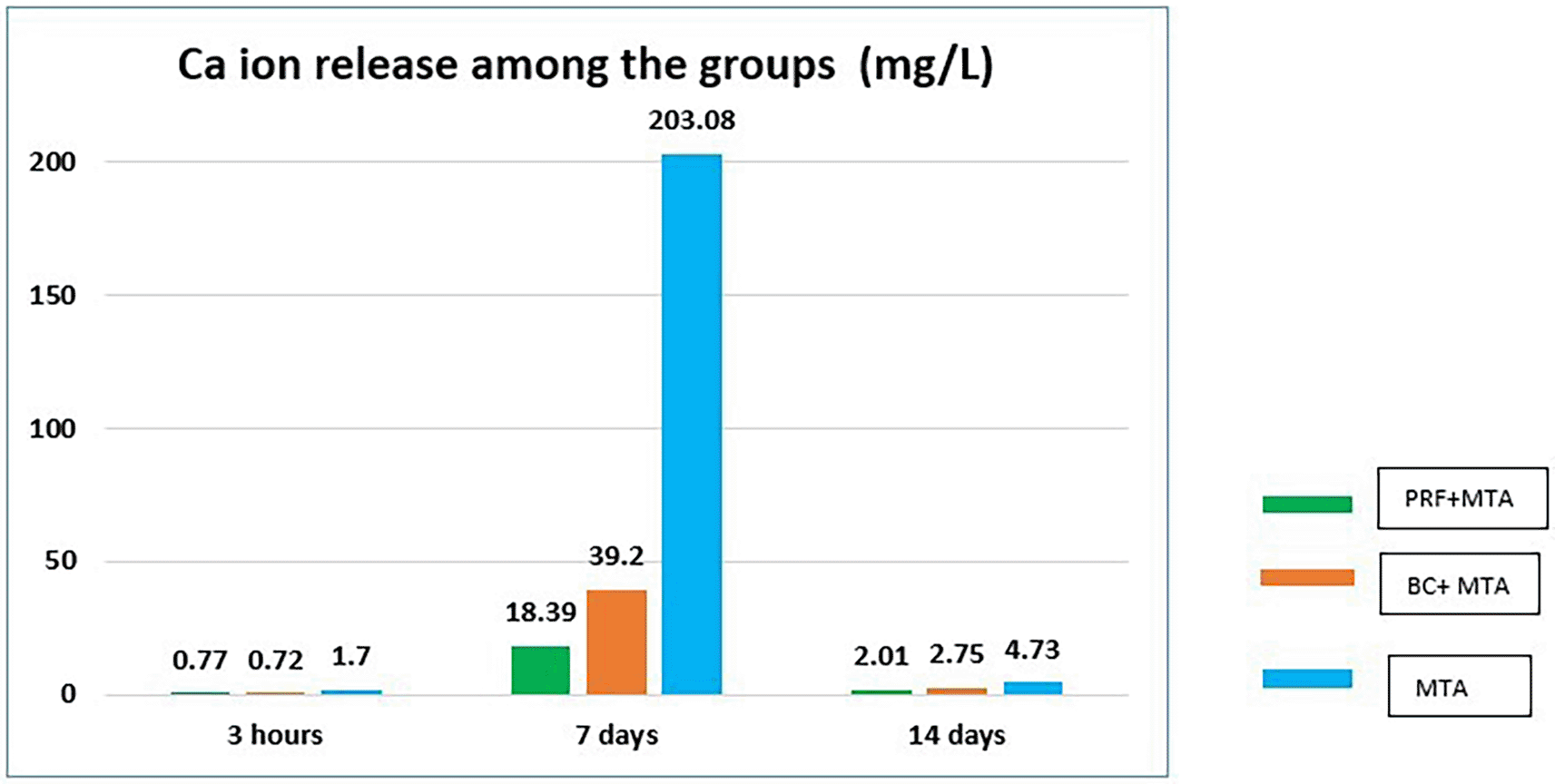
The mean Ca+ ion release of the individual groups at different time periods is given in Table 2. The mean values of Ca+ ions at 3hin the control group (Group 3) was 1.70, MTA +PRF (Group 1) was 0.77 and MTA+ BC (Group2) was 0.72. Group 3 exhibited significantly higher Ca+ ion release as compared to Group 1 and Group 2 at 3h. On the seventh day, MTA+PRF (Group 1) had a Ca+ ion value of 18.39, MTA+ BC (Group 2) recorded a value of 39.20. The control group (Group 3) recorded a maximum value of 203.08, which was significantly higher than both experimental groups. On the 14th day Ca+ion release from all groups reduced significantly. The recorded Ca+ ion release of MTA+PRF (Group 1) was 2.01, MTA+ BC (Group 2) was 2.75 and control group (Group 3) had a value of 4.73. The Ca+ ion release from Group 1 and Group 2 was not significantly different for all the experimental time periods. The control group exhibited the maximum Ca+ ion release for all the time periods as compared to the experimental groups.
The availability of calcium and hydroxyl ion depends on the dissociation of calcium hydroxide, which affects the mineralization process, and surrounding pH. The definitive objective of regenerative endodontic treatment modality for immature root apex with pulpal necrosis is continued root development.10 Immature teeth with open apex with a diameter of 1.1 mm or more respond favorably to REPs11 as it permits the migration of mesenchymal stem cells into the canal space. The revascularization probability in such cases increases by approximately 18-34%.12
The blood collected from inside the root canal was found to have up to a 600-fold increase of CD73 and CD105 markers of mesenchymal stem cells compared to systemic blood.13 Mechanically irritating the periapical tissues may cause discomfort to the patient and in some cases apical bleeding may not always be possible. Various scaffolds have been used as an alternative to (BC) which is rich in platelets, specifically PRP and PRF.
PRF contains physiological thrombin which creates symmetrical intersections in polymerized fibrin, which helps to release growth factors for up to 28 days. Additionally, their flexible fibrin network facilitates cell migration. Platelet derived growth factors (PDGF) and cytokines are abundant in platelets which play an important role in cellular differentiation. Thus, PRF scaffold has emerged as an effective biological tool in REPs.14 Blood clot makes a weak fibrin mesh as compared to PRF. The use of biomaterials along with platelet concentrates containing fibrin and growth factors could lead to a paradigm shift in how endodontic regeneration can be achieved. MTA provides signalling molecules for the maturation of stem cells.15 Ca+ ions play a crucial role in the formation of mineralized hard tissues. Therefore, the study evaluated the release of Ca+, a key element for regenerating pulp dentine complex.
An alkaline environment promotes osteogenic differentiation and bone formation. Mineralisation enzymes such as alkaline phosphatase peaks at pH 7.37 and significantly diminished under physiologic level. In vitro and in vivo research has shown that a pH above 8.0 inhibits the mineralization process. Therefore, various biological and molecular responses that influence repair and regeneration may depend on the local pH.
It is important to create an antibacterial environment at the tooth restoration interface or to control the residual bacteria in the canal space to reduce the risk of reinfection.16 Hydroxyl ions released from biomaterials thus create a hostile environment for bacterial survival and proliferation reducing this reinfection.
White MTA (WMTA) when in contact with tissue fluid, MTA dissolves and releases hydroxyl ions (OH-) increasing the pH to 11-12 and contributing to reparative dentin formation.17 Therefore the present study evaluated the influence of two different scaffolds along with MTA on the pH.
All experimental groups showed an increase in pH over period of time. The highest alkaline pH was recorded in the control group, MTA with distilled water at seven days (11.54) and the lowest in PRF/MTA group (7.78). The difference in pH between PRF and blood clot at 3h and seven days (p < 0.001) was significant, but on the 14th day these groups recorded similar values. Group 3 recorded a mean pH value of 10.76 at 3h, 11.54 on the seventh day and 11.14 on the 14th day which was significantly different from the experimental groups. This is in accordance with the study.9 In the same study, WMTA with blood recorded higher pH values in contrast to our study where the BC with MTA group recorded lower pH values. This could be because the experimental set up was different in that study: in that study, the MTA cylinder was prepared and immersed in blood whereas in our study, PRF/BC was placed over MTA and the pH recorded.
Calcium ions are known to act on osteoblasts and cementoblast cells, causing their differentiation and hard tissue mineralization. Hunter et al. (2018) reported that calcium ions released from biomaterials may influence the repair process, as they pass through the cell membranes by depolarization or activation of membrane-bound calcium channels.18 Although Ca+ ions are one of the major components released by MTA, the role of Ca ions in regenerative endodontics is largely underexplored. In the present study, the mean values for Ca+ ion release from Group1 (PRF/MTA) and Group 2 (BC/MTA) was 0.77 mg L-1 and 0.72 mg L-1 respectively at 3h, which were not significantly different from each other. At seven days, Group 1 recorded a value of 18.39 mg L-1 and Group 2 recorded a value of 39.20 mg L-1. Though the mean Ca+ ion release values of Group 2 were higher than for Group 1, it was not statistically significant at seven days (p ≤ 0.001). At day 14, there was significant reduction in the Ca+ ion release where Group 1 recorded a value of 2.01 mg L-1 and Group 2 recorded a value of 2.75 mg L-1, which were again not significantly different from each other. Overall Group1 (PRF/MTA) showed the lowest release of calcium ions compared to Group 2 (BC/MTA) in14 days. At all experimental periods, Group 3 recorded the maximum Ca+ ion release, which was statistically significant for both experimental groups. A previous study9 evaluated Ca+ ion release from WMTA with and without blood respectively, and reported that the group with WMTA and blood showed greater calcium ion release, which is contrary to what was recorded in our study. This could be because the experimental set up was different in that study, where a MTA cylinder was prepared and immersed in blood; in contrast, in our study, blood clot was placed over MTA and the ion release was recorded. To measure the release of Ca+ ions, ICP-OES equipment was used, owing to its high sensitivity and stability for inorganic analysis. It provides rapid and multi-element analysis of the solutions. In this in vitro study, both experimental groups showed diffusion of Ca+ ions from MTA through scaffolds at three hours, seven days and 14 days, though PRF/MTA allowed lower diffusion of Ca+ ion as compared to the blood clot. However, various clinical studies have suggested that PRFs induce regeneration of pulp dentine complex better than blood clot as it is a rich source of platelets and growth factors.15
There have been a limited number of studies evaluating the effect of PRF/BC with MTA on the pH and calcium ion release. In the present study, we observed that PRF and BC influenced the pH and Ca+ ion release from MTA.
Within the limitations of this in vitro study, which evaluated the influence of two scaffolds on the pH and calcium ion release from MTA over different time periods, it can be concluded that:
1. PRF and BC influenced the pH and Ca+ ion release from MTA. The values were significantly lower in the experimental groups as compared to the control group for all experimental durations.
2. Ca+ ion release in the PRF and BC group increased up to the seventh day, which reduced significantly by the fourteenth day with no significant difference between both groups.
| Views | Downloads | |
|---|---|---|
| F1000Research | - | - |
|
PubMed Central
Data from PMC are received and updated monthly.
|
- | - |
Provide sufficient details of any financial or non-financial competing interests to enable users to assess whether your comments might lead a reasonable person to question your impartiality. Consider the following examples, but note that this is not an exhaustive list:
Sign up for content alerts and receive a weekly or monthly email with all newly published articles
Already registered? Sign in
The email address should be the one you originally registered with F1000.
You registered with F1000 via Google, so we cannot reset your password.
To sign in, please click here.
If you still need help with your Google account password, please click here.
You registered with F1000 via Facebook, so we cannot reset your password.
To sign in, please click here.
If you still need help with your Facebook account password, please click here.
If your email address is registered with us, we will email you instructions to reset your password.
If you think you should have received this email but it has not arrived, please check your spam filters and/or contact for further assistance.
Comments on this article Comments (0)