Keywords
Hodgkin’s disease, wheezing, trachea, chemotherapy
This article is included in the Oncology gateway.
Hodgkin’s disease, wheezing, trachea, chemotherapy
Hodgkin's disease cases with tracheobronchial involvement are rare.1 The symptoms are not specific and may mimic many other diseases like asthma or chronic obstructive pulmonary disease (COPD), which delays the lymphoma diagnosis as well as the treatment.1,2 It is now recognised as a treatable neoplasia. Thus, it is worth mentioning this entity for clinicians especially after the recent therapeutic progress We report the case of a 38-year-old woman with a one-month history of wheezing and a non-productive cough unresponsive to bronchodilators revealing Hodgkin's disease.
A 38-year-old female presented to our department with a one-month history of dry cough and wheezing. She has never smoked. She has not any comorbidities or allergies. She is a stay-at-home spouse and is white. She has visited the emergency room several times in the last month for chest wheezing. She received beta2-mimetics nebulizations, without any improvement. The physical examination at admission revealed a good general state. Respiratory auscultation revealed bilateral diffuse wheezing. Oxygen saturation was (98 %) on room air. The examination of the ganglionar areas detected peripheral bilateral fixed supra-clavicular lymphadenopathy measuring about (2cm of diameter). There was no hepatosplenomegaly. The neurological examination was normal.
The chest x-ray showed a retractile opacity in the right chest with a hilar enlargement (Figure 1).
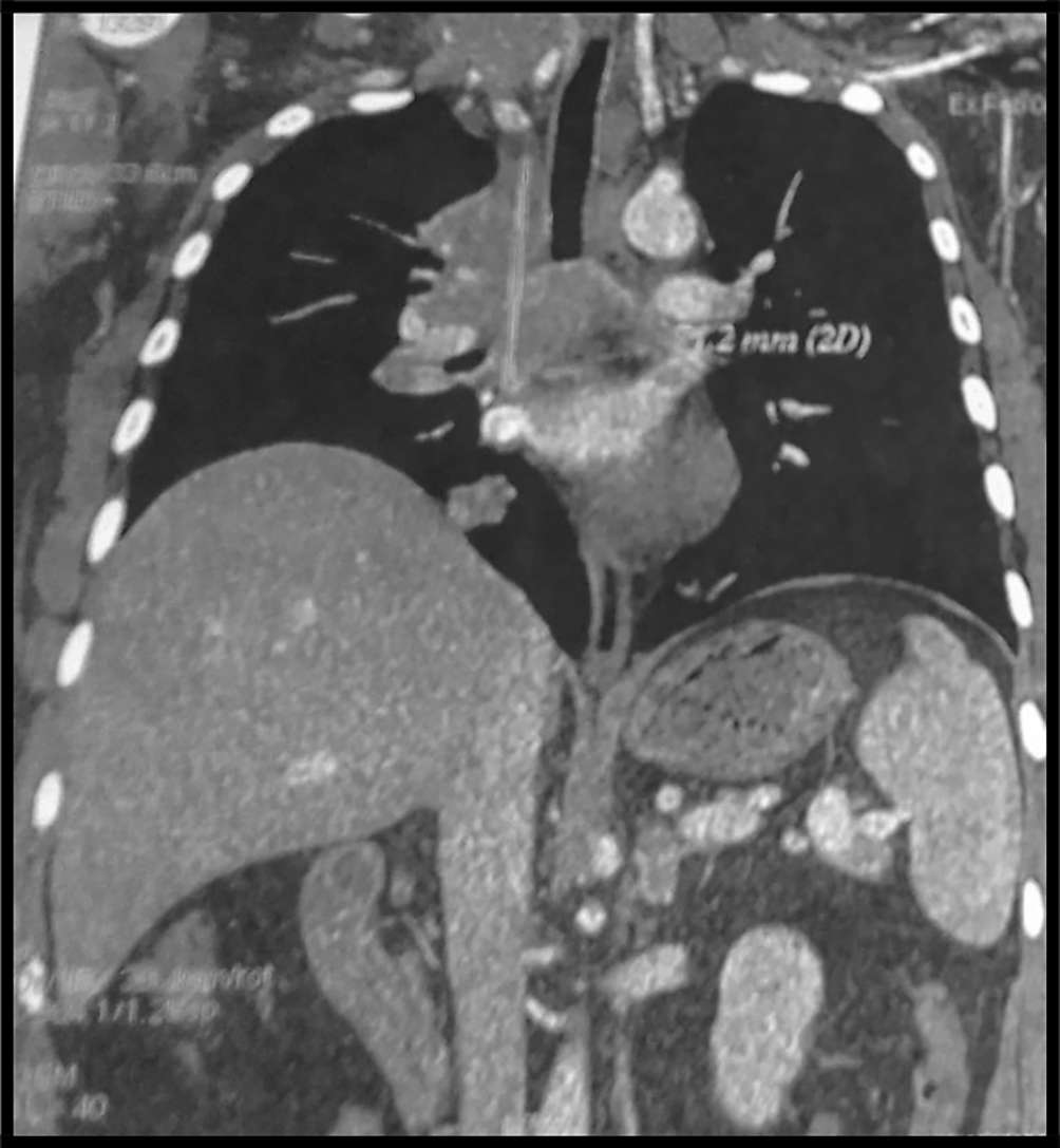
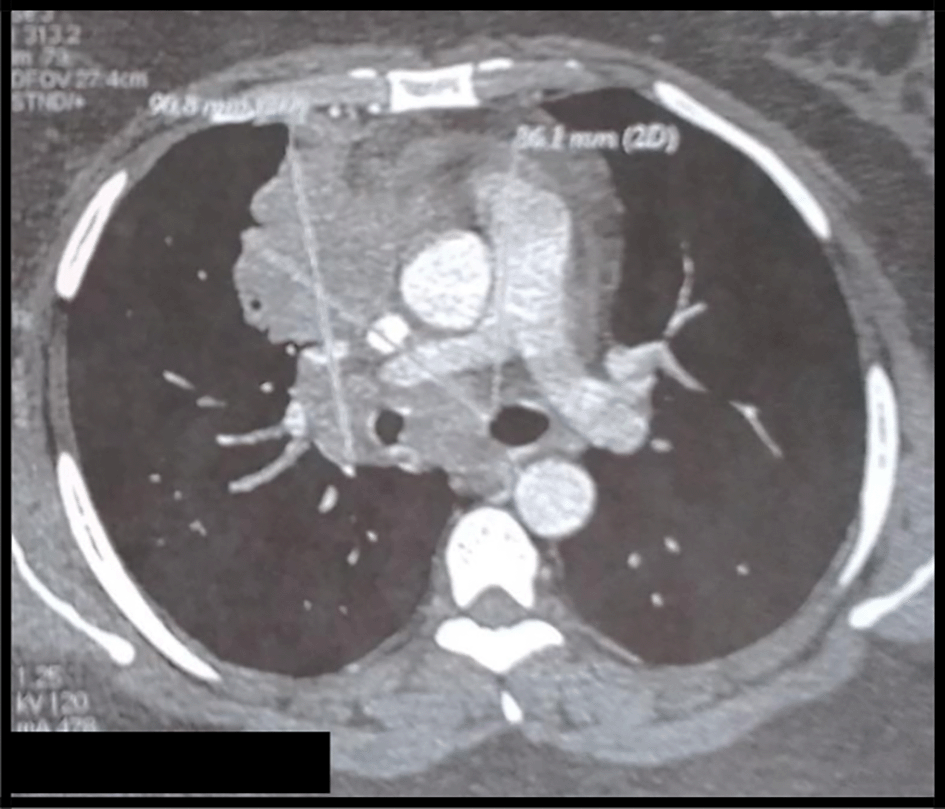
The body computed tomography (CT) scans revealed a bulky mediastino-hilar tumor in the right chest, measuring (90 x 85 x 71 mm), invading the right main bronchus, extending to the vena cava and infiltrating the pericardium with a subpleural right node (Figures 2 and 3). Bronchoscopy showed a polypoid mass located in the carina, involving the right main bronchus. Cytology revealed malignant cells. Histopathology of the bronchial biopsy did not show any malignant lesions. In addition, we performed a biopsy of the supra-clavicular adenopathy. Histopathology revealed an intense staining for CD15 and CD30 with large multinuclear reed Sternberg cells. Therefore, the diagnosis of an Hodgkin lymphoma was confirmed.


The 18-fluorodeoxyglucose-positron emission tomography (FDG-PET) showed many sites of activity of the disease, with a high metabolic fixation of the FDG in the lymph node stations (1L,3,5,6,7,8,9,10,11R) as well as subphrenic adenopathy. Besides, it revealed an intense endotracheal metabolic fixation (Figures 4, 5). Moreover, the bone marrow biopsy was negative.
The patient received 4 courses of an escalated BEACOPP chemotherapy protocol (Bleomycin, Etoposide, Adriamycin, Cyclophosphamide, Vincristine, Procarbazine, Prednisone) as detailed below (Table 1).
She was readmitted in our department 2 months after for acute respiratory failure with wheezing. Bronchoscopy revealed a bulky stenosing polypoid mass, measuring about 3 cm of diameter, rising from the carina extending to the trachea and to the main bronchus that remains partially patent. Histopathology revealed large cells associated with an inflammatory granuloma of (lymphocytes, eosinophils, neutrophils) and a positive staining for CD 15 and CD 30. Therefore, we concluded to a tumoral progression with an endotracheobronchial relapse.
The decision of the multidisciplinary medical team was to put the patient under a second-line chemotherapy: IGEV Regimen as a salvage therapy (ifosfamide, gemcitabine, vinorelbine and methylprednisolone). Moreover, injected Chest CT scans revealed a segmental pulmonary embolism. So, she received a curative anticoagulant treatment. She died one year after the initial diagnosis.
It is well known that Hodgkin lymphoma usually involves the mediastinum. However, pulmonary involvement is less common and can be observed in the lymph nodes, parenchyma, and tracheobronchial tree.1
It is worth mentioning that Hodgkin lymphoma with a tracheobronchial involvement remains extremely rare.1 Patients usually present with respiratory symptoms (such as dyspnea, wheezing and cough), fever, and weight loss.2 The diagnosis may be challenging because the symptoms are not specific. About (30%) of the patients have more than 6-months diagnosis delay.3 We should consider a tracheobronchial tumor in the differential diagnosis when the patient presents to our department with asthma symptoms without any improvement after bronchodilators. In our case, the patient complained of a dry cough and chest wheezing mimicking an asthma attack. She received Beta2-mimetics without any improvement.
Atelectasis is the most frequent radiological finding, occurring in (2/3) of patients. Besides, the chest CT scan may reveal a solitary hilar mass or an obstructive emphysema. Mediastinal or hilar lymphadenopathy are often present.4
Endoscopy may show ulceration, mucosal infiltrate, or a polypoid mass.1 Our patient had a polypoid mass rising from the trachea extended to the main bronchus.
Histopathology shows typically large Sternberg cells with positive staining for CD15 and CD30.3 The diagnosis was made thanks to a biopsy of the supra-clavicular adenopathy in this case. Recent studies suggest that endobronchial ultrasound (EBUS) may be useful in the diagnosis and staging of Hodgkin lymphoma with endobronchial involvement to avoid mediastinoscopy.5
It is worth mentioning that the PET scan is a reliable imaging tool for the diagnosis as well as for the staging of lymphoma especially in this case because mediastinal adenopathy may be due to infectious or inflammatory diseases.6,7 In our case, this exam showed an intense metabolic fixation FDG in many ganglionar areas as well as an endotracheobronchial fixation. It is also very interesting for the follow-up.
Treatment is commonly based on chemotherapy with or without radiotherapy. It depends strongly on the extent of the disease, the general state and the comorbidities.8 Complete resection of the stenotic tracheal tumor may be performed in patients with critical airway obstruction thanks to interventional procedures such as rigid bronchoscopy with a stent placement, photodynamic laser therapy, laser therapy with a neodymium: yttrium-aluminum-garnet laser (Nd-YAG) and photodynamic laser therapy.9,10
Relapsed Hodgkin lymphoma is another challenging problem for clinicians especially when it affects the tracheobronchial tree as seen in our case. Prognostic Factors associated to the relapse of the disease have been recently identified including: a poor performance status (PS), an age greater than 50 years old, failure to achieve remission after an initial therapy, anemia and an advanced lymphoma with a clinical staging (III or IV).11
To conclude, we report a very rare case of lymphoma with an endobronchial involvement. We do underline the importance of considering Hodgkin lymphoma in the differential diagnosis of asthma symptoms without any improvement after bronchodilators or in the case of a tracheal tumor. Further studies are required in order to highlight the prognostic factors in order to improve the outcomes.
Written informed consent for publication of their clinical details and/or clinical images was obtained from the family of the patient.
| Views | Downloads | |
|---|---|---|
| F1000Research | - | - |
|
PubMed Central
Data from PMC are received and updated monthly.
|
- | - |
Is the background of the case’s history and progression described in sufficient detail?
Partly
Are enough details provided of any physical examination and diagnostic tests, treatment given and outcomes?
Yes
Is sufficient discussion included of the importance of the findings and their relevance to future understanding of disease processes, diagnosis or treatment?
Partly
Is the case presented with sufficient detail to be useful for other practitioners?
Partly
Competing Interests: No competing interests were disclosed.
Reviewer Expertise: Diagnostic hematopathology; lymphoma biology
Is the background of the case’s history and progression described in sufficient detail?
Partly
Are enough details provided of any physical examination and diagnostic tests, treatment given and outcomes?
Partly
Is sufficient discussion included of the importance of the findings and their relevance to future understanding of disease processes, diagnosis or treatment?
Yes
Is the case presented with sufficient detail to be useful for other practitioners?
Yes
Competing Interests: No competing interests were disclosed.
Reviewer Expertise: My area of expertise in research is the neuronal control of the Cardiorespiratory system during exposure to hypoxia by using the transgenic animals model.
Alongside their report, reviewers assign a status to the article:
| Invited Reviewers | ||
|---|---|---|
| 1 | 2 | |
|
Version 2 (revision) 11 Sep 23 |
read | |
|
Version 1 14 Apr 23 |
read | read |
Provide sufficient details of any financial or non-financial competing interests to enable users to assess whether your comments might lead a reasonable person to question your impartiality. Consider the following examples, but note that this is not an exhaustive list:
Sign up for content alerts and receive a weekly or monthly email with all newly published articles
Already registered? Sign in
The email address should be the one you originally registered with F1000.
You registered with F1000 via Google, so we cannot reset your password.
To sign in, please click here.
If you still need help with your Google account password, please click here.
You registered with F1000 via Facebook, so we cannot reset your password.
To sign in, please click here.
If you still need help with your Facebook account password, please click here.
If your email address is registered with us, we will email you instructions to reset your password.
If you think you should have received this email but it has not arrived, please check your spam filters and/or contact for further assistance.
Comments on this article Comments (0)