ALL Metrics
-
Views
-
Downloads
Get PDF
Get XML
Cite
Export
Track
Case Report
Revised

Case Report: an unusual orbital tumor

[version 2; peer review: 1 approved, 2 approved with reservations]
PUBLISHED 27 Oct 2023
Author details Author details
OPEN PEER REVIEW
REVIEWER STATUS

Abstract

Introduction

Orbital lipoma is an extremely rare tumor, representing less than 1% of all orbital tumors. We review the literature and describe the presentation, the differential diagnosis and the management of this tumor.

Case report

We report the case of a 63-year-old patient who was referred for a diplopia with recent hemi-cranial headache. Physical examination showed no exophthalmos nor decrease in visual acuity. The patient complained of diplopia on elevation and oculomotricity examination showed limited elevation of the right eye. The Hess Lancaster test was in favor of a limited course of the right inferior rectus muscle. Magnetic resonance imaging revealed a fusiform tissue process in the right inferior rectus muscle with a fatty signal. A complete excision of the tumor was performed by a trasncunjonctival approach. Cytopathological examination was consistent with a pleomorphic lipoma. The postoperative period was uneventful. The definitive histopathologic diagnosis was a lipoma. The postoperative Magnetic resonance imaging showed the complete disappearance of the lesion. With 3 years of follow up, there is no sign of recurrence or ocular motility trouble.

Conclusion: Lipomas are rare tumors in the orbit. The clinic is variable depending on the size and the site. The clinical diagnosis is difficult to make. Only histology allows the final diagnosis.

Keywords

Lipoma, Orbit, Histopathology, MRI, Surgery.

Revised Amendments from Version 1

The appearance of pleomorphic lipoma in imaging studies is variable and not pathognomonic for diagnosis, as variation in the ratio of adipose to non-adipose components results in a wide spectrum of imaging features. MRI features such as the hyper-vascularized tissue process and the hypointense T1 and hyperintense T2 character are explained by the presence of a high proportion of non-adipose elements pleomorphic lipoma. The small size of the tumor and its early diagnosis explain the absence of exophthalmos.
In this new version, we have replied to reviewers' comments and made the following changes:
We've added a new reference (7) that also confirms our MRI findings, which show the tumor to be hypointense on T1 and hyperintense on T2.
The expression "trasncunjonctival" has been replaced by " transconjunctival".
We have replaced the expression "cytopathological" by "histopathological".
This symptom "hemi-cranial headache" has been added   in the case report section.
We've added a new expression “General examination revealed no other lipoma locations”
We've added a new expression “bilateral diplopia “

See the authors' detailed response to the review by Mehdi Hasnaoui

Introduction

Lipomas are benign tumors, rare in the orbit, representing less than 1% of all orbital tumors. They pose a differential diagnosis with a variety of other expansive orbital masses.1 We report a new case, review the literature and discuss the clinicopathological and radiological features, the differential diagnosis and the management of this entity.

Case report

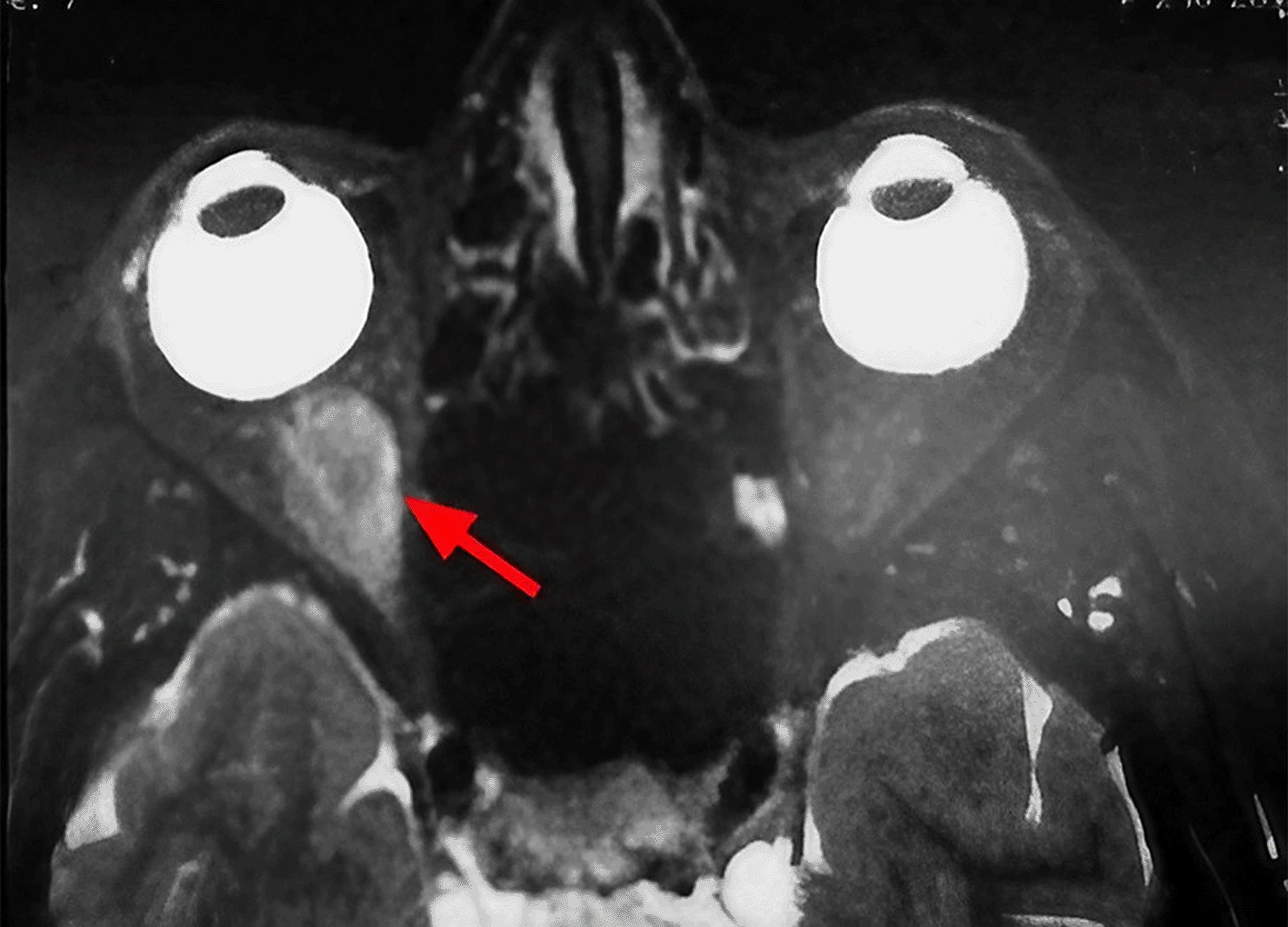
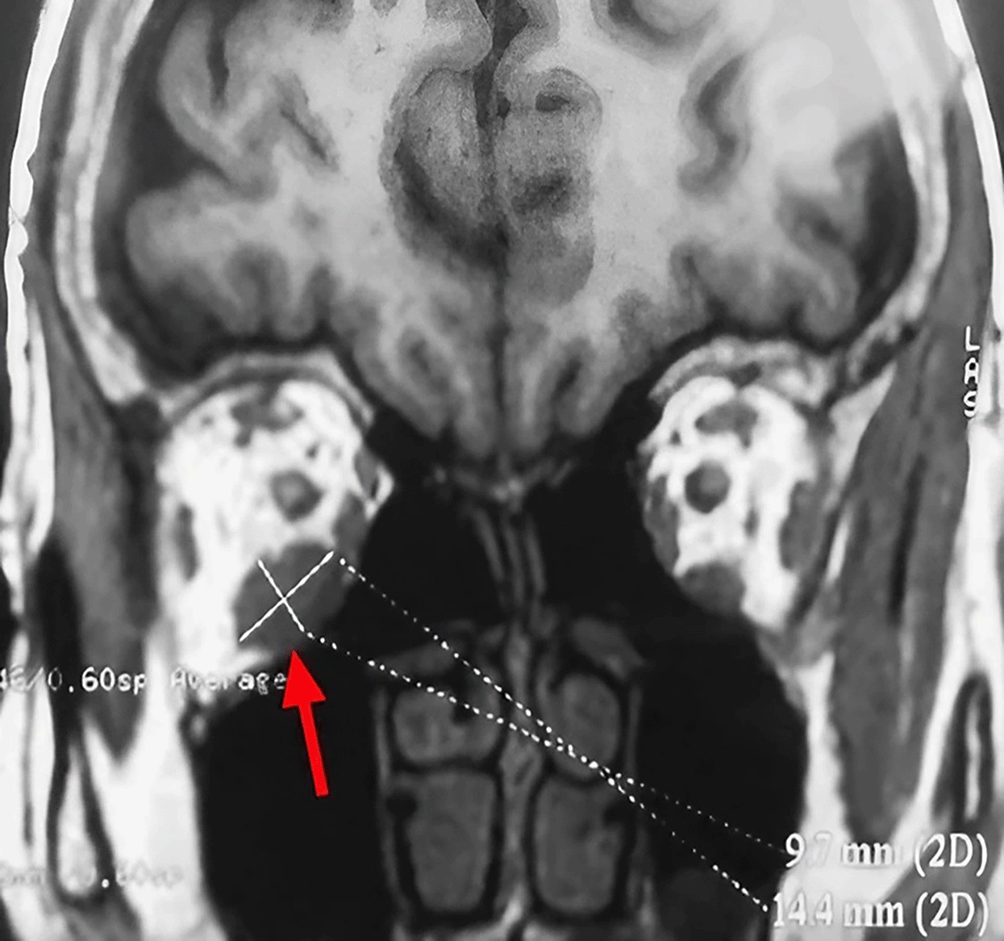
A 63-year-old unemployed Tunisian woman, with no previous personal or family pathological history, presented with a diplopia with recent hemicranial headache evolving for two weeks. Physical examination showed no exophthalmia and no decrease in visual acuity. Furthermore, it revealed bilateral diplopia on elevation. Oculomotricity examination showed limited elevation of the right eye, which was confirmed by the Hess Lancaster test that revealed a limited course of the right inferior rectus muscle. General examination revealed no other lipoma locations. Magnetic resonance imaging (MRI) showed a fusiform and hyper-vascularized tissue process located in the right inferior rectus with fatty signal. The tumor was hyperintense on spin-echo T2-weighted images (Figure 1) and hypointense on spin-echo T1-weighted images (Figure 2).

e527e0ca-eb36-4b15-bf37-396be53a3ad9_figure1.gif

Figure 1. Sagittal section of orbital MRI showing a mass with high signal on a T2 weighted image (red arrow).

e527e0ca-eb36-4b15-bf37-396be53a3ad9_figure2.gif

Figure 2. Coronal section of orbital MRI showing a mass with low signal on a T1 weighted image (red arrow).

These findings suggested various diagnosis; lipoma, inflammatory process, lymphoma and malignant tumor.

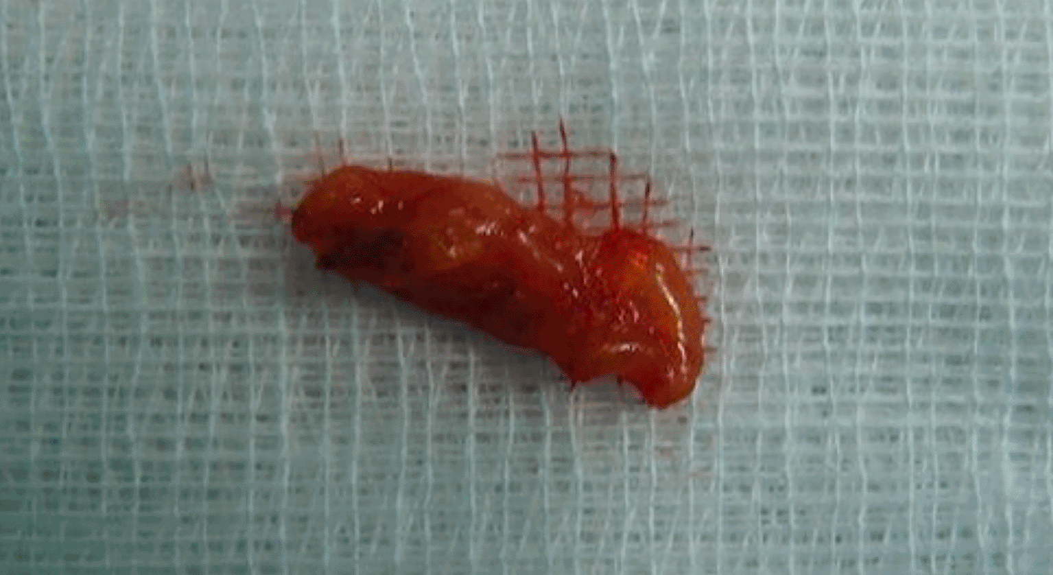
We performed a right inferior transconjunctival orbitotomy and excisional biopsy under general anesthesia. Peroperatively, we discovered an encapsulated mass of fatty tissue, thus complete excision was made. No adherences or involvement of adjacent structures occurred. The specimen was well circumscribed and slightly firmer than normal adipose tissue, with a yellow surface (Figure 3).

e527e0ca-eb36-4b15-bf37-396be53a3ad9_figure3.gif

Figure 3. Macroscopic appearance of the specimen.

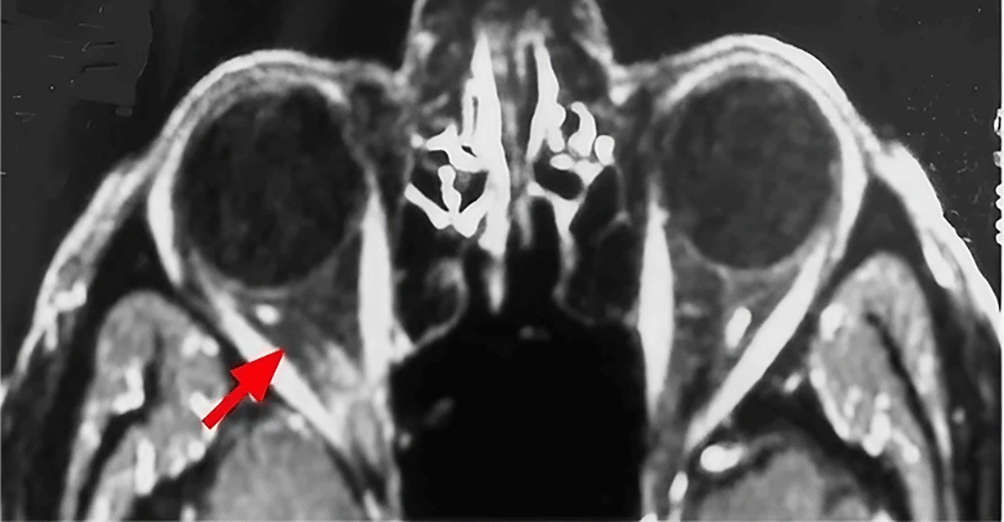
Histologic examination was consistent with a pleomorphic lipoma. The postoperative period was uneventful. Immediately after the operation, the patient reports the resolution of his diplopia. Postoperative MRI images demonstrated the complete resolution of the tumor (Figure 4). With 3 years of follow up, there is no sign of recurrence or ocular motility impairment.

e527e0ca-eb36-4b15-bf37-396be53a3ad9_figure4.gif

Figure 4. Postoperative axial MRI image demonstrating complete tumor resolution (red arrow).

Discussion

Orbital lipoma is the most common mesenchymal soft tissue tumor. However, it is rarely found in the orbit despite the presence of abundant adipose tissue in the intraorbital space.2,3 A review of the largest series of orbital tumors revealed a very low incidence of lipomas.4 Shields et al. reported only two cases of lipomas in a review of 1264 cases of orbital tumors, indicating the rarity of this entity.5 On physical examination, the diagnosis is often difficult to suggest. These tumors are often asymptomatic.

However, they can cause severe morbidity by causing progressive and painless exophthalmos, which is occasionally coupled with diplopia or ocular motility defects6 such as was observed in our patient.

Orbital lipoma exceptionally leads to a compressive neuropathy responsible for a significant decrease in visual acuity, an alteration of the afferent photomotor reflex and the visual field constriction.1 Imaging based on computed tomography (CT) scanning and MRI is essentially useful in ascertaining determining the exact seat, size and relationship to the orbit content. The fatty signal is characteristic on CT sequences. Furthermore, as was found in our patient, the tumor is hypointense on spin-echo T1-weighted images and hyperintense on spin-echo T2-weighted images.7

Histology is essential for definitive diagnosis of pleomorphic lipoma. An important histologic criterion is the presence of a mixture of fat cells, pleomorphic cells and in particular floret-like multinucleated giant cells embedded in a myxoid stroma.8 That concorded with the histological result of our case. Differential diagnosis of this tumor became more important because the number of reports about some other tumors of similar morphology, are increasing. Pleomorphic lipoma may be confused with lipomatous hemangiopericytoma, myofibroblastoma or even malignant tumors such as rhabdomyosarcoma, myxoid malignant fibrous histiocytoma and liposarcoma.9,10 Surgical excision of an orbital lipoma is not only recommended for symptomatic cases such as our patient's clinical presentation but also to exclude malignancy.11 In addition, as was noted in our patient, the long-term outcome after surgery is considered excellent.12

This case highlights the importance of orbital imaging in the context of diplopia without obvious cause to rule out an intraorbital lipoma. Nevertheless, this association remains rare and requires further documentation of cases.

Conclusion

Lipomas are benign soft tissue tumors, rarely located in the orbit. The clinical presentation is variable depends on the size and the intraorbital site. The histology makes the definitive diagnosis and may precisely identify the variant.

Consent

Written informed consent for publication of their clinical details and/or clinical images was obtained from the patient.

Comments on this article Comments (0)

Version 2
VERSION 2 PUBLISHED 19 Jun 2023
Comment
Author details Author details
Competing interests
Grant information
Copyright
Download
 
Export To
metrics
Views Downloads
F1000Research - -
PubMed Central
Data from PMC are received and updated monthly.
- -
Citations
CITE
how to cite this article
Mahmoud A, Touil H, Hann F and Messaoud R. Case Report: an unusual orbital tumor [version 2; peer review: 1 approved, 2 approved with reservations]. F1000Research 2023, 12:700 (https://doi.org/10.12688/f1000research.130056.2)
NOTE: If applicable, it is important to ensure the information in square brackets after the title is included in all citations of this article.
track
receive updates on this article
Track an article to receive email alerts on any updates to this article.

Open Peer Review

Current Reviewer Status: ?
Key to Reviewer Statuses VIEW
ApprovedThe paper is scientifically sound in its current form and only minor, if any, improvements are suggested
Approved with reservations A number of small changes, sometimes more significant revisions are required to address specific details and improve the papers academic merit.
Not approvedFundamental flaws in the paper seriously undermine the findings and conclusions
Version 2
VERSION 2
PUBLISHED 27 Oct 2023
Revised
Views
0
Cite
Reviewer Report 08 May 2024
Kirthi Koka, Orbit, Oculoplasty, Reconstructive and Aesthetic Services, Sankara Nethralaya Medical Research Foundation, Chennai, Tamil Nadu, India 
Approved with Reservations
VIEWS 0
The authors have presented an interesting case of orbital lipoma. 
The authors have reviewed literature but there is no mention of the different variants of lipomas and how one can differentiate between these.
The authors have added imaging ... Continue reading
CITE
CITE
HOW TO CITE THIS REPORT
Koka K. Reviewer Report For: Case Report: an unusual orbital tumor [version 2; peer review: 1 approved, 2 approved with reservations]. F1000Research 2023, 12:700 (https://doi.org/10.5256/f1000research.158021.r263307)
NOTE: it is important to ensure the information in square brackets after the title is included in all citations of this article.
Version 1
VERSION 1
PUBLISHED 19 Jun 2023
Views
10
Cite
Reviewer Report 16 Oct 2023
Amira Trigui, Department of Ophthalmology, University of Sfax, Sfax, Tunisia 
Approved
VIEWS 10
A 63-year-old man presented with diplopia for two weeks. MRI of the brain and orbit revealed a process in the orbit with a fatty signal. Excisional biopsy of the mass resolved the diplopia, and histology revealed a pleomorphic lipoma. This ... Continue reading
CITE
CITE
HOW TO CITE THIS REPORT
Trigui A. Reviewer Report For: Case Report: an unusual orbital tumor [version 2; peer review: 1 approved, 2 approved with reservations]. F1000Research 2023, 12:700 (https://doi.org/10.5256/f1000research.142784.r193958)
NOTE: it is important to ensure the information in square brackets after the title is included in all citations of this article.
Views
44
Cite
Reviewer Report 04 Aug 2023
Mehdi Hasnaoui, Department of Otolaryngology-Head and Neck Surgery, Tahar Sfar Hospital, Mahdia, Tunisia 
Approved with Reservations
VIEWS 44
Editorial Note from F1000Research – 14 August 2023: A COI statement has been added detailing a shared affiliation between the authors and reviewer, which was not declared at the time of the publishing of this report. The COI statement is ... Continue reading
CITE
CITE
HOW TO CITE THIS REPORT
Hasnaoui M. Reviewer Report For: Case Report: an unusual orbital tumor [version 2; peer review: 1 approved, 2 approved with reservations]. F1000Research 2023, 12:700 (https://doi.org/10.5256/f1000research.142784.r190823)
NOTE: it is important to ensure the information in square brackets after the title is included in all citations of this article.
  • Author Response 27 Oct 2023
    Anis Mahmoud, Department of Ophthalmology, Tahar Sfar University Hospital, Mahdia, 5100, Tunisia, Faculty of Medicine, University of Monastir, Monastir, Tunisia, Mahdia, Tunisia
    27 Oct 2023
    Author Response
    I would like to thank the reviewer for his interest in this work and will answer his queries in this report:

    The MRI appearance of pleomorphic lipomas is variable ... Continue reading
COMMENTS ON THIS REPORT
  • Author Response 27 Oct 2023
    Anis Mahmoud, Department of Ophthalmology, Tahar Sfar University Hospital, Mahdia, 5100, Tunisia, Faculty of Medicine, University of Monastir, Monastir, Tunisia, Mahdia, Tunisia
    27 Oct 2023
    Author Response
    I would like to thank the reviewer for his interest in this work and will answer his queries in this report:

    The MRI appearance of pleomorphic lipomas is variable ... Continue reading

Comments on this article Comments (0)

Version 2
VERSION 2 PUBLISHED 19 Jun 2023
Comment
Alongside their report, reviewers assign a status to the article:
Approved - the paper is scientifically sound in its current form and only minor, if any, improvements are suggested
Approved with reservations - A number of small changes, sometimes more significant revisions are required to address specific details and improve the papers academic merit.
Not approved - fundamental flaws in the paper seriously undermine the findings and conclusions
Sign In
If you've forgotten your password, please enter your email address below and we'll send you instructions on how to reset your password.

The email address should be the one you originally registered with F1000.

Email address not valid, please try again

You registered with F1000 via Google, so we cannot reset your password.

To sign in, please click here.

If you still need help with your Google account password, please click here.

You registered with F1000 via Facebook, so we cannot reset your password.

To sign in, please click here.

If you still need help with your Facebook account password, please click here.

Code not correct, please try again
Email us for further assistance.
Server error, please try again.