Keywords
Enterococcus faecalis, Pulp Canal Sealer (EWT), Amoxicillin, Confocal Laser Scanning Microscope, Biofilm, antimicrobial action, Obturaion, Eugenol
This article is included in the Manipal Academy of Higher Education gateway.
Enterococcus faecalis, Pulp Canal Sealer (EWT), Amoxicillin, Confocal Laser Scanning Microscope, Biofilm, antimicrobial action, Obturaion, Eugenol
In Background, "Enterococcus faecalis" and its abbreviation "E.faecalis" are corrected
Added in Results, Line 4: The mean and standard deviations between green ratio, red ratio, green to red ratio and p values for all the four groups are mentioned in Table 1.
Conclusions: Adding 10% amoxicillin to Kerr pulp canal sealer (EWT) against E. Faecalis may help in preventing post obturation infection of the periapical region.
Authors added recommendations for inclusion of antibiotics to root canal sealers for potential eradication of the bacteria in conclusion. As follows:
10% amoxicillin added to the Kerr pulp canal sealer (EWT) effectively eradicated of E. faecalis at 24 hours post-obturation. However, after seven days, plain sealer was as effective as sealer-antibiotic combination. Based on the derived conclusions, it can be recommended that inclusion of antibiotics to root canal sealers help in eradication of the bacteria.
Strong rationale of the current investigation before addressing the aim has been added to resubmitted manuscript.
Null/alternative hypothesis mentioned.
The null hypothesis of the study is there is no difference in the antimicrobial efficacy of the Kerr pulp canal sealer (EWT) with or without addition of 10% amoxicillin against E. faecalis,
Methods:
Sample size arrival: Using G*Power 3.1.9.4 software, at 95% confidence level, 90% power, 0.5 effect size, and assuming 20% loss of sample during the process, the sample size for the present study was calculated to be 60 in each group.
Inclusion and exclusion added in the manuscript.
17% EDTA is used for removing smear layer generated during instrumentation.
Dentinal blocks were infected with E.faecalis for 4 days. : study followed the protocol suggested by Flaviana Bombarda de ANDRADE et. al. (REF.28) f
Discussion:
Modified according to the suggestions given by reviewers and incorporated in the resubmitted manuscript and responded to the reviewers.
See the authors' detailed response to the review by Saravana Karthikeyan Balasubramanian
See the authors' detailed response to the review by Arindam Dutta
See the authors' detailed response to the review by Moksha Nayak
Microorganisms, biofilm and irritants have been the principal causative elements associated with the pathogenesis and progression of pulp and periapical diseases. Eradicating them is the ultimate goal of endodontic therapy. This can be achieved through the combination of asepsis chemo mechanical preparation using instruments, antimicrobial irrigating solutions, intracanal medicaments and obturate the canals system to achieve fluid tight seal.1 In many studies, Enterococcus faecalis (E. Faecalis) has been identified as the most common species associated with persistent or secondary intraradicular infections that do not respond to treatment.2–6 It has been found that E. faecalis is isolated in 23-70% of positive cultures of obturated root canals that show signs of chronic apical periodontitis.2,7–12 It is associated more with asymptomatic cases and exhibit widespread genetic polymorphisms and they often occur in monoculture.13,14
Complete elimination of microbes from root canal system is prevented by difficulty in negotiating complex anatomy, role of dentinal fluid in reducing efficacy of irrigants and intracanal medicaments. Also, high microbial virulence, biofilm formation and relative antimicrobial resistance of infecting bacteria prevent canal disinfection.15–19 In a biofilm, most antimicrobial agents and irrigants only act against microorganisms in its superficial layer, leaving those in the deeper layers unaffected.20
Root canal treatment failures can be prevented or at least minimized by following proper irrigation and obturation protocols. The obturating materials and root canal sealers should exhibit anti-microbial properties, sustained over a period of time, to prevent bacterial growth.21 These solid core obturating materials at times are unable to reach the irregularities of the root canal space such as accessory canals, apical ramifications, isthmuses, the fins, ramifications, and cul-de-sacs. Thus, root canal sealers are used in conjunction with these solid core obturating materials in order to fill these anatomical irregularities completely. The choice of a good sealer greatly affects the outcome of endodontic treatment.22
Several anti-microbials are being added to improve antibacterial properties of sealers, including antibiotics. When conventional root canal treatment alone is insufficient, antibiotics such as penicillin and amoxicillin can be prescribed for treating endodontic infection.23 Using five antibiotics, Hoelscher et al. found Kerr pulp canal sealer (PCS) EWT enhanced antibacterial activity against E. faecalis by adding antibiotics to the sealer. Their study results revealed that antibiotic such as amoxicillin, penicillin, clindamycin, and doxycycline had significantly increased antimicrobial efficacy Kerr EWT sealer.24 All of the sealer-amoxicillin combinations investigated in the study done by Sharma et al. displayed the highest zone of inhibition under both anaerobic and aerobic conditions.25 Amoxycillin at 10% volume showed the best result as an additive to PCS (EWT) sealer with the least mean apical leakage which is clinically significant.26
Antimicrobials within dentinal tubules have a greater antibacterial effect on the seventh day than it did at 24 hours according to study by Heling et al.27Antimicrobial regimens can be evaluated during and post-treatment by culture from the site of infection, blood profile and powerful microscopic examination of the histopathological section. Recently developed Confocal Laser Scanning microscopy (CLSM) has gained popularity in the field of life sciences since it can be used to view and identify single cellular structures. It facilitates immediate fixation of live and dead bacteria, which is not possible in any culture-based method.
The present study was conducted to evaluate the antimicrobial efficacy of the addition of 10% amoxicillin to PCS (EWT) against E. faecalis, at 24 h and seven days following obturation, under Confocal Laser Scanning Microscope.
The null hypothesis of the study is there is no difference in the antimicrobial efficacy of the Kerr pulp canal sealer (EWT) with or without addition of 10% amoxicillin against E. faecalis.
Prior to conducting the study, ethical clearance was obtained from Institutional Ethics Committee, Manipal College of Dental Sciences, Mangalore, Karnataka, India. Protocol approval with Ref. No. 15123. Using G*Power 3.1.9.4 software, at 95% confidence level, 90% power, 0.5 effect size, and assuming 20% loss of sample during the process, the sample size for the present study was calculated to be 15 in each group. A total of 60 freshly extracted single rooted human premolars with Type I canal anatomy and mature root apex were selected and were stored in 1% sodium hypochlorite solution for 48 h for initial sterilization, and subsequently washed in sterile distilled water. Samples were decoronated to a length of 12 mm using a carborandum disc (Mani Inc. Japan) at a speed of 250 rpm attached to a slow speed handpiece (NSK Co., Japan) under water cooling. Canal orifices were enlarged with Gates Glidden drill size #5 (Mani Inc. Japan). Working length was determined by visual examination under 2.5X dental magnifying loupes (Pierson Surgical limited, Keeler, UK) using a 10 number K file (Dentsply-Maillefer, Ballaigues, Switzerland). All canals were then enlarged till size 25 hand K files using RC prep (Premier, India) for canal lubrication. Final preparation was done using rotary ProTaper Universal files (Dentsply, York, PA, USA, till File F2, using X-SMARTTM Electric Endo Motor. Throughout the instrumentation, 5.25% sodium hypochlorite was used to irrigate the canals, followed by irrigation with normal saline and 17% EDTA for removing smear layer generated during instrumentation. Red nail varnish (Maybelline) was applied in double layers, to cover the outer surface of the samples except 3 mm from the apical end of roots. Samples were dried for 24 hours before being autoclaved (MELAG Euroklav 23 VS-S, Russia).
Infection of the dentin blocks: The Enterococcus faecalis strain ATCC 29212 (ATCC, USA) was cultured and reactivated in Brain Heart Infusion broth (BHI, Difco, Kansas City, MO, USA), which was maintained at 37°C for 24 hours. Broth was transferred to another BHI flask and incubated again for another 24 hours to achieve exponential growth and adjusted to McFarland standard No. 1 (3 × 10⁸ CFU/mL).
The present study followed the protocol suggested by Flaviana Bombarda de ANDRADE et al. for in vitro intratubular dentinal bacterial contamination for antimicrobial endodontic tests for confocal laser scanning microscopy.28 On the first day, all dentinal blocks were infected over a period of 4 days. On the second day, following incubation, samples were agitated in a vortex for 10 s, and then inocula from the microtubes were discarded. One mL of sterilized BHI broth was inserted, following which a centrifuge cycle of 3,600 g for 5 min at 25°C was done. The microtubes were incubated again at 37°C for 24 hours. On the third day, a new inoculum of E. faecalis was inserted into the sample tubes, at exponential growth phase after seven hours of subculture in BHI broth. The centrifugation protocol was repeated twice at each speed, at 25°C. Procedures were repeated on the fourth day as described for the second day. On the fifth day, the samples were removed from the microtubes. Half of the samples were sectioned and then were stained with SYTO9 stain and the other half maintained in the incubator for another 7 days.
Sealer-antibiotic combination was prepared manually by mixing the antibiotic to the sealer 10% by weight using an electronic weighing scale (Sartorious Lab balance, Germany).29 The sealer was mixed according to manufacturer’s specifications for obturation. Root canals were irrigated with 17% EDTA for one minute and dried with paper points (Diadent, S. Korea) Sealer-antibiotic paste was lightly coated on to the canal walls twice with gutta-percha and canals were obturated with gutta-percha points (Diadent, S. Korea) with lateral compaction technique. Access cavity was restored with composite resin (3M ESPE, USA). All specimens were incubated at 37°C in humid conditions.
Prior to obturation, the samples were randomly distributed into two groups of 30 specimens each. After the root filling, the samples were subdivided into two more groups of 15 teeth each, based on the time of assessment into the following:
Group 1: S - sealer group (24 h)
Group 2: SA - sealer antibiotic group (24 h)
Half if the samples sectioned on the seventh day were designated number ‘7’
Group 1 a: S7 - Seventh day sealer group
Group 2 a: SA7 - Seventh day sealer antibiotic group
Low-speed hand pieces with small round burs were used to fracture each cylindrical dentin block by making thin vertical grooves in the middle (Dentsply Maillefer, Ballaigues, Switzerland). Following this, specimens were fractured using a chisel and mallet into two semi cylindrical halves. The outer convex surface was ground using low speed handpiece (NSK Co., Japan) attached to a water cooler and a fine carbide bur (Dentsply-Maillefer, Ballaigues, Switzerland) at 300 rpm, to achieve a standard thickness of 1 mm. Thirty specimens from each experimental group were sectioned 24 hours post-obturation and the rest on the seventh day and stained for analysis under a confocal microscope.
The sectioned dentin pieces were stained with SYTO 9 Green Fluorescent Nucleic Acid Stain (ThermoFisher Scientific, USA) and propidium iodide (Himedia, India), according to manufacturer’s instructions for 20 minutes in the dark at room temperature, and then rinsed with phosphate buffered saline for one minute. Each sample was then air-dried briefly and transferred into a micro centrifuge until used. Images were captured at magnification of 40×. In the case of SYTO9, the excitation/emission wavelengths were 480/500 nm, whereas in the case of propidium iodide, they were 490/635 nm. Image acquisition and analysis using CLSM at a resolution of 1024×1024 was carried out using Leica Application Suite (Leica Microsystems, Germany). Borders of the root canal were focused and two images per sample were obtained randomly. Background noise was then reduced in the images (Leica Application Suite software) and further quantitative analysis was carried out using ImageJ software (V ImageJ: Rasband, W.S., ImageJ, U. S. National Institutes of Health, Bethesda, Maryland, USA).30,31
The results were interpreted as:
Green ratio was calculated by dividing the area of the living cells by the size of the entire magnified field of view at 40×.
Red ratio was calculated by dividing the area of the dead cells by the size of the entire magnified field of view at 40×.
Green to red ratio: the value obtained by dividing the green ratio with the red ratio.
The mean and standard deviations between green ratio, red ratio, green to red ratio and p values for all the four groupes are mentioned in Table 1.
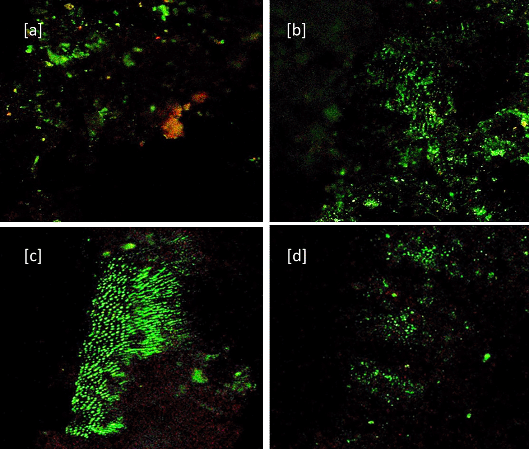
CLSM images (40×) for 24 hours groups showed: [a] Baseline for Sealer group [b] Sealer group showing viable cells (in green color) at 24 h. [c] Baseline for Sealer-antibiotic group [d] Sealer-antibiotic group showing viable cells at 24 h (in green color) (Figure 1) After 7 days: [a] Baseline for Sealer group [b] Sealer group showing dead cells (Red color) and viable cells (in green color) [c] Baseline for Sealer-antibiotic group [d] sealer-antibiotic group showing dead cells (Figure 2).

[a] Baseline for Sealer group. [b] Sealer group showing viable cells (in green color) at 24 hrs. [c] Baseline for Sealer-antibiotic group. [d] Sealer-antibiotic group showing viable cells at 24 hrs (in green color).
[a] Baseline for Sealer group. [b] Sealer group showing dead cells (Red color) and viable cells (in green color). [c] Baseline for Sealer-antibiotic group. [d] Sealer-antibiotic group showing dead cells.
The first bar graph shows individual rd and green ratio of groups S7, SA7, SA, S (Figure 3). The second bar graph showthe green to red ratios of all groups along the x axis, while they axis shows different groups (Figure 4).


Bar graph showing in X axis green to red ratios of all groups. In Y axis showing different groups.
In the present study both, highest green and red ratio were seen in the S group followed by SA7, SA and least in S7 (Figure 3). There was no statistical difference among the groups in red ratio, but green ratio of S group showed statistically significant difference with other groups (Table 1). Ratio of green to red also shows that the mean value of S group was highest with lowest antibacterial effect on E. Faecalis followed by SA, SA7 and least in S7 with no statistically significant difference (Figure 4). The ratios were comparable between SA and SA7 with a slightly better ratio in the SA group. The SA, SA7, S7 showed marked reduction in the green ratio and live bacteria when compared with the S group. Comparison of the red ratio using Post hoc Tukey test between different groups showed no statistically significant difference (Table 2). Comparison of the green ratio using Post hoc Tukey test showed no statistically significant difference between different groups except between S7 and S showing significant reduction in live bacteria in S7 compared to S (Table 2).
E. faecalis was chosen as study microbe since it is found especially in failed to endodontic infections. It is because of its virulence factors such as aggregation substance, surface adhesins, sex pheromones, lipoteichoic acid, extracellular superoxide production, the lytic enzymes gelatinase and hyaluronidase, and the toxin cytolysin.32 The present study used CLSM for evaluating antimicrobial efficacy od sealer since it is one of the most commonly used fluorescence microscopic techniques recently in endodontics, particularly in three-dimensional (3D) studies involving biological cells and tissues. It is a flexibile approach, which makes it suitable for use in fast imaging of dynamic processes in living cells, sensitive morphological analysis of tissues.33 The S group (sealer without antibiotics at 24 hours) proved to have little antibacterial effect against E. faecalis at 24 hours compared to baseline. Even though highest red ratio is recorded for this group it could not outnumber the green ratio (Figure 1 [a] Baseline, [b] after 24 h) The reason for the highest red ratio in S group could be due to increase in the free eugenol release from freshly mixed sealer or cytotoxicity expressed within the first few hours. This hypothesis is supported by in vitro study, which showed inhibition of E. faecalis only within the first five hours of incubation due to the release of free eugenol.34 Eldeniz et al., also found that the antibacterial activity was higher at the 13th hour. Cytotoxicity of ZOE based sealers have been shown to sharply reduce after 24 hours of setting.35 The green ratio was highest in this group when compared to other groups because of the decrease in the release of eugenol from set sealer at 24 hours, allowing E. faecalis to grow. The results are in agreement with Zhang et al who evaluated the in vitro antibacterial activity of similar sealers and found no significant antibacterial activity one day after setting.36 According to Baer et al., sealers without amoxicillin did not inhibit the growth of E. faecalis. Additionally, they found no statistical difference between fresh mixed samples and those that had been set (p > 0.05).37 This is in agreement with the present study. Pizzo et al. in 2006 evaluated in vitro antimicrobial action of root canal sealers and found ZOE sealer to be equally effective in inhibiting bacterial growth until 24 hours after mixing.38 It is likely that the set materials released eugenol that contributed to this.39
The SA group (sealer with antibiotics at 24 hours) combination showed a marked positive inhibitory effect on E. faecalis. (Figure 1 – [c] Baseline, [d] after 24 h) A possible hypothesis is the alkaline pH of the combination enhancing the antimicrobial activity against E. faecalis and its susceptibility towards amoxicillin. The results are in agreement with Baer et al. who evaluated in vitro, the antibacterial effect of amoxicillin when added to different sealers and found that sealers mixed with amoxicillin were significantly more effective than without.37 According to Binoy et al., the reported, pH of the combination was 8.55, this high alkalinity might have deleterious effects on microorganisms in obturated canals. Vidulasri et al. stated that the anti-microbial activity against E. faecalis was improved when antibiotics like amoxicillin and clindamycin were added to zinc oxide eugenol sealer. Amoxicillin is beta-lactam bactericidal broad-spectrum antibiotic that acts by inhibiting bacterial cell wall synthesis.29 According to a previous study, E. faecalis is more sensitive to the antibiotics amoxicillin, amoxicillin-clavulanic acid, benzyl penicillin, vancomycin, and doxycycline while being less sensitive to the antibiotics erythromycin and azithromycin.40 An antibiotic-enhanced sealer that alters the environment of the microorganism and retains bactericidal qualities after setting time may be crucial for the success of initial endodontic therapy and for avoiding re-infection.41,42
The S7 group (sealer without antibiotics after seven days) showed significant improvement in antimicrobial effect compared to S group with a reversal in the green to red ratio. [Figure 2 – [a] Baseline, [b] after seven days] (p < 0.01). A possible hypothesis is a bactericidal effect of eugenol due to its sustained release on microbes causing protein denaturation. The results are in agreement with Pizzo G et al., who found that after seven days from mixing, ZOE containing sealer, still exerted antibacterial activity.38 Heling demonstrated the antibacterial property of Pulp Canal Sealer (EWT) at seven days owing to the bactericidal effect of eugenol as a part of the liquid.27 Hasheminia et al. stated that eugenol is a potent antibacterial agent, acting on microbes by protein denaturation.43 The findings of the present study demonstrating antibacterial activity of EWT after seven days are justified by the above mentioned studies. The results are contradicted by Smadi et al. who tested nine sealers against E. faecalis and found that most sealers had antibacterial properties immediately after mixing, but these properties deteriorated over time.44 Zhang et al. also did not see any antibacterial effect of eugenol based sealer beginning on the third day.36 This could be because of the use of different formulations of sealers or use of different microbiological techniques of assessment. In our study results were contrary to these findings probably because of the use of confocal laser scanning microscope for analysis.
The SA showed good antibacterial efficacy in 24 hours which could be because of the presence of antibiotics and maintained its efficacy even after seven days in the SA7 group (sealer with antibiotic after seven days) (Figure 2 – [c] Baseline, [d] after seven days) with no significant statistical difference with the S7 and SA. The results are in agreement with Baer et al.37 who concluded that the antimicrobial properties and inhibition of the E. faecalis growth even after seven days can be demonstrated in sealers combined with amoxicillin. However, a slight increase in the viable cell count is seen when compared with the 24 hour group, which could probably be because of multiplication of cell adjacent to the zone of killing or slow release of amoxicillin from a viscous mix.45 Our results are in agreement with Binoy et al. who reported the combination to have greater viscosity when compared with other antibiotic combinations. This finding further explains the reason for no enhancement in the antimicrobial property of 10% amoxicillin sealer combination after seven days when compared with 24 hours.39 However, more longitudinal clinical studies are needed to confirm the outcome of addition of different antibiotics to various sealers.
Within the limitations, the outcome of the current study concluded that adding antibiotics 10% amoxicillin to zinc oxide eugenol sealer increases its antibacterial effect against E. faecalis within 24 hours, while plain ZOE sealer demonstrate good antibacterial property against E. faecalis only after seven days.
Study recommends to evaluate the antibacterial efficacy of other endodontic sealers by adding various antibiotics. Additionally, investigations pertaining to the sealer properties such as shrinkage, setting time, flow, sealability, diffusability and substantivity of the sealer-antibiotic combination need to be conducted before we use these combinations clinically.
• Adding 10% amoxicillin to Kerr pulp canal sealer (EWT) against E. Faecalis may help in preventing post obturation infection of the periapical region.
• Since amoxicillin is reported as the drug of choice for endodontic infections in most countries, 10% amoxicillin is added to sealer in the present research, to prevent reinfection.
• Clindamycin and erythromycin can be alternative drugs for patients allergic to penicillin.
| Views | Downloads | |
|---|---|---|
| F1000Research | - | - |
|
PubMed Central
Data from PMC are received and updated monthly.
|
- | - |
Provide sufficient details of any financial or non-financial competing interests to enable users to assess whether your comments might lead a reasonable person to question your impartiality. Consider the following examples, but note that this is not an exhaustive list:
Sign up for content alerts and receive a weekly or monthly email with all newly published articles
Already registered? Sign in
The email address should be the one you originally registered with F1000.
You registered with F1000 via Google, so we cannot reset your password.
To sign in, please click here.
If you still need help with your Google account password, please click here.
You registered with F1000 via Facebook, so we cannot reset your password.
To sign in, please click here.
If you still need help with your Facebook account password, please click here.
If your email address is registered with us, we will email you instructions to reset your password.
If you think you should have received this email but it has not arrived, please check your spam filters and/or contact for further assistance.
I thank the editorial team for helping us to expedite the process of review and for bringing out the publication.
I thank the editorial team for helping us to expedite the process of review and for bringing out the publication.