Keywords
Apocrine carcinoma, Axillary mass, rare presentation, apocrine neoplasms, solid lesion, lymph node excision, malignant, surgical excision.
This article is included in the Oncology gateway.
This article is included in the Datta Meghe Institute of Higher Education and Research collection.
Apocrine carcinoma, Axillary mass, rare presentation, apocrine neoplasms, solid lesion, lymph node excision, malignant, surgical excision.
I have made several changes to the article to improve its clarity and readability.
Firstly, I reorganized the content to ensure more additions to the history and treatment plan. Additionally, Biopsy findings and metastatic workup were added. I also incorporated relevant information from the patient, which is up to date, to provide the most accurate and current information possible. Overall, these changes aim to make the article more informative and engaging for the reader.
See the authors' detailed response to the review by Nour Kibbi
See the authors' detailed response to the review by Rehan Zahid
Apocrine carcinoma of the axilla is a seldom-seen form of breast cancer that originates in the axillary sweat glands. This disease has been reported to be an invasive ductal carcinoma subtype, the most prevalent form of breast cancer among women. From a surgical perspective, apocrine carcinoma of the axilla may present as a palpable mass or a non-palpable abnormality on imaging studies, such as mammography or ultrasound. Treatment typically involves surgical excision of the tumor with clear margins, which may be followed by radiation therapy and/or systemic chemotherapy depending on the stage and biology of the cancer. From a pathological standpoint, Large, pleomorphic cells with an abundance of eosinophilic cytoplasm and prominent nucleoli are seen in axillary apocrine carcinomas. Apocrine carcinoma of the axilla is uncommon, but it can still be a clinically relevant diagnosis that has to be treated promptly and effectively to provide the patient the best chance of survival.
In 2022, a male aged 48, hailing from South Asian origins and employed as a carpenter, visited the surgery department due to the presence of a lump (Figure 1) in the axilla. The patient initially discovered a painless, indurated nodule in their right axilla around a year back. Over time, this nodule increased in size until the time of their presentation that is the size of 8 × 8 × 2.3 cm. The patient attempted ayurvedic treatment for a year, but the lesion continued to progress. On local examination, signs included inflammation of the skin covering the area, occasional discharge of a serosanguinous nature, and mild pain in the affected axilla. The patient did not experience any constitutional symptoms such as fever, weight loss, night sweats, or loss of appetite. The patient had no family history of malignancy. A physical evaluation and USG for both breasts did not find any abnormalities. USG of the right axilla suggested a well-defined solid isoechoic lesion with multiple microcalcifications with prominent vascularity. Additionally, the patient had a preexisting condition of hypertension and was taking antihypertensive medications. Patient revealed his past habit of smoking bidi once a day. He did not consume alcohol.
A Tru Cut Biopsy was carried out which revealed cells showing moderate amount of eosinophillic to pale vaccuolated cytoplasm, Eccentric nuclei showing mild hyperchromasia and significant mitotic activity. Another population of cells seen with squamoid feature intervening the stroma showing desmoplastic reaction. Biopsy findings were suggestive of Malignancy of Adnexal origin.
Patient underwent wide local excision of the right axillary lesion with right axillary lymphadenectomy up to level III was carried out. A sufficient clearance margin of 1-1.5 cm was taken, and primary closure of defect was achieved.
Seven axillary lymph nodes were isolated, the largest lymph node was measured to be 4×2×1.5 cm.
Grossly (Figure 2), the tumour mass was white, firm in consistency and measured 4×4×3.5 cm. On the cut section, solid, homogenous blackish areas were identified with the involvement of overlying skin.
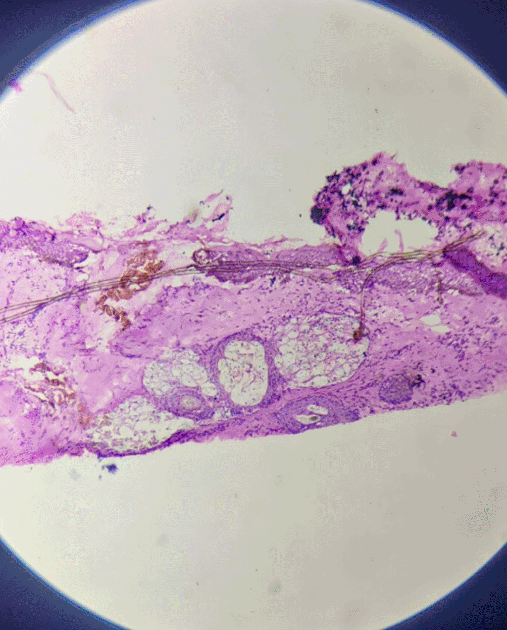
Microscopically, sections from superior, anterior and posterior margins showed unremarkable squamous lining epithelium with unremarkable deeper tissue and adnexal structure with few distended ducts of histopathology. Section from the inferior margin (Figure 3) was positive for infiltration by malignant epithelial cells. Sections also show fibro-collagenous areas with minimal scattered inflammatory infiltrate. Sections from the tumour were also positive for perineural and lymphovascular invasion.

Section from all seven lymph nodes shows histopathological features suggestive of metastatic deposits of epithelial malignancy.
The epidermis is visible in some portions of the tumour, which has a dermis with mostly papillary cystic architecture. Focal inflammation with numerous benign apocrine glands were noted.
The tumour with papillary architecture (Figures 4–6) was found to have fibrovascular cores lined by eosinophilic epithelial cells. The patient's recuperation after surgery proceeded without any complications. The patient’s case was discussed in the hospital’s interdisciplinary tumour board and he was considered for adjuvant radiotherapy. Considering the metastatic involvement in the lymph nodes and to exclude the possibility of distant metastasis, a comprehensive whole-body PET-CT scan was performed, revealing no signs of distant metastatic spread.
A thorough follow-up plan was advised to the patient, but he did not follow through.
Apocrine carcinoma is an extremely uncommon adnexal malignancy with limited data on histologic prognostic factors and patient outcomes.1 The axilla and adjacent medial upper arm are the most typical sites for apocrine carcinoma.2 The tumour, which derives from pleuripotent adenexal cells capable of eccrine and follicular development, had first been discovered by Goldstein in 1982.3 Reddish-purple subcutaneous nodules and solid or cystic masses are common characteristics of these tumours. Skin ulceration may be a comorbid condition, and they are frequently locally advanced when diagnosed. This tumour has a sluggish rate of growth, is locally invasive, and has the potential to spread to nearby lymph nodes, the lungs, the liver, the bone, and the brain.4 At the time of diagnosis, lymph node metastases were present in over fifty per cent of all reported individuals suffering from apocrine carcinoma. The recommended treatment for these lesions is wide local excision.5 Apocrine gland carcinomas and eccrine carcinomas are the two primary subtypes of sweat gland carcinomas. Apocrine carcinomas appear as hard, rubbery, cystic, solitary or numerous, non-tender masses with red to purple overlaying skin. Eccrine gland carcinomas lack distinguishing clinical characteristics, rendering gross examination diagnosis nearly difficult. They often only affect older individuals and present as quasi-tender, subcutaneous nodules.6 Although the tumour often arises de novo, it can potentially result from previously present benign tumours like apocrine hyperplasia or apocrine adenoma.7
Wide local excision is the preferred method of management of primary cutaneous ductal apocrine carcinoma. In addition to excision, chemotherapy as well as radiotherapy have been utilised, but they haven’t significantly reduced mortality or morbidity among patients with either localised or metastatic disease. The total number of reported instances and the amount of follow-up data that currently exists, both seem insufficient for determining the prognosis. Therefore, there is a need for further case accumulation.
The rare tumour referred to as apocrine carcinoma has a characteristic but non-specific histological appearance. For determining the prognosis and formulating particular therapy recommendations, the reported cases and follow-up data appear to be insufficient. Hence, additional cases needs to be collected.
Written informed consent for publication of their clinical details and clinical images was obtained from the patient.
All data underlying the results are available as part of the article and no additional source data are required.
| Views | Downloads | |
|---|---|---|
| F1000Research | - | - |
|
PubMed Central
Data from PMC are received and updated monthly.
|
- | - |
Is the background of the case’s history and progression described in sufficient detail?
Yes
Are enough details provided of any physical examination and diagnostic tests, treatment given and outcomes?
Yes
Is sufficient discussion included of the importance of the findings and their relevance to future understanding of disease processes, diagnosis or treatment?
Yes
Is the case presented with sufficient detail to be useful for other practitioners?
Yes
Competing Interests: No competing interests were disclosed.
Reviewer Expertise: Rare tumors
Is the background of the case’s history and progression described in sufficient detail?
Partly
Are enough details provided of any physical examination and diagnostic tests, treatment given and outcomes?
No
Is sufficient discussion included of the importance of the findings and their relevance to future understanding of disease processes, diagnosis or treatment?
Partly
Is the case presented with sufficient detail to be useful for other practitioners?
Partly
Competing Interests: No competing interests were disclosed.
Reviewer Expertise: Plastic and Reconstructive Surgery
Alongside their report, reviewers assign a status to the article:
| Invited Reviewers | |||
|---|---|---|---|
| 1 | 2 | 3 | |
|
Version 3 (revision) 08 Nov 23 |
read | read | |
|
Version 2 (revision) 27 Sep 23 |
read | ||
|
Version 1 10 Jul 23 |
read | ||
Provide sufficient details of any financial or non-financial competing interests to enable users to assess whether your comments might lead a reasonable person to question your impartiality. Consider the following examples, but note that this is not an exhaustive list:
Sign up for content alerts and receive a weekly or monthly email with all newly published articles
Already registered? Sign in
The email address should be the one you originally registered with F1000.
You registered with F1000 via Google, so we cannot reset your password.
To sign in, please click here.
If you still need help with your Google account password, please click here.
You registered with F1000 via Facebook, so we cannot reset your password.
To sign in, please click here.
If you still need help with your Facebook account password, please click here.
If your email address is registered with us, we will email you instructions to reset your password.
If you think you should have received this email but it has not arrived, please check your spam filters and/or contact for further assistance.
Comments on this article Comments (0)