Keywords
Uniprot ID P02649, APOE, Apolipoprotein E, antibody characterization, antibody validation, Western Blot, immunoprecipitation
This article is included in the Cell & Molecular Biology gateway.
This article is included in the YCharOS (Antibody Characterization through Open Science) gateway.
Uniprot ID P02649, APOE, Apolipoprotein E, antibody characterization, antibody validation, Western Blot, immunoprecipitation
In this new version, Figure 1 has been updated to repeat Western blot testing for antibodies ab1907* and MA5-15852* to determine whether they would successfully target ApoE when using a dilution of 1/200 rather than 1/1000. Table 1 was also updated. The title was also amended.
See the authors' detailed response to the review by Nathalie Macrez
See the authors' detailed response to the review by Leonard Kritharides and Maaike Kockx
Apolipoprotein E, or APOE, is a 299 amino acid transcribed and secreted to regulate lipid homeostasis by controlling the uptake of cholesterol and lipoproteins via receptor-mediated endocytosis.1,2 Three major isoforms of APOE exist, apoE4, apoE3, and apoE2.1 Despite only differing by single amino acid substitutions, their functionalities are altered at both the cellular and molecular levels.1 Accordingly, the binding affinity of APOE to its ligand, the LDL receptor (LDLR), varies depending on the isoform.2 ApoE3 and apoE4 bind to LDLR with high affinity while apoE2 binds with low affinity.3
Association studies have demonstrated apoE4 to be a genetic risk factor for cardiovascular disease, as it can cause high levels of circulating cholesterol, in the form of LDL as well as a genetic risk factor for late-onset Alzheimer’s disease.4–7 The critical role of APOE in health and disease highlights the need for additional research into the protein’s mechanism of action and potential for therapeutic strategies.8 Mechanistic studies would be greatly facilitated with the availability of validated and high-quality antibodies.
Here, we compared the performance of a range of commercially-available antibodies for Apolipoprotein E and validated several antibodies for Western Blot and immunoprecipitation, enabling biochemical and cellular assessment of Apolipoprotein E properties and function.
Our standard protocol involves comparing readouts from wild-type (WT) and knockout (KO) cells.9–11 The first step was to identify a cell line(s) that expresses sufficient levels of Apolipoprotein E to generate a measurable signal. To this end, we examined the DepMap transcriptomics database to identify all cell lines that express the target at levels greater than 2.5 log2 (transcripts per million “TPM” + 1), which we have found to be a suitable cut-off (Cancer Dependency Map Portal, RRID:SCR_017655). Commercially available HAP1 cells expressed the APOE transcript at RNA levels above the average range of cancer cells analyzed. Parental and APOE knockout HAP1 cells were obtained from Horizon Discovery (Table 1).
| Institution | Catalog number | RRID (Cellosaurus) | Cell line | Genotype |
|---|---|---|---|---|
| Horizon Discovery | C631 | CVCL_Y019 | HAP1 | WT |
| Horizon Discovery | HZGHC005366c001 | CVCL_SC97 | HAP1 | APOE KO (16bp deletion) |
Apolipoprotein E is predicted to be a secreted protein. Accordingly, we collected concentrated culture media from both WT and APOE KO cells and used the conditioned media to probe the performance of the antibodies (Table 2) side-by-side by Western Blot and immunoprecipitation.10,11 The profiles of the tested antibodies are shown in Figures 1 and 2.
| Company | Catalog number | Lot number | RRID (Antibody Registry) | Clonality | Immunogenic region | Clone ID | Host | Concentration (μg/μL) | Vendors recommended applications |
|---|---|---|---|---|---|---|---|---|---|
| GeneTex | GTX635889* | 44195 | AB_2909916 | monoclonal | proprietary information | GT27711 | mouse | 1.0 | Wb |
| GeneTex | GTX635891* | 44195 | AB_2909917 | monoclonal | proprietary information | GT1627 | mouse | 1.0 | Wb |
| Abcam | ab52607** | GR33789797 | AB_867704 | recombinant-mono | proprietary information | EP1374Y | rabbit | 0.1 | Wb, IP, IF |
| Abcam | ab51015** | GR19880919 | AB_867703 | recombinant-mono | proprietary information | EP1373Y | rabbit | 0.14 | Wb, IP, IF |
| Abcam | ab1907* | GR33619625 | AB_302669 | monoclonal | polymorphic amino acid 158 | E6D7 | mouse | 1.0 | IF |
| Cell Signaling Technology | 13366** | 4 | AB_2798191 | recombinant-mono | proprietary information | D7I9N | rabbit | n/a | Wb, IP, IF |
| Aviva Systems Biology | ARP54283 | QC56479-160608 | AB_10640958 | polyclonal | N-terminal | - | rabbit | 0.5 | Wb |
| Thermo Fisher Scientific | 701241** | 2477346 | AB_2532438 | recombinant-mono | amino acids 240-251 | 16H22L18 | rabbit | 0.5 | Wb, IF |
| Thermo Fisher Scientific | MA5-41148** | XH3670137 | AB_2898902 | recombinant-mono | C-terminal | SC0536 | rabbit | 1.0 | Wb |
| Thermo Fisher Scientific | MA5-15852* | XH3669852 | AB_11153583 | monoclonal | recombinant fragment | 1H4 | mouse | n/a | Wb |
| Bio-Techne | MAB41441* | ZRQ0318021 | AB_2289763 | monoclonal | recombinant fragment | 395004 | rat | 5.0 | Wb |
| Bio-Techne | NB110-60531* | COEN01-2 | AB_920623 | monoclonal | proprietary informationn | WUE-4 | mouse | 1.0 | Wb, IP |
| Proteintech | 18254-1-AP | 68183 | AB_2878525 | polyclonal | fusion protein Ag13070 | rabbit | 0.4 | Wb | |
| Proteintech | 66830-1-Ig* | 10008911 | AB_2882173 | monoclonal | fusion protein Ag28186 | 1B2C9 | mouse | 2.1 | Wb, IF |

HAP1 WT and APOE KO were cultured in serum free media. Media were collected, concentrated, and 30 μg of protein were processed for Western Blot with the indicated Apolipoprotein E antibodies. The Ponceau stained transfers of each blot are shown. Antibody dilutions were chosen according to the recommendations of the antibody supplier. Exceptions were given for antibodies ab1907*,13366**,18254-1-AP, MA5-15852* and 66830-1-Ig* which were titrated to the concentrations listed below, as the signals were too weak when following the supplier’s recommendations. Antibody dilutions used: GTX635889* at 1/200, GTX635891* at 1/200, ab52607** at 1/200, ab51015** at 1/1000, ab1907* at 1/200, 13366** at 1/500, ARP54283 at 1/1000, 701241** at 1/200, MA5-41148** at 1/200, MA5-15852* at 1/200, MAB41441* at 1/200, NB110-60531* at 1/200, 18254-1-AP at 1/200, 66830-1-Ig* at 1/200. Apolipoprotein E predicted band size: 36 kDa. *Monoclonal antibody, **Recombinant antibody.
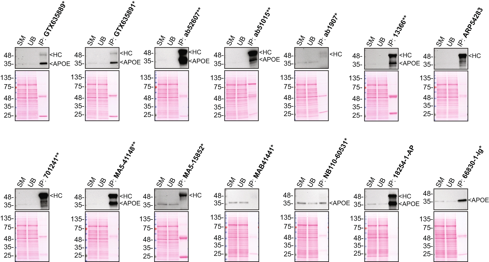
Immunoprecipitation was performed on 0.9 mg concentrated culture media from HAP1 WT, and using 2.0 μg of the indicated Apolipoprotein E antibodies pre-coupled to protein G or protein A magnetic beads. Samples were washed and processed for Western Blot with the indicated Apolipoprotein E antibody. Antibody 13366** was used at 1/500 for all Western Blots. The Ponceau stained transfers of each blot are shown. SM=3% starting material; UB=3% unbound fraction; IP=immunoprecipitate; HC=heavy chain; *Monoclonal antibody, **Recombinant antibody.
In conclusion, we have screened Apolipoprotein E commercial antibodies by Western Blot and immunoprecipitation and identified several high-quality antibodies under our standardized experimental conditions. The underlying data was previously uploaded to an open access repository, Zenodo.12,13
All Apolipoprotein E antibodies are listed in Table 2, together with their corresponding Research Resource Identifiers (RRID), to ensure the antibodies are cited properly.14 All antibodies tested detect Human ApoE. Peroxidase-conjugated goat anti-mouse and anti-rabbit antibodies are from Thermo Fisher Scientific (cat. number 65-6520 and 62-6120).
HAP1 WT and APOE KO cell lines used are listed in Table 1, together with their corresponding RRID, to ensure the cell lines are cited properly.15 Cells were cultured in DMEM high-glucose (GE Healthcare cat. number SH30081.01) containing 10% fetal bovine serum (Wisent, cat. number 080450), 2 mM L-glutamate (Wisent cat. number 609065), 100 IU penicillin and 100 μg/mL streptomycin (Wisent cat. number 450201). Cells were starved in DMEM high-glucose containing L-glutamate and penicillin/streptomycin.
HAP1 cells WT and APOE KO were washed 3× with PBS 1× and starved for ~18 hrs. Culture media were collected and centrifuged for 10 min at 500× g to eliminate cells and larger contaminants, then for 10 min at 4500× g to eliminate smaller contaminants. Culture media were concentrated by centrifuging at 4000× g for 30 min using Amicon Ultra-15 Centrifugal Filter Units with a membrane NMWL of 10 kDa (MilliporeSigma cat. number UFC901024).
Western Blots were performed as described in our standard operating procedure.16 Midi precast 4-20% Tris-Glycine polyacrylamide gels from Thermo Fisher Scientific (cat. number WXP42012BOX) were used and proteins were transferred on nitrocellulose membranes. Proteins on the blots were visualized with Ponceau S staining (Thermo Fisher Scientific, cat. number BP103-10) which is scanned to show together with individual Western Blot. Blots were blocked with 5% milk for 1 hr, and antibodies were incubated overnight at 4°C with 5% bovine serum albumin (BSA) (Wisent, cat. number 800-095) in TBS with 0.1% Tween 20 (TBST) (Cell Signaling Technology, cat. number 9997). Following three washes with TBST, the peroxidase conjugated secondary antibody was incubated at a dilution of ~0.2 μg/mL in TBST with 5% milk for 1 hr at room temperature followed by three washes with TBST. Membranes were incubated with Pierce ECL from Thermo Fisher Scientific (cat. number 32106) or with Clarity Western ECL Substrate from Bio-Rad (cat. number 1705061) prior to detection with the iBright™ CL1500 Imaging System from Thermo Fisher Scientific (cat. number A44240).
Immunoprecipitation was performed as described in our standard operating procedure.17 Antibody-bead conjugates were prepared by adding 2 μg or 20 μL of antibody at an unknown concentration to 500 μL of Pierce IP Lysis Buffer from Thermo Fisher Scientific (cat. number 87788) in a 1.5 mL microcentrifuge tube, together with 30 μL of Dynabeads protein G - (for Mouse and rat antibodies) and protein A - (for rabbit antibodies) from Thermo Fisher Scientific (cat. number 10003D and 10002D, respectively). Pierce IP Lysis Buffer (25 mM Tris-HCl pH 7.4, 150 mM NaCl, 1 mM EDTA, 1% NP-40 and 5% glycerol) was supplemented with the Halt Protease Inhibitor Cocktail 100X from Thermo Fisher Scientific (cat. number 78446) at a final concentration of 1×. Tubes were rocked for ~1 hr at 4°C followed by two washes to remove unbound antibodies. Starved HAP1 WT culture media were concentrated as described above. 0.6 mL aliquots at 1.5 mg/L of protein were incubated with an antibody-bead conjugate for ~1 hr at 4°C. The unbound fractions were collected, and beads were subsequently washed three times with 1.0 mL of IP Lysis Buffer and processed for SDS-PAGE and Western Blot on precast midi 4-20% Tris-Glycine polyacrylamide gels. Prot-A: HRP (MilliporeSigma, cat. number P8651) was used as a secondary detection system at a concentration 0.4 μg/mL.
| Views | Downloads | |
|---|---|---|
| F1000Research | - | - |
|
PubMed Central
Data from PMC are received and updated monthly.
|
- | - |
Provide sufficient details of any financial or non-financial competing interests to enable users to assess whether your comments might lead a reasonable person to question your impartiality. Consider the following examples, but note that this is not an exhaustive list:
Sign up for content alerts and receive a weekly or monthly email with all newly published articles
Already registered? Sign in
The email address should be the one you originally registered with F1000.
You registered with F1000 via Google, so we cannot reset your password.
To sign in, please click here.
If you still need help with your Google account password, please click here.
You registered with F1000 via Facebook, so we cannot reset your password.
To sign in, please click here.
If you still need help with your Facebook account password, please click here.
If your email address is registered with us, we will email you instructions to reset your password.
If you think you should have received this email but it has not arrived, please check your spam filters and/or contact for further assistance.
Comments on this article Comments (0)