Keywords
Tongue Disease, Glossitis, Prevalence Study, Stomatognathic System Abnormalities
This article is included in the Manipal Academy of Higher Education gateway.
Tongue Disease, Glossitis, Prevalence Study, Stomatognathic System Abnormalities
The tongue is a prominent muscular organ of the oral cavity that plays a vital role in speech, mastication and digestion. The integrity of the tongue mucosa frequently reflects the oral and systemic health of an individual.1 Oral cavity, being the portal of entry to various infectious and carcinogenic agents, mucosa of the tongue is easily exposed to pathologic changes.2 Numerous oral and systemic diseases affect the tongue, impairing its structure and function and owing to its strategic position, those tongue changes can be easily appreciated. Notable structural alterations including the color, texture, size, shape, appearance of the tongue can occur due to a variety of local causes or systemic conditions. Functional abnormalities of the tongue include inability/restriction of the tongue movements, taste disturbances and/or discomfort. This can often occur in neurological conditions (palsy), oral submucous fibrosis, malignancy affecting the base of the tongue, neuromuscular disorders and post-radiotherapy. The tongue changes can either be due to local factors like trauma, contact allergy, benign, reactive or malignant growth or due to underlying systemic conditions like anemia nutritional deficiencies, infections, hormonal disturbances, immune-mediated diseases and metabolic conditions.3 Even though tongue manifestations can be the early and easily identifiable diagnostic clue, it is often overlooked during dental examination unless the patients report of specific tongue complaints.
Various studies have assessed the prevalence of tongue abnormalities in different population groups, however the clinical correlation with the changes noted in the tongue is seldom looked into.1,3 There are no population-specific studies that have assessed both the structural and functional abnormalities of the tongue. The present study intended to assess the prevalence of structural and functional abnormalities of the tongue in patients visiting a tertiary care hospital and its possible association with the patient’s history and the symptoms reported. This study will help in the understanding of the commonly encountered symptomatic and asymptomatic tongue abnormalities in patients. The present study also assessed the physiological range of tongue protrusion in the study population as such data is unavailable in the literature.
This cross-sectional study included the outpatients visiting the various units of Kasturba Medical College & Hospital and the Dental outpatient department of Manipal College of Dental Sciences, Manipal for a period of 20 weeks from September 1st, 2021. Kasturba Medical College and Kasturba Hospital Institutional Ethics Committee approved the study protocol (IEC 409/2021; approved on 8th August 2021). All research projects carried out in the Health Sciences division at the Manipal Academy of Higher Education come under the purview and scrutiny of the Institutional Ethics Committee, which is named as Kasturba Medical College and Kasturba Hospital Institutional Ethics Committee. This project was registered with the Clinical Trials Registry India (CTRI/2021/09/036996; registered on 30th September 2021).
The inclusion criteria were all patients above 18 years of age who visited the dental outpatient department. Patients with severe restriction in mouth opening and those who were not willing to participate were excluded from the study. Informed written consent was obtained from individuals who were willing to participate in the study. The tongue examination was performed on patients and the following relevant data parameters were written down for each patient in the specially designed proforma by the principal investigator: age, gender and the participants were asked regarding any symptoms or abnormalities noticed pertaining to tongue. Also, the medical history, drug history and oral abusive habit history were recorded for each patient in a specially designed proforma.
The clinical examination of the oral cavity and tongue was performed in accordance with WHO guidelines (Geneva, 1997) in a well-lit, noise and crowd free room on a dental chair with artificial lighting and a mouth mirror under aseptic conditions.4 Any structural abnormality of tongue related to color, texture, size, shape and appearance were noted. The tongue was checked for depapillation, coating or inflammation, any ulcers, white lesions, red patches, pigmentation, varices and growth/swelling.
If any of the aforementioned changes were observed, the location, extent and surface characteristics of these lesions were recorded. The oral mucosal pathologies were clinically diagnosed based on their characteristic history and presenting features. The diagnosis of lichen planus was given when bilateral white lacy pattern was observed with characteristic Wickham’s striae. When scrapable white patches were seen in medically compromised patients leaving behind erythematous surfaces on rubbing a diagnosis of candidiasis was recorded. Leukoplakia is defined as “white plaques of questionable risk having excluded (other) known that carry no increased risk for cancer”. It was diagnosed by excluding other causes of raised non-scrapable white diseases or disorders patches especially in patients with strong habit history. Erythroplakia appeared as raised red velvety patches that could not be attributed to any other disease. Oral submucous fibrosis is typically diagnosed in the presence of fibrotic bands and marbled appearance of the buccal mucosa. Erythema multiforme presents as multiple oral ulcers with crusting of the lips. Allergic stomatitis is established when multiple acute onset ulcers/erythema with the history of known triggering agents.5 Suspicious growth with induration of base is diagnosed as malignancy and were sent for histopathologic examination for definitive diagnosis.
Also, subjective symptoms and functional abnormalities like dysgeusia, burning tongue, taste loss, and tongue protrusion interferences were noted. Tongue movements were evaluated and maximal tongue protrusion was measured. This was performed by asking the patient to protrude the tongue to the maximum, and measurement was done using a sterile steel scale by measuring the distance between the maxillary incisal tip and the tip of the tongue (Figure 1). Any deviation of the tongue during protrusion or difficulty in protrusion was also recorded.
All analysis was done using SPSS (RRID:SCR_002865) version 18 (SPSS Inc. Released 2009. PASW Statistics for Windows, Version 18.0. Chicago: SPSS Inc.). A p-value of <0.05 was considered statistically significant. Categorical variables were compared using Fisher's exact test or Chi-squared test.
The study population included 1,143 patients, of which 564 were male (49.3%) and 579 were female (50.7%). The mean age of the subjects was 38.59 years old with an age range of 18–85 years old (Standard Deviation; SD= 13.88). The distribution of various structural tongue abnormalities is presented in Table 1.15
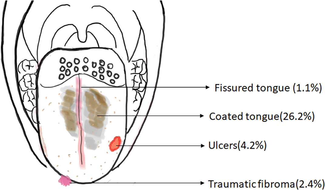
The schematic representation of the common structural abnormalities of the tongue observed in the present study is shown in Figure 2.

Among the various comorbidities observed, 10.5% of the study population presented with diabetes mellitus followed by hypertension (8.9%), thyroid disorders (5.0%), immunocompromised state (2.8%) and anemia (0.8%). Oral abusive habits were reported by 25.7% of the subjects. Tobacco consumption (14.6%) was more commonly reported than alcohol (11.1%). Subjective symptoms including pain (7.0%) and burning sensation (8.2%) were also reported by the study population.
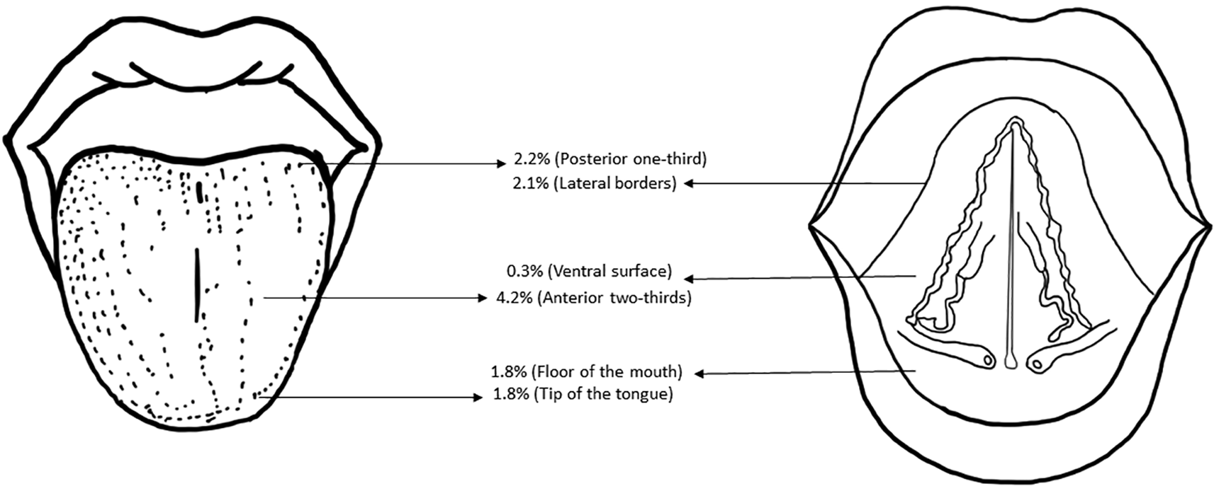
Coral pink colored physiologic appearance was noted in 91.9% of participants, 5.9% had red tongue and 0.9% had pale tongue. Overall, 91.3% of them had a normal texture of the dorsal surface of the tongue and 7.1% had smooth bald tongues. Figure 3 shows the topographic distribution of the tongue abnormalities observed in the study population. Figure 4 shows the various structural abnormalities observed in the present study.

Functional abnormalities of tongue were noted in 8.7% of the study participants, of which 3.7% had restricted tongue movement (Table 2).
| Functional abnormalities | Present, n (%) |
|---|---|
| Deviation | 5 (0.4) |
| Taste dysfunction | 53 (4.6) |
The range of tongue protrusion observed in the study was 15 mm to 54 mm with a mean of 40.5 mm. Table 3 depicts the range of tongue protrusion in the study population.
| S.No | Distribution of tongue protrusion, mm | Males, n | Females, n | Total, n (%) |
|---|---|---|---|---|
| 1 | 50-60 | 07 | 15 | 22 (1.9) |
| 2 | 40-50 | 266 | 278 | 544 (47.6) |
| 3 | 30-40 | 245 | 258 | 503 (44) |
| 4 | 20-30 | 30 | 28 | 58 (5.1) |
| 5 | 10-20 | 16 | 0 | 16 (1.4) |
| 6 | 0-9 | 0 | 0 | 0 |
Using Fischer’s exact test, the association between tongue abnormalities and tobacco associated habits was assessed. There was a positive association of tobacco related habits with burning, pain, oral submucous fibrosis, verrucous carcinoma, squamous cell carcinoma, coated tongue, candidiasis, restricted tongue movement and taste dysfunction (Table 4).
| Structural lesions | N (%) | N (%) | P-value | |
|---|---|---|---|---|
| Burning | Absent | 922 (94.5) | 127 (76.0) | <0.001* |
| Present | 54 (5.5) | 40 (24.0) | ||
| Pain | Absent | 933 (95.6) | 130 (77.8) | <0.001* |
| Present | 43 (4.4) | 37 (22.2) | ||
| Coral Pink | Absent | 76 (7.8) | 17 (10.2) | 0.296 |
| Present | 900 (92.2) | 150 (89.8) | ||
| Red | Absent | 921 (94.4) | 155 (92.8) | 0.431 |
| Present | 55 (5.6) | 12 (7.2) | ||
| Pale | Absent | 969 (99.3) | 164 (98.2) | 0.169 |
| Present | 7 (0.7) | 3 (1.8) | ||
| Coarse tongue | Absent | 73 (7.5) | 26 (15.6) | 0.001* |
| Present | 903 (92.5) | 141 (84.4) | ||
| Smooth | Absent | 915 (93.8) | 147 (88.0) | 0.008* |
| Present | 61 (6.2) | 20 (12.0) | ||
| Fissured tongue | Absent | 967 (99.1) | 163 (97.6) | 0.108 |
| Present | 9 (0.9) | 4 (2.4) | ||
| Enlarged | Absent | 965 (98.9) | 142 (85.0) | <0.001* |
| Present | 11 (1.1) | 25 (15.0) | ||
| Depapillation | Absent | 923 (94.6) | 148 (88.6) | 0.003* |
| Present | 53 (5.4) | 19 (11.4) | ||
| Coating on tongue | Absent | 741 (75.9) | 102 (61.1) | <0.001* |
| Present | 235 (24.1) | 65 (38.9) | ||
| Ulcers | Absent | 943 (96.6) | 152 (91.0) | 0.001* |
| Present | 33 (3.4) | 15 (9.0) | ||
| Candidiasis | Absent | 967 (99.1) | 160 (95.8) | 0.005* |
| Present | 9 (0.9) | 7 (4.2) | ||
| Leukoplakia | Absent | 973 (99.7) | 165 (98.8) | 0.157 |
| Present | 3 (0.3) | 2 (1.2) | ||
| Lichen planus | Absent | 965 (98.9) | 160 (95.8) | 0.01* |
| Present | 11 (1.1) | 7 (4.2) | ||
| Oral Submucous fibrosis | Absent | 972 (99.6) | 147 (88.0) | <0.001* |
| Present | 4 (0.4) | 20 (12.0) | ||
| Verrucous carcinoma | Absent | 976 (100) | 165 (98.8) | 0.001* |
| Present | 0 (0) | 2 (1.2) | ||
| Squamous Cell Carcinoma | Absent | 976 (100) | 163 (97.6) | <0.001* |
| Present | 0 (0) | 4 (2.4) | ||
| Geographic tongue | Absent | 964 (98.8) | 163 (97.6) | 0.274 |
| Present | 12 (1.2) | 4 (2.4) | ||
| Pemphigus | Absent | 973 (99.7) | 167 (100) | 0.473 |
| Present | 3 (0.3) | 0 (0) | ||
| Erythroplakia | Absent | 975 (99.9) | 167 (100) | >0.99 |
| Present | 1 (0.1) | 0 (0) | ||
| Erythema multiforme | Absent | 974 (99.8) | 165 (98.8) | 0.104 |
| Present | 2 (0.2) | 2 (1.2) | ||
| Allergy | Absent | 971 (99.5) | 163 (97.6) | 0.031* |
| Present | 5 (0.5) | 4 (2.4) | ||
| Erythematous candidiasis | Absent | 975 (99.9) | 166 (99.4) | 0.271 |
| Present | 1 (0.1) | 1 (0.6) | ||
| Varices | Absent | 901 (92.3) | 145 (86.8) | 0.019* |
| Present | 75 (7.7) | 22 (13.2) | ||
| Growth/Swelling | Absent | 963 (98.7) | 152 (91) | <0.001* |
| Present | 13 (1.3) | 15 (9.0) | ||
| Deviation | Absent | 974 (99.8) | 164 (98.2) | 0.024* |
| Present | 2 (0.2) | 3 (1.8) | ||
| Restriction of tongue movement | Absent | 963 (98.7) | 138 (82.6) | <0.001* |
| Present | 13 (1.3) | 29 (17.4) | ||
| Taste dysfunction | Absent | 952 (97.5) | 138 (82.6) | <0.001* |
| Present | 24 (2.5) | 29 (17.4) | ||
Tongue abnormalities are commonly encountered in dental practice but often overlooked, if asymptomatic. The tongue may be affected by a wide range of local and systemic conditions. Structural and functional tongue abnormalities can be easily recognized on careful and detailed clinical examination. The present study evaluated the prevalence of structural and functional abnormalities of the tongue in a tertiary care hospital-based population. There is limited literature pertaining to the measurements of normal protrusive tongue movements. Understanding the physiologic range of tongue protrusion in a normal population aids in early identification of tongue restriction. Restriction in the tongue protrusion occurs in various conditions like oral submucous fibrosis, carcinomas of the base of the tongue, scleroderma and in neurological disorders.
In the present study, overall tongue abnormalities were present in 66.5% of the subjects. A Turkish study by Avcu et al. (2003) showed prevalence of tongue lesions in 52.2% of the study subjects.6 Whereas, other studies from Turkey, India and Libya showed a low prevalence of 4.9%, 2.8% and 9.2%, respectively.7,8,9
The anatomic distribution of tongue lesions was majorly noted on the anterior two thirds of the tongue (4.2%) and least seen in the ventral surface of tongue (0.3%). A study on the Libyan population showed that 90% of the lesions were on the dorsum, followed by lateral border (7%), posterior one third (2%). The least common site was the ventral surface of tongue (1%), which is in accordance with the present study.9
The most prevalent comorbidity in the study population was diabetes mellitus (10.5%), followed by hypertension (8.9%). Bhattacharya et al. (2016) estimated the prevalence of diabetes mellitus to be 3.1%, which is lower than the observations from the current study.3 A similar study on the Libyan population by Byahatti et al. (2010) found an even lower prevalence of subjects affected with diabetes mellitus (0.4%), followed by hypertension (0.3%).9 Patil et al. (2013) conducted a study in which the prevalence of anemia was 3.8%, which was higher in comparison to the present study.10
In the present study, 5.9% had red erythematous tongue and 0.9% had pale tongue. Physiologic melanin pigmentation of the tongue was noted in 4.8% of the study subjects, whereas other studies by Mumcu et al.11 (2005) and Shinde et al.8 (2017) noted 6.9% and 2.1%, respectively. There is no literature available that has assessed the prevalence of other color variations on the tongue.
A positive oral abusive habit history was noted in 25.7% of the study population, of which 14.6% were tobacco users and 11.1% had alcohol consumption. In a study done by Motallebnejab et al. (2008), 54.7% of patients smoked tobacco and 35.3% had alcohol habits.12 Other studies had estimated the prevalence of tongue lesions in smokers to be 1.93%.9 A positive association between smoking and presence of hairy tongue was found by Darwazeh et al. (2010).13
There was a positive association between tobacco related habits and burning sensation of the tongue in the present study. Tobacco related habits were also associated with coated tongue, ulcers, increased incidence of candidiasis, oral lichen planus, oral submucous fibrosis, verrucous carcinoma, squamous cell carcinoma, restricted tongue movement and taste dysfunction. Similarly, Bhattacharya et al., found a positive association between oral lichen planus and oral abusive habit.3 Avcu et al., estimated the correlation between tongue lesions and smoking in the Turkish population and concluded that tongue lesions (hairy tongue, fissured tongue and coated tongue) were increased in smokers and heavy smokers than non-smokers.6 Among the structural tongue abnormalities, coated tongue was the most prevalent finding (26.2%), followed by depapillation (6.3%), ulcers (4.2%), enlarged tongue (3.1%), oral submucous fibrosis (2.5%) in the present study. Similarly, coated tongue was the most prevalent finding (30.6%) followed by fissured tongue (20.1%)3 in a study conducted by Bhattacharya et al., on the Indian population. The prevalence of coated tongue was 28.0% in an Indian study10 by Patil S et al. Shinde et al., (2017) estimated the prevalence of fissured tongue (51.7%) was high, followed by coated tongue (14.3%), geographic tongue (10%).8 A higher prevalence of fissured tongue (48.4%) was observed in a study in the Libyan population.9 By contrast, a very low prevalence of 1.1% with fissured tongue was noted in our study population. Lingual varicosity was noted in 8.5% of our subjects. Whereas, varicosity was the least common finding (0.4%) in an Indian population by Bhattacharya PT.3
To the best of our knowledge, the present study was the first to evaluate the presence of functional abnormalities of the tongue that included restriction of tongue protrusion and taste dysfunction. There is no literature that has addressed the various functional abnormalities that affect the tongue in a large population. Taste abnormalities were reported in 4.6% of the study population; 6.5% of the study subjects had tongue protrusion less than 30 mm. This is the first study that has evaluated the functional as well as structural abnormalities affecting the tongue. Kotlow et al., (1999) graded ankyloglossia into four classes: Class I- Mild (12-16 mm), Class II- Moderate (8-11 mm), Class III- Severe (3-7 mm), Class IV- Complete (less than 3 mm).14 Considering the tongue protrusion range observed in the present study population and considering the classification of ankyloglossia, we propose a new grading for tongue protrusion, as depicted in Table 5.
Limitations of the present study include the observational nature due to which the contributing factors like oral hygiene, oral abusive habits, medications and systemic disorders could not be associated with the findings on the tongue. Multicentric studies from diverse populations shall aid in deeper understanding about the abnormalities that can affect the tongue.
The results from the present study reflected a high prevalence of abnormalities affecting the tongue. Careful and detailed evaluation of the tongue examination is mandated in routine dental checkup. This shall help in the prompt identification of various etiological factors causing structural and functional abnormalities of the tongue. Understanding the normal tongue protrusion range will aid in the early detection of restricted tongue movements.
We confirm that we have obtained permission to use images and data from the participants included in this work.
Mendeley Data: Structural and functional abnormalities of the tongue: An epidemiological study from a tertiary center with a proposed grading system for tongue protrusion. https://doi.org/10.17632/k3vx49rbfn.1. 15
Data are available under the terms of the Creative Commons Attribution 4.0 International license (CC-BY 4.0).
| Views | Downloads | |
|---|---|---|
| F1000Research | - | - |
|
PubMed Central
Data from PMC are received and updated monthly.
|
- | - |
Is the work clearly and accurately presented and does it cite the current literature?
Partly
Is the study design appropriate and is the work technically sound?
Partly
Are sufficient details of methods and analysis provided to allow replication by others?
No
If applicable, is the statistical analysis and its interpretation appropriate?
Yes
Are all the source data underlying the results available to ensure full reproducibility?
Yes
Are the conclusions drawn adequately supported by the results?
Partly
Competing Interests: No competing interests were disclosed.
Reviewer Expertise: oral and maxillofacial surgery, oral medicine, head and neck oncology, clinical dentistry, oral and maxillofacial radiology
Is the work clearly and accurately presented and does it cite the current literature?
Yes
Is the study design appropriate and is the work technically sound?
Yes
Are sufficient details of methods and analysis provided to allow replication by others?
Yes
If applicable, is the statistical analysis and its interpretation appropriate?
Yes
Are all the source data underlying the results available to ensure full reproducibility?
Yes
Are the conclusions drawn adequately supported by the results?
Yes
Competing Interests: No competing interests were disclosed.
Reviewer Expertise: dentistry
Alongside their report, reviewers assign a status to the article:
| Invited Reviewers | ||
|---|---|---|
| 1 | 2 | |
|
Version 2 (revision) 16 Feb 24 |
read | read |
|
Version 1 13 Jul 23 |
read | read |
Provide sufficient details of any financial or non-financial competing interests to enable users to assess whether your comments might lead a reasonable person to question your impartiality. Consider the following examples, but note that this is not an exhaustive list:
Sign up for content alerts and receive a weekly or monthly email with all newly published articles
Already registered? Sign in
The email address should be the one you originally registered with F1000.
You registered with F1000 via Google, so we cannot reset your password.
To sign in, please click here.
If you still need help with your Google account password, please click here.
You registered with F1000 via Facebook, so we cannot reset your password.
To sign in, please click here.
If you still need help with your Facebook account password, please click here.
If your email address is registered with us, we will email you instructions to reset your password.
If you think you should have received this email but it has not arrived, please check your spam filters and/or contact for further assistance.
Comments on this article Comments (0)