Keywords
Periodontal disease, Chronic periodontitis, Residual periodontal pockets, Photodynamic therapy, Laser Therapy, Indocyanine Green, Maintenance therapy, Photosensitizers.
This article is included in the Manipal Academy of Higher Education gateway.
Periodontal disease, Chronic periodontitis, Residual periodontal pockets, Photodynamic therapy, Laser Therapy, Indocyanine Green, Maintenance therapy, Photosensitizers.
Amendments from version 1
In the Treatment protocol section
Treatment subheading
The first sentence was modified:
At day zero, patients fulfilling the inclusion criteria were subjected to full mouth SRP. According to the randomisation the test sites were then treated with PDT which is explained as follows. Immediately after full mouth SRP, the test sites were properly dried using air syringe to receive the photosensitizer. 0.5ml of the dye was then the photosensitizer (0.5 mL) was the photosensitizer (0.5 mL) was applied in the test site using a syringe.
The Last sentence was modified:
All the patients were monitored for 3 months. They were blinded throughout the procedure and study period to maintain the integrity of the assignment.
Results Section
Clinical Parameters subheading
The values were modified :
There was statistical significance from baseline to 3 months regarding PD with values of 5.57±0.21mm 1.73±0.42mm and 3.33±0.31mm 1.73±0.42 mm and 1.22±0.43 mm respectively for the test group and 5.57±0.24mm 1.70±0.46mm and 1.28±0.42mm2.84±0.44mm 1.70±0.46 mm and 1.28±0.42 mm respectively for the control group (P value <0.05). Similarly, for gain in CAL in the test group, there was a statistically significant difference from baseline (5.57±0.21mm1.77±0.40 mm) to 3 months (3.37±0.33mm1.27±0.41mm) and in the control group from baseline (5.57±0.24mm1.73±0.46 mm) to 3 months (3.14±0.55mm1.30±0.41 mm) (P value <0.05).
Data from Table 3 has been modified.
Microbiological parameters
Table 4 showing detection frequency of organisms at baseline and 3 months using Mc Nemar P test has been added.
See the authors' detailed response to the review by Anurag Satpathy
Periodontitis is a result of an exaggerated reaction of the host tissue in response to the microbial challenge which ultimately leads to loss of hard and soft tissues of the periodontium.1 With this sort of a continuous microbial challenge, the periodontal tissues are subjected to toxic bacterial (virulence) factors that have the potential to alter various host cell operations. Periodontal therapy is primarily aimed at removal of these hard tissue and soft tissue deposits from the root surface to prevent progression of the disease. This is achieved by means of scaling and root planing (SRP) by hand and/or power driven instruments.2 However, in spite of being the standard of care for treatment of periodontal disease, SRP may not be sufficient to completely remove all periodontal pathogens.3 The varied clinical presentations such as deep pockets, presence of furcations or intrabony defects may render mechanical debridement inadequate for disruption of the tenacious biofilm which is the primary niche of putative periodontal pathogens.
LASER technology have been around for quite some time now and is currently in the 4th decade in the dental field. It holds a promising non-invasive mode of treatment when it comes to periodontal therapy.4 As discussed previously, periodontal disease causing bacteria not only accumulates on the hard tissue but also can invade the soft tissues of the periodontium. In this regard, Diode lasers have proven to be a boon because they are nothing but soft tissue lasers which have an affinity for pigment containing cells for example black pigmented bacteria (spirochetes).5 LASER also exerts an antibacterial effect which may be further improved by the addition of a photosensitizing dye. This phenomenon is known as photodynamic therapy (PDT).6 Indocyanine green is one such photosensitizer which absorbs wavelength at a range of 750-950 nm with a maximum value of 810 nm and exerts its main action by photo-thermal effect which induces cell damage by increasing intracellular temperature.7 PDT includes a photochemical reaction mediated by oxygen which gets activated on application of light in the presence of a photosensitizing compound that leads to generation of highly reactive nascent oxygen species that are toxic to the microorganisms. The Major advantages of antimicrobial PDT (aPDT) are that they explicitly target cells, causing no collateral damage to non-pigmented host tissue cells and is only initiated when laser light is irradiated. It also lacks the development of resistant bacterial species, which is common with injudicious use of antibiotics.6
Residual pockets may contain periodontal pathogens persistently and may even re-harbour these microorganisms which are incompatible with periodontal health.8 Therefore, such sites need special professional attention. Re-instrumentation repeatedly in these sites may cumulatively cause damage to the hard tissues, thereby calling for maintenance protocols for residual pockets which are efficient even after repeated application.9
Recent body of evidence have shown that adjunctive use of PDT with SRP for treatment of residual periodontal pocket may provide additional improvement and better outcomes. However, there is no available literature which have evaluated the efficacy of the treatment protocol using indocyanine green as a photosensitizer in photodynamic therapy in maintenance patients. Therefore, the purpose of this study is to assess the changes in clinical and microbiological parameters following multiple applications of PDT using indocyanine green and diode laser along with SRP in treating patients enrolled in periodontal maintenance programme.
The present split-mouth, double-blinded, randomized controlled clinical trial comprised of 25 patients suiting the inclusion criteria. They were recruited from Department of Periodontology, Manipal College of Dental Sciences, Mangalore, India from December 2021 to January 2023.
The study protocol was reviewed and approved by Institutional Research Ethics Committee and was conducted in accordance with Helsinki Declaration 1975, as revised in 2013(registered at Clinical Trial Registry India as CTRI/2023/01/61447).
Informed consent was taken from all participants intimating them the nature, potential risks, and benefits of their participation in the study. They were free to withdraw from the study at their will. The information sheet and consent form can be found as Extended data.
Reference of the present study was taken from Grzech lesniak et al.10 An effect size of 0.2 was considered with a 95% confidence interval and 80% power. Thus, a sample size of 25 was calculated for this split mouth design.
The subjects were arbitrarily allocated to either of the interventions: A) (SRP) with antimicrobial photodynamic therapy (aPDT) on right side; SRP on left side or B) SRP right side and aPDT on left side. Selected sites were randomly assigned to one of the two treatment modalities by a computer generated block randomization (allocation ratio of 1:1) before each intervention procedure. A third staff member (RS) who wasn’t directly involved in the study carried out the randomization. Opaque sealed envelopes were used to conceal the allocation sequence. The primary investigator, outcome assessor, biostatistician as well as the patients were unaware of the details of the series of the study. As and when a patient came and fit the criteria for the study, the envelope was opened and treatment protocol was followed as described below.
Treatment protocol
Periodontal measurements
All 25 Patients underwent full mouth clinical periodontal assessment around all teeth by a single calibrated examiner (DK) following the CONSORT guidelines (Figure 1: consort flow chart).11 The measurements were taken at four sites per tooth (distal, mesial, facial/buccal, lingual/palatal) using a University of North Carolina No.15 periodontal probe (Hu-friedy™, Chicago IL) and Customized acrylic stents were prepared to guide the probe placement in the same plane. Clinical variables included the measurements of plaque index (PI), modified sulcular bleeding index (mSBI), probing pocket depth (PD) and clinical attachment level (CAL). PD was measured as the distance between the gingival margin and the base of the pocket and CAL was calculated as the distance from the CEJ and the bottom of the sulcus or of the periodontal pocket. The primary outcome being probing depth and clinical attachment level based on the number of sites and secondary outcome being plaque index and bleeding index.

Preparation of indocyanine green
Under aseptic circumstances a fresh 5 mg/mL solution with the photosensitizer dye Indocyanine green (Aurogreen®, Aurolabs, Madurai, India) was prepared. In 5ml of sterile water, ICG is dissolved to prepare an initial 5 mg/mL ICG stock solution. This solution is further diluted in saline solution at the ratio of 1:5 to achieve the final ICG concentration of 5 mg/mL.12 The aqueous solution becomes unstable on coming in contact with the environment and hence should be utilised on the same day within 10 hours.
Laser parameters
The laser system used in the study is Diode laser (Picasso AMD) with wavelength of 810 nm. It was applied in a continuous mode, circumferentially around the tooth, with a power of 2 W for 60 s. Total energy produced is 6 J/cm2.
Microbiological assessment
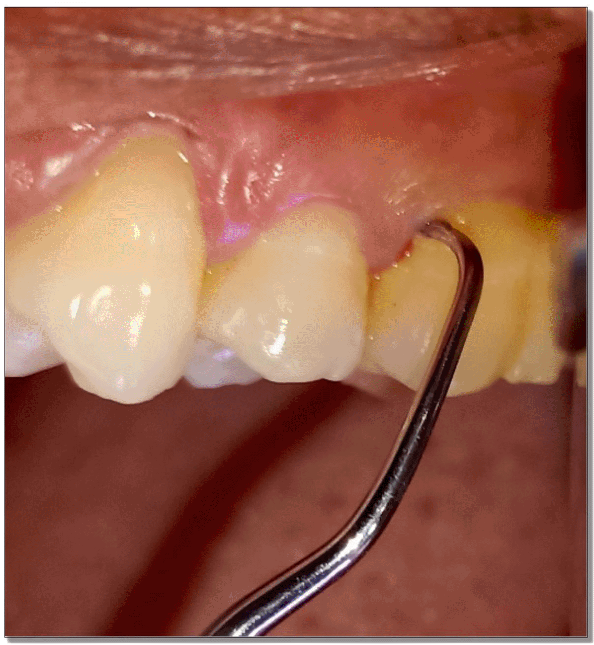
Microbiological samples were collected through plaque from the deepest periodontal pockets at baseline and three months after therapy using sterile curettes (Figure 2). They were then transferred to Eppendorf tubes containing 500 μL of sterile phosphate buffer saline (PBS). Microbiological analysis consisted of a molecular test for the detection of four periodontal pathogens. The test was performed with Multiplex PCR (polymerase chain reaction) method. Bacterial DNA was extracted using DNA thermal cycler (ProFlex™ 3× 32-well PCR System, Applied Biosystems, Foster City, CA) following the instructions of the manufacturer (Table 1).
The following periodontal pathogenic species were analysed:
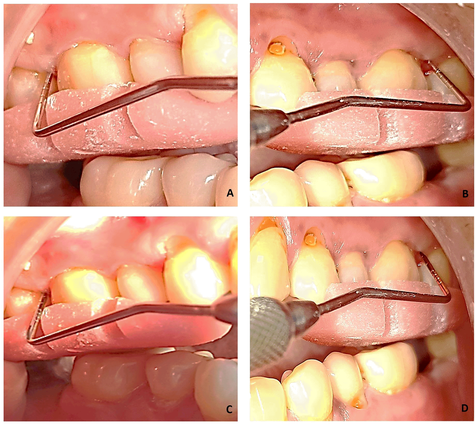
At day zero, patients fulfilling the inclusion criteria were subjected to full mouth SRP. According to the randomisation the test sites were then treated with PDT which is explained as follows. Immediately after full mouth SRP, the test sites were properly dried using air syringe to receive the photosensitizer. 0.5 ml of the dye was then applied in the test site using a syringe. To avoid ‘carry-across’ effect, a conscious effort was made to not spill the photosensitizer in other parts of the oral cavity. Anyhow, the dye is not known to get activated without laser light. Distilled water was used after 2 minutes to wash out the free photosensitizer. The 810 nm diode soft tissue laser was brought in contact with the gingival tissues below the gingival margin and light was applied for 60 seconds in a circumferential manner around the tooth. The laser fibre tip was kept in contact with the soft tissue wall constantly (Figure 3). The strokes addressed small sections and were systematically overlapping. The fibre tip was continually inspected to remove any accumulated debris with water moistened gauze. aPDT was repeated after 14 days.

The contralateral site received sham LASER which imitated the irradiation without it being turned on. All the patients were monitored for 3 months. They were blinded throughout the procedure and study period to maintain the integrity of the assignment.
Statistical analysis was done for all the clinical parameters and microbiological parameters. Mean and standard deviation was calculated at baseline and at 3 months’ follow-up. Data was analysed using paired ‘t’ test for plaque index, gingival index, clinical attachment level and probing depth at baseline and at 3 months’ follow-up. Mc Nemar P test and Fischer exact P test were used for microbiological parameters. Statistical Package for Social Sciences (SPSS), version 17 (SPSS Inc, Chicago IL) was the used software. A P value of <0.05 was considered significant.
This split mouth, single centre, randomised controlled trial was done for a period of 3 months. There were no unwanted events reported by any patient throughout the study. Out of 25 subjects initially recruited, one was lost to drop out and subsequently 24 patients were carried forward for analysis. 10 participants were females and 14 were males, aged between 30 and 78 years. The age and sex distribution of both groups were uniform (P>0.05) (Table 2). A total of 130 teeth were examined, 66 in test group and 64 in control group, with 171 intervention sites: 86 in test group and 85 in control group.
The participants in the study showed decreased full mouth plaque score (FMPS) and full mouth bleeding score (FMBS) that is <25%. This signifies that all of them had maintained an optimum standard of oral hygiene throughout the study period. The results also showed a substantial reduction of PI, mSBI throughout the study period in both groups with P value of <0.000 (Table 3).
| Clinical parameters | Groups | Baseline | 3 months | Baseline vs 3 months, P value |
|---|---|---|---|---|
| PI | Test group | 1.7±0.25 | 1.14±0.23 | 0.000* |
| Control group | 1.65±0.32 | 1.16±0.18 | 0.000* | |
| P value | 0.5† | 0.6† | ||
| SBI | Test group | 1.82±0.24 | 1.05±0.26 | 0.000* |
| Control group | 1.69±0.42 | 1.01± 0.15 | 0.000* | |
| P value | 0.2† | 0.6† | ||
| PPD | Test group | 5.57±0.21 | 3.33±0.31 | 0.000* |
| Control group | 5.57±0.24 | 2.84±0.44 | 0.000* | |
| P value | 0.2† | 0.6† | ||
| CAL | Test group | 5.57±0.21 | 3.37±0.33 | 0.000* |
| Control group | 5.57±0.24 | 3.14±0.55 | 0.000* | |
| P value | 0.8† | 0.6† |
There was statistical significance from baseline to 3 months regarding PD with values of 5.57±0.21 mm and 3.33±0.31 mm respectively for the test group and 5.57±0.24 mm and 2.84±0.44 mm respectively for the control group (P value <0.05). Similarly, for gain in CAL in the test group, there was a statistically significant difference from baseline (5.57±0.21 mm) to 3 months (3.37±0.33 mm) and in the control group from baseline (5.57±0.24 mm) to 3 months (3.14±0.55 mm) (P value <0.05). However, intergroup comparison of PD and CAL did not show any statistical significance at any given period (P>0.05) (Table 3).
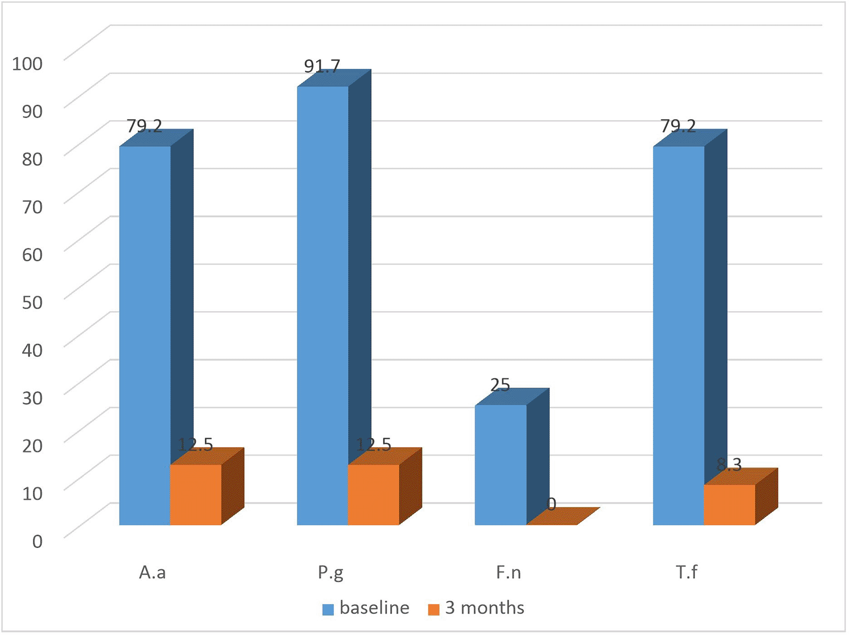
All groups exhibited a substantial decrease in the detection frequency of A.a, P.g, T.f, F.n from baseline till the termination of the study (Figure 4: test) and (Figure 5: control). Both the groups showed equally greater reduction in detection frequency of P.g and T.f at 3 months (Table 4).

| Pathogen | Time interval | Test group | Control group |
|---|---|---|---|
| Frequency of organisms present (%) | Frequency (%) | ||
| Aggregatibacter Actinomycetemcomitans* | baseline | 20 (83.3) | 19 (79.2) |
| 3 months | 5 (20.8) | 3 (12.5) | |
| Porphyromonas gingivalis* | baseline | 22 (91.7) | 22 (91.7) |
| 3 months | 5 (20.8) | 3 (12.5) | |
| Fusobacter nucleatum** | baseline | 7 (29.2) | 6 (25) |
| 3 months | 0 | 0 | |
| Tannerella forsythia* | baseline | 19 (79.2) | 19 (79.2) |
| 3 months | 2 | 2 (8.3) |
The corner-stone of cause-related therapy is considered to be non-surgical periodontal therapy (NSPT) which encompasses the removal of supragingival and subgingival biofilm and calculus.13 Yet, a number of ‘residual pockets’ often remain after non-surgical periodontal therapy.14 Residual pockets, particularly >4 mm, have shown to be a positive predictor of clinical loss of attachment over the years and hence represents a clinical scenario which is more difficult to treat than a patient of untreated periodontitis15.16 Photodynamic therapy has been used in periodontal treatment for over a quarter of a century now. They are being increasingly used as adjunctive treatment with NSPT and eliminates disadvantage such as bacterial resistance seen with systemic or local antibiotics.17
This study focussed on evaluating the clinical and microbiological effects of antimicrobial photodynamic therapy using indocyanine green in patients enrolled in maintenance therapy, having residual pockets. The use of a split mouth design in the present study provided an advantage over parallel design study where the same subject served as control, hence comparison of all treatment methods could be done under similar and optimally standard healing periods. A statistically significant improvement was seen at 3 months for plaque scores and bleeding scores in both the intervention groups without any intergroup differences at any given point. There was a significant reduction in the probing pocket depth and considerable gain in clinical attachment level at the end of 3 months in both groups (Figure 6). However, the difference between the two groups were not statistically significant. There are studies which are in agreement with the present study6,18.19 A systematic review by Azarpazhooh et al. (2010) demonstrated that PDT as a treatment modality whether stand-alone or as adjunct, was not superior to SRP therapy alone.20
However, a systematic review of four RCTs by Xue et al(2017) stated that additional clinical improvement were observed in SRP+PDT group compared to SRP alone for residual pockets for supportive periodontal therapy.21 As none of the studies used Indocyanine green as the photosensitizer, no clear-cut conclusion can be drawn. With that, coming to the next aspect- the photosensitizer, majority of available literature on PDT demonstrate the use of conventional photosensitizers that are methylene blue or toluidine blue. The present randomised controlled trial is the first to determine the outcome of PDT using Indocyanine Green as the photosensitizing agent in treatment of residual pockets in maintenance therapy patients. However, the outcomes in the current study, though favourable at 3 months, did not prove added benefits of ICG compared to SRP alone. This is not commensurate with previous studies utilising ICG as photosensitizing agent. A systematic review conducted by Bashir et al (2021) concluded that ICG mediated PDT in treatment of periodontitis significantly improved the clinical outcomes compared to SRP alone.22 This disparity may be due to the fact that there is no standardised protocol for use of soft tissue diode LASER and the frequency of antimicrobial PDT application for treatment of periodontal disease. Lack of standardisation eliminates many studies for analysis in systematic reviews. Also, the utilisation of ICG in the field of periodontitis is relatively new hence it should be acknowledged that there may have been studies which are yet in the process of trial, or have gone through publication.
Regarding microbiological analysis, the detection frequencies of the 4 major putative periodontal pathogens were analysed. The detection frequencies of P.g, A.a, T.f, F.n were significantly lower at 3 months (Figure 7). Even though the detection frequency lowered at the end of the study for all the pathogens, there was no statistical significance in any of the groups from baseline to 3 months. This may be due to the fact that the sample size for the present study was limited and for analysis of non-parametric variable such as the absence or presence of micro-organisms, a bigger sample size is necessary to demonstrate the effectiveness of the treatment modality.

No intergroup association was observed with respect to the presence of bacteria. Similar results were reported by Chondros et al. (2009),18 Polansky et al. (2009),23 De Micheli et al. (2011)24 and Giannopoulou et al. (2012).25 Polansky and group tested for P. gingivalis, T. forsythia and T. denticola at baseline and at 3 months. The levels of prevotella species (P.g) had significantly come down in both the groups. No significant reductions of T. forsythia and T. denticola were observed in either groups. Moreover, DeMicheli and group (2011) did not observe any additional reduction in the bacterial species in the test group (diode laser with SRP). Similar to the present study where the detection frequency of each organism tested, reduced significantly at 3 months, a systematic review of 17 randomised controlled clinical trials, by Akram et al. (2016)26concluded that adjunctive PDT application led to a reduction of the following species A.a, P.g, T. f, and T.d equal to the control group (SRP alone). This was in line with another study by Cappuyns et al. (2011)19 who demonstrated that detection frequency of P.g, T.f and T.d significantly reduced 2 and 6 months following intervention in all the groups that is SRP, DSL and PDT and that one procedure was not superior to the other.
The present study employed two sittings of PDT application similar to an earlier study by Segarra (2017) that have shown at least two applications of PDT is necessary to achieve the expected outcome.27 On the contrary a study by Müller Campanile et al. (2015) demonstrated that the number of applications is not proportional to better outcomes of PDT.28 Although there are not many systematic reviews to authenticate the statement, most of the available literature regarding the number of sittings of PDT emphasize of multiple sittings of atleast 2 or more than 2 to attain the desired effect29.30 Maybe future trials can implement more than 2 sittings for treating residual pockets.
It is noteworthy to consider other limitations of the present study. Firstly, a bigger sample size is necessary to validate the use of indocyanine based PDT in maintenance patients. The time period taken as 3 months for the duration of the study provided a short term overview about the effect of different treatment modalities. A longer study period can be implied in future studies. There could be chances that the periodontopathogens could translocate from one niche to another within the oral cavity- which is known as intraoral translocation. This may explain the variable results obtained within the same patients from different treatment modalities.
To date, successful application of antimicrobial photodynamic therapy remains conflicting and insufficient. This is because of the variation in methodology, poorly defined treatment parameters and the lack of a standard protocol in form of dosimetry or appropriate illumination devices. The present study does not show additional benefits of PDT over SRP; however, it does not negate the role of PDT with indocyanine green. Improvement was seen in the test and control group alike at the termination of the study period, though not statistically significant. Hence, future investigations should aim at standardising the treatment protocols of LASER, utilising indocyanine green in treatment of residual pockets, in a larger population. Furthermore, a quantitative analysis of the micro-organisms should be intended which would give us an enhanced understanding and prove the efficacy of ICG based antimicrobial photodynamic therapy.
| Views | Downloads | |
|---|---|---|
| F1000Research | - | - |
|
PubMed Central
Data from PMC are received and updated monthly.
|
- | - |
Provide sufficient details of any financial or non-financial competing interests to enable users to assess whether your comments might lead a reasonable person to question your impartiality. Consider the following examples, but note that this is not an exhaustive list:
Sign up for content alerts and receive a weekly or monthly email with all newly published articles
Already registered? Sign in
The email address should be the one you originally registered with F1000.
You registered with F1000 via Google, so we cannot reset your password.
To sign in, please click here.
If you still need help with your Google account password, please click here.
You registered with F1000 via Facebook, so we cannot reset your password.
To sign in, please click here.
If you still need help with your Facebook account password, please click here.
If your email address is registered with us, we will email you instructions to reset your password.
If you think you should have received this email but it has not arrived, please check your spam filters and/or contact for further assistance.
Comments on this article Comments (0)