Keywords
Melanoma, Gastric metastasis, Multi-organ metastases, Digestive endoscopy, Histopathology
This article is included in the Oncology gateway.
Melanoma, Gastric metastasis, Multi-organ metastases, Digestive endoscopy, Histopathology
In this revised version of the manuscript, we have made few changes in response to the reviewer’s valuable feedback. To avoid any remaining ambiguity and improve clarity, we have modified the title to: “Overlooked Skin Melanoma Unveiled by Gastric Metastasis.” This new title preserves the underlying message of the reviewer’s earlier suggestion while ensuring that the clinical content is conveyed more directly and clearly to the reader.
As requested, we have deleted the sentence that described cytological atypia and pleomorphism as typical features of melanoma, acknowledging that these features are indicative of malignancy in general rather than specific to melanoma.
Additionally, we have maintained the point about the potential for metastatic melanoma to undergo phenotypic dedifferentiation and lose immunohistochemical marker expression. For accuracy and transparency, we have ensured that references 12 and 13 are appropriately cited to support this statement, as the reviewer recommended.
See the authors' detailed response to the review by Hanen Jaziri
See the authors' detailed response to the review by Gerardo Ferrara
The incidence of melanoma is on the rise, particularly in developed countries with lighter skin populations, representing 1.7% of global cancer cases.1 It also constitutes 10% of all skin cancers and remains the primary cause of death among these malignancies.2 In fact, melanoma embodies a heterogeneous tumour group of distinct precursor cells, biological signature and presentations.1,2 Nodular melanoma is notably a malignant subtype, known for its aggressive behaviour and accounting for 16% to 25.6% of invasive cutaneous melanoma.2
The most frequent metastatic sites include the skin, lungs, liver, central nervous system and bones. Less frequently, metastases occur in the kidneys, adrenal glands, and gastrointestinal (GI) tract, with gastric involvement being particularly rare.3
In this report, we present a rare case of gastric metastasis from melanoma that led to the discovery of the initially unnoticed primary skin lesion. We describe the endoscopic and histopathological findings, as well as the nonspecific clinical symptoms that prompted the diagnostic investigation.
A 62-year-old Caucasian male patient, with a medical history of hypertension and coronary artery disease, presented with diffuse abdominal pain, along with dyspepsia, anorexia and weight loss for the past 6 months. Physical examination revealed abdominal tenderness and multiple fixed and hard nodules of the thoracic and abdominal walls, as well as in the Douglas pouch on rectal examination, suggestive of carcinomatosis. An enlarged, firm, and tender liver was also noted. No jaundice or palpable lymph nodes were observed.
Laboratory findings were within the normal range, notably for complete blood count and liver function tests.
Abdominal ultrasound (US) showed a non-dysmorphic liver with a heterogenous lobulated hypoechoic mass, vascularised on Doppler, measuring 83 × 60 mm, associated with omental adipose tissue nodule of 37 mm, sharing the same characteristics. On the CT scan ( Figure 1), the hepatic mass was isodense, poorly defined, spanning segments V, VII and VIII, showing weak enhancement with contrast, measuring 100 × 60 mm. There were also multiple scattered tissue masses enhanced with contrast, located in the peritoneum, in the retroperitoneum, and sub-peritoneal, as well as in the left adrenal gland. The pancreas was normal. Plus, multiple solid pulmonary nodules were observed, involving all segments, alongside mediastinal and abdominal lymphadenopathies.
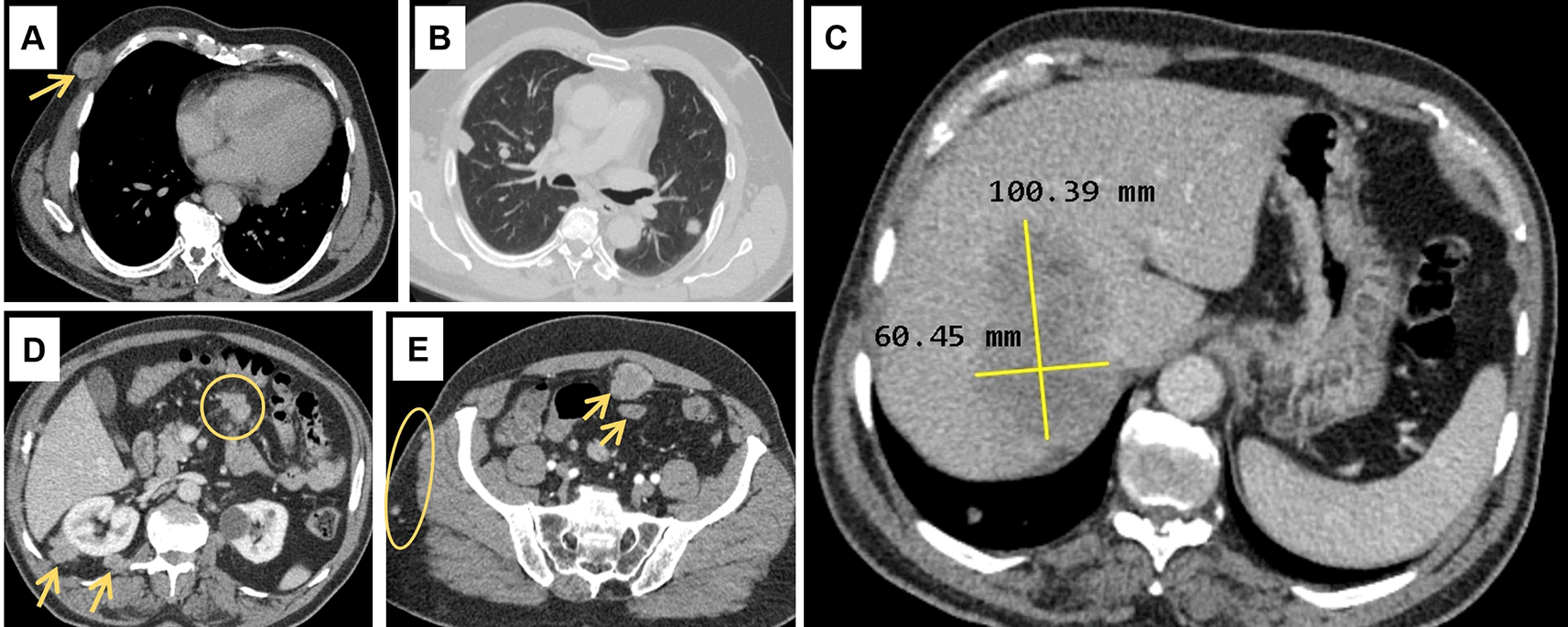
(A): Subcutaneous soft tissue nodule of the anterior right thoracic wall (arrow). (B): Bilateral pulmonary nodules. (C): Hypodense hepatic mass in the right lobe on the portal phase, measuring 100 × 60 millimetres. (D): Intraperitoneal adenomegalies (circle) and heterogeneous nodules in the right perirenal retroperitoneal region. (E): Heterogeneous peritoneal nodules (arrows) and small subcutaneous soft tissue nodules in the right lateral abdominal wall (circle).
At this point, widespread metastases were the leading considered diagnosis based on the clinical context and radiological presentation. Alpha-fetoprotein and prostate specific antigen levels were normal.
In the investigation for the primary cancer, and given the recent dyspepsia and epigastric pain, an upper gastrointestinal endoscopy was performed revealing a polypoid lesion at the antro-fundic junction measuring 20 mm in diameter, with a depressed and ulcerated centre, containing dark pigmentation ( Figure 2). The histological examination showed a tumour proliferation extensively invading the antro-fundic mucosa, composed of discohesive cells, with deposits of brownish pigments. Immunohistochemistry analysis revealed positive staining for melanocytic markers Melan-A, S100 protein and HMB45 ( Figure 3). Other markers were negative, namely pancytokeratin, TTF 1, CD 45, chromogranin, and synaptophysin. These findings were consistent with a gastric localisation a melanoma.

Retroflexion shows an elevated lesion at the antrum-fundus junction with a depressed centre, resembling a volcano appearance, with brown pigments.
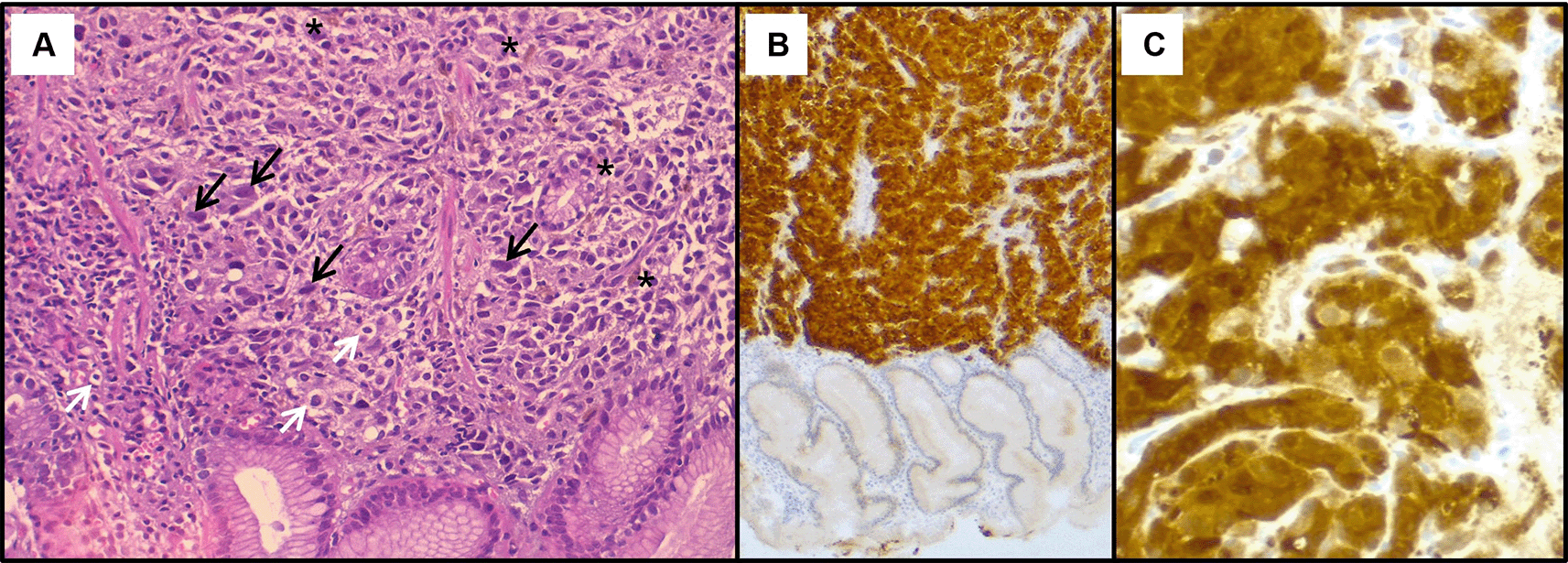
(A): Haematoxylin & eosin staining on x200 magnification shows malignant proliferation infiltrating the gastric mucosa. Tumour cells are pleomorphic and disaggregated, exhibiting marked nuclear atypia with hyperchromatic and sometimes monstruous nuclei (black arrows). Some larger cells with clear cytoplasm were present (white arrows). Deposits of brown pigments are also seen (*). Immunohistochemistry demonstrated positive staining with Melan-A (B) and PS 100 (C).
With these results in mind, a thorough skin examination was thus undertaken unveiling a cutaneous lesion in the back, manifesting as a circumscribed pink nodule of 8 mm with brown discoloration ( Figure 4).

It appears as a circumscribed dark pink nodule measuring 8 millimetres in diameter with areas of brown discoloration. The lesion also has a slightly irregular surface and a surrounding area of erythema.
Histological assessment of the skin lesion biopsy confirmed nodular melanoma, with immunohistochemistry showing positive staining for Melan-A, HMB45, and p16 ( Figure 5).

(A): Haematoxylin & eosin staining on x100 magnification shows malignant cellular proliferation (*) in the dermis, composed of vaguely naevoid cells and larger cells with abundant eosinophilic cytoplasm, atypical pleomorphic nucleolated nuclei, and no junctional activity. On immunohistochemistry, tumour cells exhibit positive staining for HMB45 (B), Melan-A (C), and p16 (D).
Ultimately, the diagnosis of malignant cutaneous melanoma with diffuse multi-organ metastases, particularly involving the stomach, was confirmed. Subsequently, the patient’s general condition deteriorated, precluding the initiation of planned palliative chemotherapy. Supportive care was provided, including pain management, with a documented follow-up of 6 months.
The stomach is an unusual metastatic site, accounting for 2.6% of secondary lesions, particularly from breast, lung, and melanoma. Melanoma frequently metastasizes to the GI tract, with cutaneous melanoma primarily affecting the upper GI tract, while uveal melanoma tends to spread to the liver.4,5 An autopsy study of 216 patients with advanced malignant melanoma found the GI system to be the second most common metastatic site, after lymph nodes (73.6%) and lungs (71.3%).6 In this series, GI metastases were often part of multiple-organ involvement (95%), with secondary gastric melanoma in 22.7% of cases. Liver metastases were observed in 58.3% of cases. Other GI metastases included the peritoneum (42.6%), pancreas (37.5%), small bowel (35.6%), spleen (30.6%), colon (28.2%), oral cavity, oesophagus (9.3%), and biliary tract (8.8%). A similar autopsy study of 100 patients with cutaneous melanoma found gastric involvement in 26%.7 Despite these findings, GI metastases seem to be often subclinical, with clinical series showing much lower incidence, such as intestinal metastases in only 1% to 7% of cases.3 Consistent with these reports, our patient presented with disseminated melanoma affecting multiple organs, including the lungs, liver, stomach, subcutaneous tissue, adrenal gland, peritoneum, and lymph nodes.
Also, gastric metastases from melanoma may be present at diagnosis or develop years later, indicating recurrence.8
A recent systematic review by Reggiani et al.9 included 113 patients with gastric melanoma metastases, predominantly from skin primary lesions (62%). Most patients were male (64%) with a median age of 63 years. Only 10% were asymptomatic, while common symptoms included bleeding (34.5%), abdominal pain (34.5%), anorexia and weight loss (23%), and nausea/vomiting (17.7%). Dyspepsia was noted in 5.3%. Gastric metastases were single in 42.5% and mostly located in the gastric body (60.2%). However, variability in lesion descriptions across studies prevented the identification of consistent endoscopic features.
Nelson et al.10 proposed an endoscopic classification for GI melanoma metastases with three types: 1) melanotic nodules on normal folds with ulcerated tips; 2) elevated submucosal lesions with ulcerated centres, sometimes with visible melanin, often described as volcano-like or donut-shaped,9 as seen in our case; and 3) mass lesions with necrosis and melanosis. Such protruding lesions, especially with brown deposits, should raise suspicion for gastric melanoma. Other descriptions include polypoid masses, small nodules, and black spots.5,8,11
GI melanoma metastases can resemble primary gastric cancer or other gastric metastases,5,9,11 making histopathological examination and immunohistochemistry essential for diagnosis. These metastases typically show positive staining for melanocytic markers,12 as seen in both our gastric and skin lesions. However, phenotypic switching during tumour progression can cause loss of typical immunophenotypic features, such as Melan-A, HMB45, S100, and SOX 10.12,13 In such cases, molecular testing for melanoma-related mutations like BRAF, NRAS, and NF1 may be necessary.13 For our patient, the diagnosis was confirmed by histopathological findings and the typical immunohistochemical profile, without the need for further tests.
Metastatic melanoma has a poor prognosis, with distant metastatic burden being a key prognostic factor. Non-pulmonary visceral metastases and elevated serum lactate dehydrogenase are linked to worse survival, with a one-year survival rate of 33%.14,15 In the above-mentioned systematic review of gastric melanoma metastases, the median survival was 3 months, with 16% and 4% survival rates at 1 and 2 years, respectively.9 Survival was notably lower with multiple gastric lesions compared to a single metastasis.
GI resections have been performed in localised metastasis, with evidence of possible prolonged remission, or as an emergency procedure for complicated cases like GI bleeding or perforation.8,16 However, our patient presented extensive metastatic disease and hence was not a proper candidate for surgery. Systemic treatments, particularly immunotherapy, are now the preferred first-line therapy for metastatic melanoma, showing promising survival benefits.17 Unfortunately, immunotherapy was unavailable at the time, and the patient’s rapid decline precluded chemotherapy.
GI metastases, particularly in the stomach, are rare, with melanoma being the most common cancer to spread to this site. This case highlights how a gastric metastasis revealed an underlying malignant cutaneous melanoma. A protruding gastric lesion with dark pigmentation during endoscopy should raise suspicion of melanoma, warranting a thorough skin examination for potentially overlooked lesions, which is also crucial in widespread metastatic disease. Histopathological and immunohistochemical analysis remain the gold standards for diagnosis. In cases of widespread metastases, palliative systemic treatment, including chemotherapy or immunotherapy, is preferred, with surgery reserved for selected oligometastatic cases. Despite treatment, the prognosis remains poor.
| Views | Downloads | |
|---|---|---|
| F1000Research | - | - |
|
PubMed Central
Data from PMC are received and updated monthly.
|
- | - |
Provide sufficient details of any financial or non-financial competing interests to enable users to assess whether your comments might lead a reasonable person to question your impartiality. Consider the following examples, but note that this is not an exhaustive list:
Sign up for content alerts and receive a weekly or monthly email with all newly published articles
Already registered? Sign in
The email address should be the one you originally registered with F1000.
You registered with F1000 via Google, so we cannot reset your password.
To sign in, please click here.
If you still need help with your Google account password, please click here.
You registered with F1000 via Facebook, so we cannot reset your password.
To sign in, please click here.
If you still need help with your Facebook account password, please click here.
If your email address is registered with us, we will email you instructions to reset your password.
If you think you should have received this email but it has not arrived, please check your spam filters and/or contact for further assistance.
Comments on this article Comments (0)