Keywords
Spinal meningioma; pediatric meningioma; back pain; gait disturbance
This article is included in the Oncology gateway.
Pediatric meningiomas are rare tumors originating from the meninges’ arachnoid cap cells, representing a small fraction of all meningiomas. The clinical presentation of pediatric meningiomas varies widely depending on their location within the central nervous system. We report a case of pediatric spinal meningioma revealed by gait disturbance. We emphasize clinical and imaging features, as well as therapeutic management of spinal meningiomas.
We present the case of a 15-year-old boy with a one-year history of chronic thoracic back pain, worsening gait disturbances, and difficulty walking. Neurological examination revealed a spastic gait with increased muscle tone and bilateral pyramidal syndrome. Magnetic resonance imaging (MRI) demonstrated a posterior intradural extramedullary mass at the T6-T7 level, causing dorsal spinal cord compression. The patient underwent complete resection of the tumor, and histological examination confirmed the diagnosis of a WHO grade I psammomatous spinal meningioma.
This case highlights the clinical and imaging characteristics of spinal meningiomas in pediatric patients. MRI remains the gold standard for diagnostic imaging, effectively narrowing down differential diagnoses. However, a definitive diagnosis requires histological examination. The primary therapeutic approach for pediatric meningiomas is surgical resection.
Spinal meningioma; pediatric meningioma; back pain; gait disturbance
1. Spinal pediatric meningiomas are rare and can mimic various conditions, making diagnosis particularly challenging.
2. MRI plays a crucial role in differentiating between potential diagnoses. However, histopathological confirmation is essential for diagnosing meningiomas and guiding appropriate management.
3. Detailed history, thorough physical examination, and genetic screening are crucial to rule out associations with genetic conditions, particularly NF2, which necessitates a different management approach and genetic counseling.
4. Surgery aiming for total tumor resection is the first-line treatment for spinal pediatric meningiomas. Unfortunately, the post-operative course in this population is often complicated by higher recurrence rates and additional complications.
Gait abnormalities are common in children and can result from various conditions, including cerebral palsy, neuromuscular disorders, and spinal cord tumors, such as spinal meningiomas. Spinal meningiomas account for less than 10% of central nervous system tumors in the pediatric population.1
Pediatric meningiomas (PMs) are an even rarer entity, accounting for less than 5% of all meningiomas, and spinal PMs are estimated to be exceedingly rare.2
Spinal meningiomas are characterized by their intradural, extramedullary growth,1 often resulting in a mass effect on the spinal cord that can lead to significant spinal cord compression and related complications.3
The management of pediatric meningiomas is challenging, with a higher incidence of preoperative and postoperative complications.3,4 Surgical resection remains the first-line treatment, similar to adult cases. On the other hand, radiotherapy (RT) is considered on a case-by-case basis due to its potential risks in the pediatric population although it is often required, particularly in NF2-associated cases, where tumors are more aggressive and recurrent.3,4
We report a case of pediatric spinal meningioma revealed by gait disturbance. We emphasize clinical and imaging features, as well as therapeutic management of spinal meningiomas.
A 15-year-old male presented with a one-year history of progressively worsening back pain, described as dull, persistent, and localized to the thoracic spine. The pain had an insidious onset and was non-radiating. The patient also reported an unsteady gait, difficulty ambulating, and lower extremity weakness, limiting his walking distance to approximately 100 meters. He denied any history of trauma, fever, malignancy, bowel or bladder dysfunction, seizures, or headaches. Family history was unremarkable, with no known cases of neurofibromatosis, malignancy, Gorlin syndrome, or familial meningiomatosis.
On physical examination, the patient exhibited a gait disturbance with increased muscle tone, though muscle strength testing was within normal limits. Neurological examination revealed abolition of abdominal cutaneous reflexes, bilateral symmetric hyperreflexia in the lower limbs, and a positive Babinski sign. No pain was elicited upon percussion of the spinous processes. He had no meningeal signs, spinal limitation, or arthritis.
Cardiovascular and pulmonary examinations were unremarkable, and no cutaneous lesions were noted.
C-reactive protein, calcium level, liver tests, and renal function were within normal ranges. The complete blood count was without abnormalities.
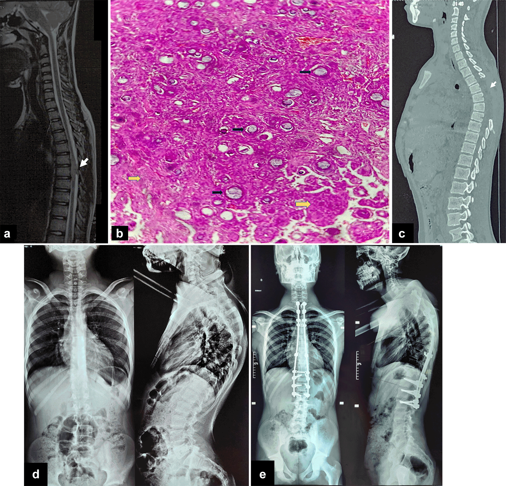
Given the presence of neurological signs, a spine MRI was performed showing a posterior intradural mass at the T6 and T7 vertebral levels, characterized by an extramedullary, well-circumscribed, homogeneous, and lobulated appearance. The lesion measured approximately 1.8 × 1.2 × 0.9 cm and exhibited iso-intensity on T1-weighted images and hyperintensity on T2-weighted images, with homogeneous and marked enhancement after contrast administration. The mass exerted a local mass effect, resulting in dorsal spinal cord compression. These findings were initially suggestive of a thoracic glioma, with differential diagnoses including low-grade ependymoma or astrocytoma [Figure 1a, 1c].

The patient underwent posterior spinal surgery, including a wide laminectomy spanning T5, T6, and T7. Upon opening the dura mater, a whitish, firm mass compressing the adjacent spinal cord was observed. The tumor displayed a well-defined cleavage plane with the dura mater, with only minor adhesions to the adjacent arachnoid. Complete resection of the tumor was achieved.
Histopathological examination revealed a proliferation of meningothelial cells with poorly defined cytoplasmic borders, creating a syncytial appearance and exhibiting cytological pleomorphism. Numerous psammoma bodies were observed throughout the specimen, with no evidence of anaplasia or tumor necrosis [Figure 1b]. These findings were consistent with a WHO Grade 1 psammomatous meningioma.
Unfortunately, the postoperative course was complicated by the onset of hyperalgesic back pain two months later, along with the development of dorsal hyperkyphosis [Figure 1d]. This complication was attributed to the extensive laminectomy. A posterior thoracic arthrodesis was performed [Figure 1e]. The patient is currently undergoing motor rehabilitation with favorable clinical progress.
We present the case of a pediatric spinal meningioma revealed by an insidious onset of gait disturbance, thoracic back pain, and lower limb weakness. Meningiomas are typically slow-growing tumors originating from the meninges.3
Similarly to intracranial meningiomas, spinal meningiomas arise from arachnoid cap cells. Abnormal proliferation of these cells leads to the formation of intradural extramedullary masses. Spinal meningiomas represent 5 to 10% of all meningiomas and constitute 25 to 45% of all intraspinal tumors.1,2 Spinal meningioma typically occurs in the thoracic spine since arachnoid cap cells are predominantly located at this spinal level.3,4
Pediatric meningiomas are scarce, accounting for less than 5% of pediatric spinal tumors, and an even a smaller fraction of overall pediatric meningiomas.2
Unlike in adults, pediatric meningiomas seem to be less influenced by hormonal factors, which may explain the similar gender distribution observed in this population.2–4
Meningiomas are graded by the World Health Organization (WHO) into three grades based on their histological features, such as mitotic rate, cellular atypia, and brain invasion. Grade I meningiomas are benign characterized by a low mitotic rate (<4 per 10 high-power fields) and the absence of brain invasion, and include a psammomatous subtype,4 as seen in our patient. The same grading system applies to PMs.
In contrast to adults, an association between pediatric meningiomas and type 2 neurofibromatosis (NF2) has been reported in 13% of cases and even in more than 50% of patients with intracranial meningiomas.5
The NF2 gene mutation is responsible for abnormal cell proliferation and tumor formation.
NF2-related meningiomas are characterized by an earlier onset, more rapid growth, atypical histological features, and higher WHO grades (II and III).2–5
Pediatric meningiomas may also be associated with other genetic syndromes, such as Gorlin syndrome (basal cell nevus syndrome) and familial meningiomatosis. However, our patient had no personal or family history of these conditions. In a study of CT scans from 82 patients with Gorlin syndrome, 5% showed radiological features suggestive of meningioma.6
Spinal meningiomas are characterized by their intradural, extramedullary growth, typically affecting the thoracic spine,3,4 as seen in our patient. While asymptomatic spinal meningiomas usually exhibit a slow, linear growth pattern, symptomatic tumors are prone to an exponentially accelerating growth rate,3 leading to varying degrees of spinal cord compression, ranging from insidious motor and sensory dysfunction to gait disturbances, and difficulty walking as observed in our patient. Symptoms generally worsen progressively over time as the tumor continues to expand, exerting increased pressure on the spinal cord. In advanced stages, severe spinal cord compression can disrupt autonomic functions, resulting in bowel and bladder disturbances, which are reported in approximately 36% of patients, and irreversible neurological damage.2–3 Chronic cases often present with persistent back pain, as was evident in our patient, which may be associated with radiculopathy or evolve into a more debilitating condition. Longstanding tumor presence can also contribute to spinal deformities, such as kyphosis.3
In addition, both pediatric and adult meningiomas share similar basic MRI features, such as a well-circumscribed, intradural extramedullary location, the characteristic dural tail sign, and comparable signal intensities on T1- and T2-weighted images. However, spinal pediatric meningiomas tend to exhibit more aggressive patterns. Indeed, they may appear larger at diagnosis, involve longer spinal segments, and cause more significant compression of the spinal cord compared to those in adults.1–4
Upon clinical presentation and examination, distinguishing pediatric spinal meningiomas from other etiologies is challenging due to their ability to mimic various conditions. In this case, the patient’s insidious gait disturbances and progressive lower body weakness raised clinical suspicion of spinal cord compression. Both the patient and family denied any recent trauma, and the physical examination showed no signs of bruising.
Neoplastic etiologies are among the most frequent causes of spinal cord compression in the pediatric population, commonly involving ependymomas, astrocytomas, gliomas, and meningiomas. Clinically, distinguishing between these neoplasms is often impossible. MRI is crucial for differential diagnosis and guiding surgical planning, Although definitive diagnosis relies on histopathological examination for accurate confirmation and management.
Contrary to spinal meningiomas, which are typically extramedullary, these tumors have an intramedullary growth pattern. Therefore, the characteristic “dural tail sign”,3,4,6 an enhancement extending along the dura adjacent to the tumor on post-contrast images, is absent in intramedullary tumors.
Gliomas and astrocytomas typically present with poorly defined margins, often involving multiple spinal segments and causing spinal cord expansion, in contrast to the well-defined and circumscribed appearance of meningiomas. Ependymomas, although intramedullary-like gliomas, are also well-defined and circumscribed, often featuring a “cap sign” on T2-weighted images, corresponding to a rim of hemosiderin. A cystic component and syringomyelia can be associated with ependymomas, which is a distinguishing feature from meningiomas.7
Tethered cord syndrome can mimic spinal meningioma due to gait abnormalities and progressive motor dysfunction of the lower limbs. However, no foot deformities or cutaneous markers such as sacral dimples, lipomas, or hairy patches along the midline of the back were observed on examination. MRI ruled out this diagnosis, as it did not reveal a low-lying conus medullaris, thickened filum terminal, or evidence of spinal cord tension.8
Given the presentation of spasticity, lower limb weakness, and hyperreflexia, spinal meningioma could also be mistaken for transverse myelitis, which accounts for 20% of spinal cord compression cases in the pediatric population, most commonly occurring between 10 and 19 years of age. However, the absence of a rapid onset over hours to days and the lack of sensory or autonomic dysfunctions made this diagnosis less likely in our patient. Moreover, MRI did not demonstrate the characteristic spinal cord lesions of transverse myelitis, and laboratory results were within normal ranges.9
Management of meningioma is largely dependent on its symptomatic status.3 Asymptomatic meningiomas can often be managed with routine observation and imaging.3,4
Conversely, symptomatic and rapidly enlarging meningiomas, typically require surgical resection as the primary approach.3,4
Complete surgical excision of meningiomas is the gold standard.2–4
Adjuvant radiotherapy (RT) may be recommended, in cases of recurrent tumors, non-resectable, growing lesions, or when gross total resection is not achievable.3,4 However, RT is generally avoided in pediatric patients due to the heightened risk of secondary malignancies and long-term neurocognitive sequelae.2–5
Unfortunately, the postoperative course in pediatric patients is marked by a higher incidence of spinal instability, especially following extensive laminectomies,2–4,10 necessitating spinal fusion or stabilization procedures, as was required in our patient.
Recently, emerging surgical techniques, such as endoscopic endonasal approaches, are being investigated for the resection of midline anterior skull base tumors, offering a minimally invasive alternative.4
To date, novel pharmacotherapies, including multikinase inhibitors targeting vascular endothelial growth factor receptors and bevacizumab, have shown promising results; however, consensus guidelines have yet to be established.10 Interestingly, radionuclide therapies targeting somatostatin receptors are under exploration for the management of refractory meningiomas, demonstrating great potential.10
Regular follow-ups with MRI are recommended every 6 months for the first 5 years post-treatment, followed by annual assessment.3,4,10
The diagnosis of spinal tumors should be considered in children with gait disturbances.
Pediatric meningiomas present with a spectrum of symptoms, ranging from back pain to severe spinal cord compression and even spinal deformities. MRI typically shows a well-defined and circumscribed extramedullary appearance with the characteristic dural tail sign on post-contrast images. A total resection is considered the gold standard. Radiotherapy is reserved for specific scenarios, including recurrence, subtotal resection, inoperability, or high-grade tumors.
Consent: Written informed consent for publication of their clinical details was obtained from the patient’s legal guardians.
| Views | Downloads | |
|---|---|---|
| F1000Research | - | - |
|
PubMed Central
Data from PMC are received and updated monthly.
|
- | - |
Is the background of the case’s history and progression described in sufficient detail?
Partly
Are enough details provided of any physical examination and diagnostic tests, treatment given and outcomes?
Partly
Is sufficient discussion included of the importance of the findings and their relevance to future understanding of disease processes, diagnosis or treatment?
Partly
Is the case presented with sufficient detail to be useful for other practitioners?
Yes
Competing Interests: No competing interests were disclosed.
Reviewer Expertise: Vascular neurosurgery and skull base neurosurgery
Alongside their report, reviewers assign a status to the article:
| Invited Reviewers | |
|---|---|
| 1 | |
|
Version 2 (revision) 19 Aug 25 |
|
|
Version 1 30 Oct 24 |
read |
Provide sufficient details of any financial or non-financial competing interests to enable users to assess whether your comments might lead a reasonable person to question your impartiality. Consider the following examples, but note that this is not an exhaustive list:
Sign up for content alerts and receive a weekly or monthly email with all newly published articles
Already registered? Sign in
The email address should be the one you originally registered with F1000.
You registered with F1000 via Google, so we cannot reset your password.
To sign in, please click here.
If you still need help with your Google account password, please click here.
You registered with F1000 via Facebook, so we cannot reset your password.
To sign in, please click here.
If you still need help with your Facebook account password, please click here.
If your email address is registered with us, we will email you instructions to reset your password.
If you think you should have received this email but it has not arrived, please check your spam filters and/or contact for further assistance.
Comments on this article Comments (0)