Keywords
Scleral lens, front surface eccentricity, higher order aberrations, refractive surgery, Small Incision Lenticule Extraction
This article is included in the Eye Health gateway.
Scleral lens, front surface eccentricity, higher order aberrations, refractive surgery, Small Incision Lenticule Extraction
Details regarding the follow-up period, indications for scleral lenses with varying front surface eccentricities (FSEs), and the probable mechanisms underlying higher-order aberration correction have been incorporated. In addition, a table summarizing the specifications and details of the trial lenses has been included.
See the authors' detailed response to the review by Suraj Kumar Chaurasiya
Small incision lenticule extraction (SMILE) is a keraratorefractive procedure widely used as an effective surgical treatment for common refractive errors.1 The central corneal curvature is modified, making it flatter to correct myopia and steeper to correct hyperopia. Thereby, influencing the corneal optical quality and producing aberrations leading to distorted images. SMILE may increase the total higher-order aberrations (HOAs), which are thought to be responsible for poor quality of vision, even with visual acuity (VA) of 20/25 or better.2 Many visual symptoms like starbursts, shadowing, reduction in contrast sensitivity and ghost images postoperatively may be secondary to an increase in the HOAs.3 Total HOAs induced after Wavefront-guided (WFG) keratorefractive procedures are similar to SMILE.2 Scleral contact lenses (SCLs) are large-diameter lenses that vault over the entire cornea, mask the corneal irregularity, and enhance vision in irregular corneas.4 They are known to effectively neutralize HOAs induced by the anterior corneal surface.5 The use of different eccentricities on the front surface of SCLs has been shown to improve the visual performance in eyes with irregular cornea by reducing the HOAs.6 This case demonstrates the impact of different FSEs in the Boston Sight Scleral (BSS) lens (Boston, Massachusetts, USA) on ocular aberrations and visual acuity, and also hypothesizes how eccentricity of SCLs can be calculated according to the asphericity (Q - value) of the posterior corneal surface.
A female in her 20s presented with complaints of diminution of vision for both near and distance for the last 4 months associated with shadowing and ghosting of images after keratorefractive surgery. She had undergone SMILE surgery for myopic refractive error of -4.50 Dioptre Sphere (DS) in the right eye (RE), and -8.00 DS in the left eye (LE) 6 months back.
Her uncorrected high contrast distance visual acuity (HCDVA) recorded under room illumination using the COMPlog chart (COMPlog Clinical Vision Measurement Systems Ltd, London, UK) was 20/60 and 20/40 in RE and LE respectively. The near VA using the English Chart under room illumination was noted to be N6 @ 25 cms in both eyes (BE). After subjective refraction, her HCDVA improved to 20/20p in RE and 20/25 in LE. With pinhole, VA in LE improved to 20/20. Even with the best-corrected vision, her complaints of shadowing of optotypes, and ghosting of images persisted.
On slit-lamp biomicroscopy, the cornea in BE revealed a well-demarcated ring-shaped area of ablation with faint scarring at the anterior stroma. Intra-ocular pressure and posterior segment were within normal limits in BE.
Corneal topography (Allegro Oculyzer, WaveLight, GmbH, Erlangen, Germany) ( Figure 1A and B) showed average flat and steep keratometric readings. The front and back surface asphericity (@6 mm) for RE was 0.47 and 0, and for LE was 1.57 and -0.03. iTrace (Tracey Technologies Corp. TX, USA) performed under scotopic conditions keeping the fixed scan of diameter 3.2 mm showing HOAs of 0.230 μ in RE and 0.237 μ in LE. Anterior segment optical coherence tomography (AS-OCT) (Optovue, Inc., Fremont, CA) showed epithelial irregularity bilaterally. With complaints of ghosting of images, and unwillingness to use spectacles, the patient was advised for a contact lens trial.
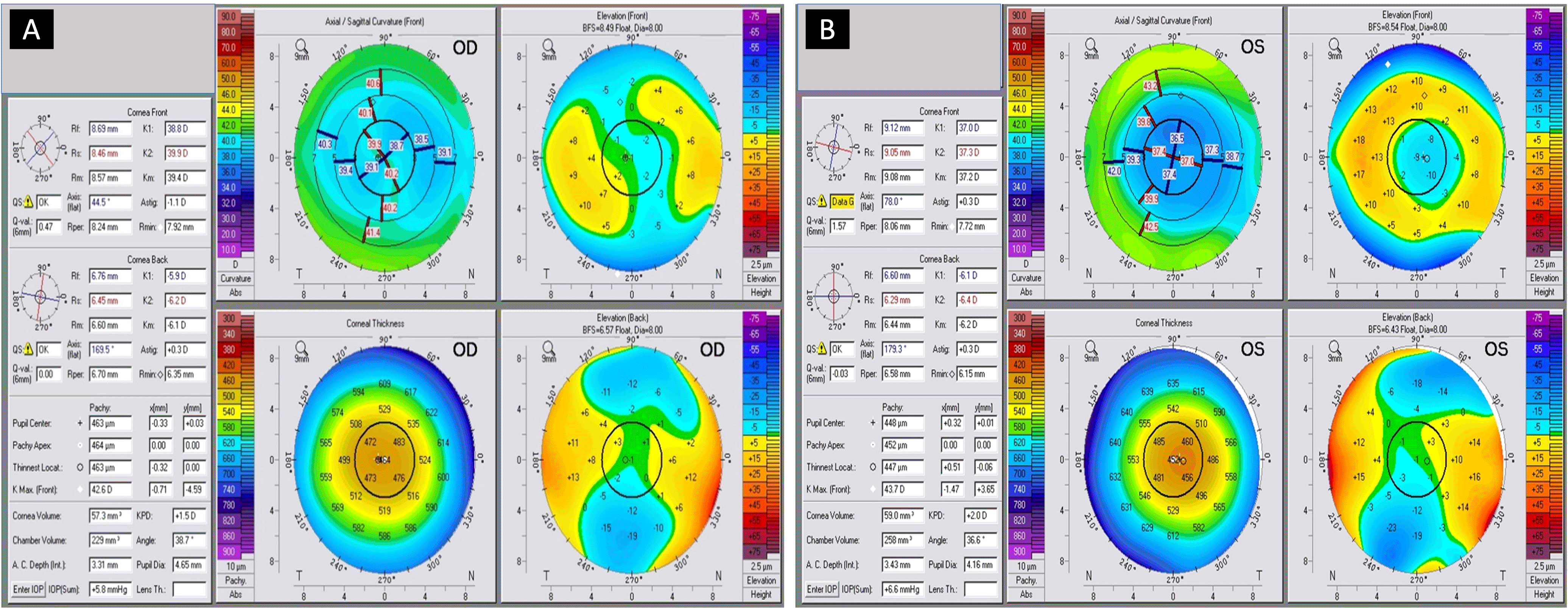
It shows the posterior surface asphericity (Q-value) for the right (0.00) and the left eye (-0.03).
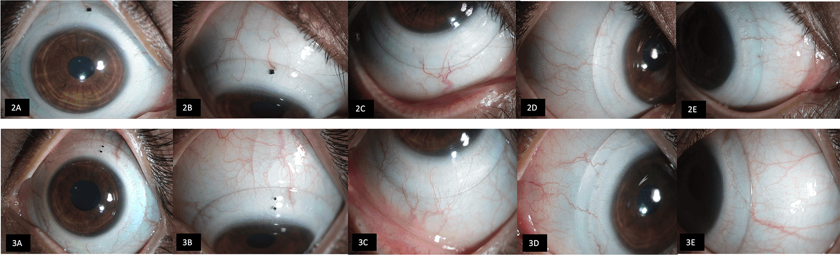
Both conventional RCL (Purecon, New Delhi, India) and Rose K2 lenses (Menicon Co. Ltd, Nagoya, Japan) were tried with no significant improvement in visual quality. Consequently, the patient was advised BSS lens trial. The initial trial lens was selected based on the company’s guidelines to achieve optimal fitting characteristics, which included a central vault ranging from 200-400 microns, and a limbal vault ranging from 50-100 microns with well-aligned lens haptics resting on the sclera and overlying conjunctiva. The specifications of the selected BSS lenses were 18.5 mm diameter, 2.80 mm sagittal height, 8.00 mm base curve, with standard haptics. All trial devices shared similar parameters except their eccentricity, which varied from 0, 0.6 and 0.8 ( Table 1). Initially, a lens with 0.6 eccentricity (FSE 1) was tried, followed by 0 (FSE 0) and lastly, 0.8 (FSE 2), while keeping all other parameters constant. The patient was given an adaptation time of 15 minutes after the application of each lens, after which visual acuity was noted, followed by over-refraction. Figures 2 and 3 illustrate the final SCL fit prescribed for the patient. Ocular aberrometry was performed using iTrace with SCLs of different eccentricities. Between two consecutive lenses, a gap of 10 minutes was given. All these measurements were recorded with a lens in situ under scotopic conditions with a scan size diameter of 3.2 mm. The best corrected HCDVA of 20/20 and a subjective reduction in image ghosting were observed with an FSE 0 lens. Table 2 summarizes the impact of various FSEs on HOAs and HCDVA. Since both the quality of vision and HOAs improved with the FSE 0 lens, these lenses were dispensed. Preservative-free 0.9% normal saline was given to fill the vault, and the patient was advised to replenish it every 4-6 hours of lens wear. The patient received comprehensive training on lens insertion, removal and maintenance. She was advised to use a Boston Simplus® Multi-Action solution (Bausch & Lomb, NY, USA) for lens cleaning, disinfection and storage. The patient was followed up at 1 month and then at a 3-month visit. During this period, visual acuity remained stable, and no adverse events were noted. She reported high satisfaction with visual quality, especially under mesopic conditions.
| Diameter (mm) | Base curve (mm) | Haptic design | Sagittal height (mm) | Front surface eccentricity (FSE) | |
|---|---|---|---|---|---|
| 1 | 18.5 | 8.0 | Standard | 2.80 | FSE 1 (0.6) |
| 2 | 18.5 | 8.0 | Standard | 2.80 | FSE 0 (0.0) |
| 3 | 18.5 | 8.0 | Standard | 2.80 | FSE 2 (0.8) |
While keratorefractive surgeries are designed to reduce pre-existing aberrations, they can also induce newer aberrations affecting visual quality and contribute to patient dissatisfaction.7 Although WFG enhancement surgeries may mitigate these aberrations, many patients are either reluctant to undergo a repeat surgery or deemed unfit due to the associated risk of post-surgical ectasia.
RCLs have been widely used for visual rehabilitation in patients with irregular corneas. The tear film between the lens and the cornea effectively masks corneal irregularity. They not only correct refractive errors but also eliminate third to fifth-order and total HOAs.8 Fitting corneal lenses can be particularly challenging in oblate corneas. Reverse geometry lenses can give a better fit, however, the continuous gliding movement of these lenses on the cornea with blinking can lead to instability and discomfort. In our case, the RCLs neither tolerated nor improved visual quality.
SCLs offer superior comfort and tolerability compared with RCLs. Kumar et al demonstrated that SCLs can reduce HOAs by approximately 70–80% in eyes following penetrating keratoplasty, radial keratotomy, and in post-LASIK ectasia.9 Hastings et al reported that WFG - SCLs provide enhanced visual and optical rehabilitation compared with conventional SCL designs.10 However, the implementation of these customised SCLs requires advanced technical expertise and sub-micron precision lathe-cutting technology, posing manufacturing challenges and potentially limiting their widespread clinical availability.
Jagadeesh et al reported that an FSE of 0.60 maximised visual performance and reduced HOAs in moderate keratoconus.11 Hussion et al also observed enhanced high and low-contrast visual acuity with different eccentricities.12 Similarly, Badrinarayanan et al noted improvements in HOAs, corrected distance VA and contrast sensitivity in keratoconus patients with varying eccentricities.13 Despite these findings, an optimal FSE value has not been established.
HOAs originating from the anterior corneal surface are neutralised by an SCL, whereas those arising from the posterior corneal surface persist.14 In this context, FSEs may compensate for aberrations attributable to the posterior cornea. Additionally, FSEs facilitate improved alignment between the ocular optical axis and the optical zone of the SCL, which may further contribute to enhanced visual performance.12 SCL with different FSE can be considered in cases with suboptimal improvement in visual quality, particularly in eyes with penetrating keratoplasty, refractive surgery ectasia, and asymmetric ablation profile.
The thickness of the fluid reservoir,15 the lens material,16 and the polymer coating17 have minimal impact on HOAs. Considering the irregularities of the corneal anterior surface, and the anterior and posterior asphericity of the cornea, we hypothesize that a lens with FSE closely matching the eccentricity (Ɛ) of the posterior surface of the cornea should be selected as the first lens for trial. In our case, the posterior asphericity (Q) was noted to be 0 and -0.03 in RE and LE, respectively. The formula Q = –Ɛ2 can be used to convert asphericity into eccentricity,18 resulting in the values of 0.00 for RE and 0.17 for LE. Consequently, the selected SCL for the trial was chosen based on the closest FSE, which was 0 for both eyes.
In our case, all lens designs resulted in measurable alterations in HOAs. However, the patient’s functional ability to resolve the visual target must be considered alongside objective aberration metrics. Although a greater reduction in HOAs was achieved with an FSE of 0.6, the patient subjectively reported superior visual perception with an FSE of 0. This finding can be illustrated through the simulation of the “E” optotype, demonstrating differences in the point spread function.
Further investigations involving larger sample sizes and corneas with a wider range of posterior surface eccentricities are required to validate our hypothesis. Additionally, the development of a structured algorithm to guide the selection of FSE for different patient subgroups is warranted. Until robust correlations are established, a pragmatic approach would be to try lenses with varying eccentricities before finalizing the lens design. Failure to achieve pinhole visual acuity and persistent subjective complaints of visual distortion despite optimal refraction may serve as clinical indicators for incorporating alternative FSEs into the SCL. Moreover, quantitative assessment of ocular aberrations is essential, as it not only improves understanding of patients’ subjective visual symptoms but also enables objective comparison of the optical effects associated with different FSEs.
This case illustrates the influence of different FSEs of BSS devices on visual outcomes following SMILE surgery. SCL with varying FSEs helps to further mitigate these aberrations, thereby enhancing optical quality. The insights gained from this case may facilitate the selection of the first trial lens, considerably reduce the chair time and guide in prescribing lenses with appropriate FSE in post-keratorefractive surgery.
| Views | Downloads | |
|---|---|---|
| F1000Research | - | - |
|
PubMed Central
Data from PMC are received and updated monthly.
|
- | - |
Provide sufficient details of any financial or non-financial competing interests to enable users to assess whether your comments might lead a reasonable person to question your impartiality. Consider the following examples, but note that this is not an exhaustive list:
Sign up for content alerts and receive a weekly or monthly email with all newly published articles
Already registered? Sign in
The email address should be the one you originally registered with F1000.
You registered with F1000 via Google, so we cannot reset your password.
To sign in, please click here.
If you still need help with your Google account password, please click here.
You registered with F1000 via Facebook, so we cannot reset your password.
To sign in, please click here.
If you still need help with your Facebook account password, please click here.
If your email address is registered with us, we will email you instructions to reset your password.
If you think you should have received this email but it has not arrived, please check your spam filters and/or contact for further assistance.
Comments on this article Comments (0)