Keywords
Guillain Barre Syndrome (GBS), Acute Motor- Sensory Axonal Neuropathy (AMSAN), Posterior Reversible Encephalopathy Syndrome (PRES), Plasma Exchange, Post Partum, Intrauterine Death
This article is included in the Manipal Academy of Higher Education gateway.
Guillain Barre Syndrome (GBS), Acute Motor- Sensory Axonal Neuropathy (AMSAN), Posterior Reversible Encephalopathy Syndrome (PRES), Plasma Exchange, Post Partum, Intrauterine Death
The incidence of Guillian Barre Syndrome (GBS) is relatively low during pregnancy. The risk of developing GBS increases after delivery, particularly during the first two weeks of puerperium.1
Here, we present a case of Acute Motor- Sensory Axonal Neuropathy (AMSAN), he rare and most severe form of GBS, complicating the postpartum period in our patient. Acute Motor Sensory Axonal Neuropathy (AMSAN)-type GBS is characterized by sensory and motor fiber axonal degeneration.2
Prevention of severe axonal damage in the early stages of the disease stays top priority, it being a major impediment to attaining a favourable long-term outcome.3 Preganglionic sympathetic axonal demyelination or postganglionic axonal degeneration causes blood pressure fluctuations in GBS patients.4
Our patient with AMSAN-type GBS developed severe dysautonomia and accelerated hypertension, which ultimately led to Posterior Reversible Encephalopathy Syndrome (PRES). The contribution of a multidisciplinary team towards her care cannot be overstated.
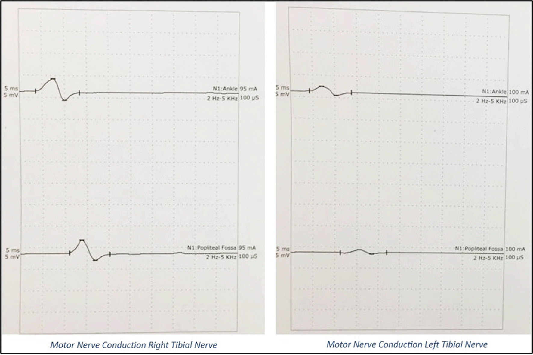
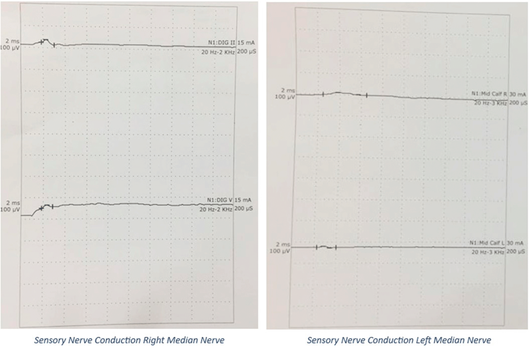
A 20’s postpartum female with antepartum stillbirth, following spontaneous expulsion, with no existing comorbidities or prior neurological disease, presented to the emergency department on post-partum day 8 with chief complaints of inability to move both lower limbs for 5 days, inability to raise her arms for 3 days, and difficulty in breathing since that morning. Her symptoms started immediately post-partum and had rapidly progressed over the last five days. On examination, she was conscious, well-oriented, and afebrile, with stable vitals. Her oxygen saturation was 98% with room air and a single breath count of 13, with the use of accessory respiratory muscles. Neurological examination demonstrated flaccidity in all upper and lower limb muscles with a power of 2/5 for the proximal muscles, 4/5 for the distal muscles of the upper limb, and 1/5 power for both proximal and distal muscles of the lower limb, bilaterally, as per the Medical Research Council scale grading for muscle strength. The patient also demonstrated areflexia with mute plantar reflex. There were features suggestive of bilateral LMN facial and bulbar palsy. The patient had a history of symmetrical glove and stocking-type pin prick sensation in the distal extremities and a loss of temperature sensation but had normal proprioception and vibration senses. No features of autonomic disturbances were observed. At the end of the examination, GBS with ascending diaphragmatic involvement was suspected and confirmed by Nerve Conduction Velocity (NCV) studies. NCV, as shown in Figure 2 and Figure 3, revealed the AMSAN variant of GBS, with reduced amplitudes of CMAPs and SNAPs, and absence of non-anatomical conduction blocks. CSF analysis results were normal.
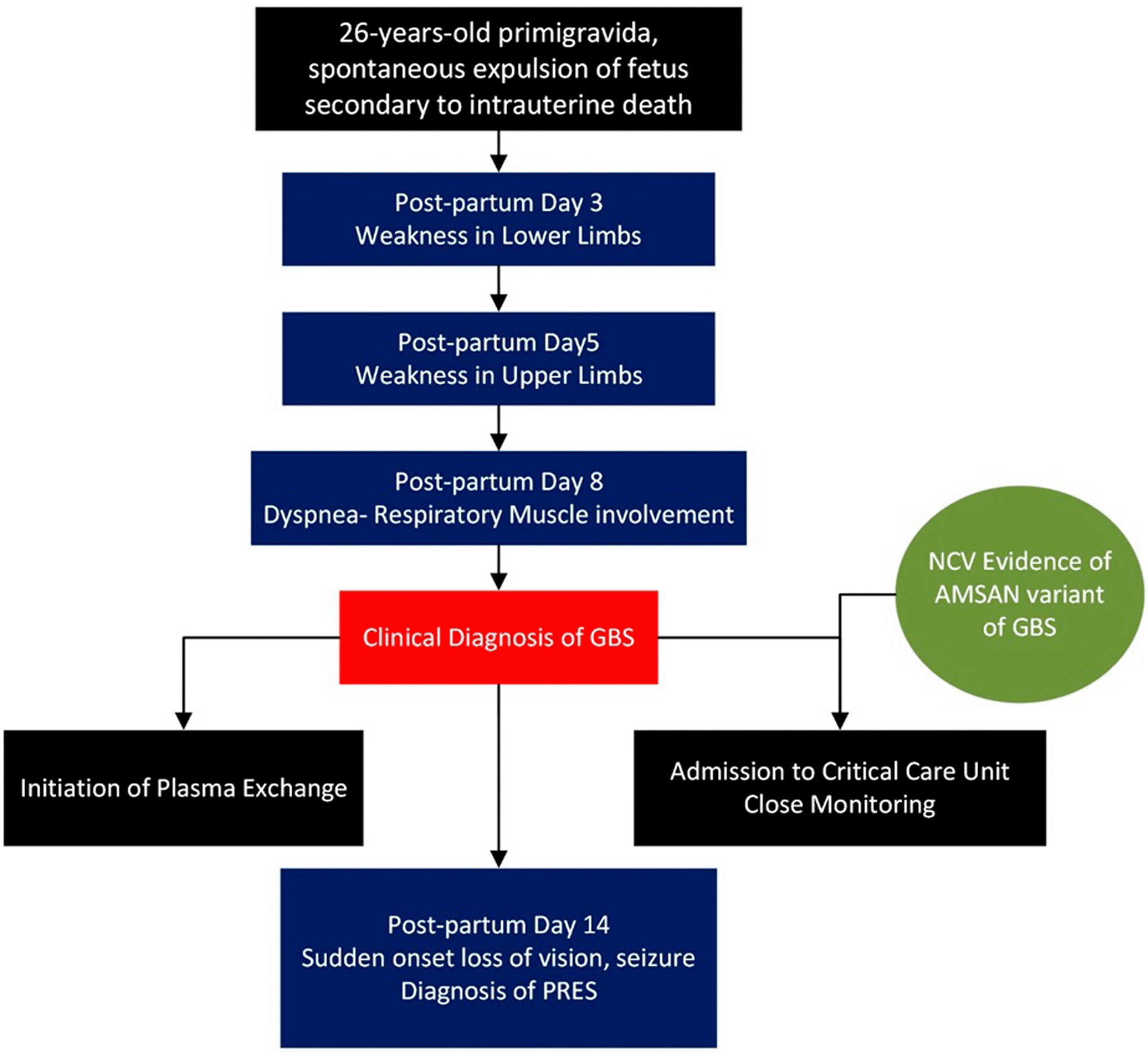
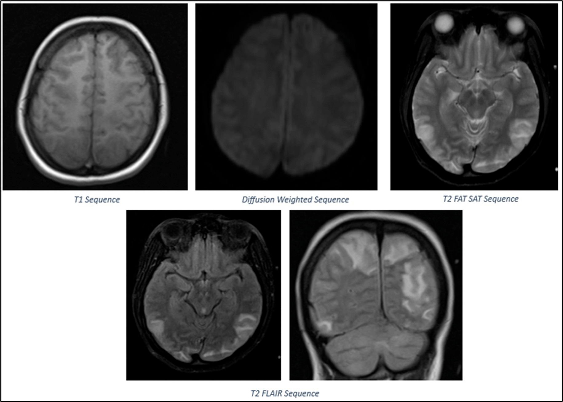
The patient was started on emergency therapeutic plasmapheresis and further managed in the intensive care unit. The patient was intubated, and lung-protective ventilation was initiated in view of worsening respiratory distress. Cabergoline was initiated to suppress lactation. On postpartum day 14, she developed sudden onset loss of vision, headache, raised blood pressure of 200/110 mmHg and two episodes of generalized tonic- clonic seizures. Ophthalmic examinations revealed normal extraocular movements, reactive pupils, papilledema, and absent perception of light bilaterally. Brain MRI revealed cortical and subcortical T2/FLAIR hyperintensities with edema in the bilateral high frontal, posterior parietal, occipital regions, and bilateral cerebellar hemispheres, consistent with PRES, as shown in Figure 4. She was treated with anticonvulsants and antihypertensives. The patient regained vision over the next two days. The patient underwent two more cycles of plasmapheresis. Simultaneously, the patient underwent limb and chest physiotherapy. The patient was discharged after one month of hospitalization. The patient was able to walk with support. The timeline of the patient’s course in the hospital is presented in Figure 1.
GBS is a set of autoimmune diseases characterized by polyradiculoneuropathy. It is typically preceded by infectious diseases or immunological stimulation that triggers autoimmune reactions in peripheral nerves.5 Our patient did not report infection days or weeks prior to symptom onset. Her symptoms developed post-delivery, and there was no history or records during the antenatal period that were suggestive of GBS prior to delivery. Moreover, there is no evidence to suggest that our patient had GBS prior to delivery, which may have resulted in antepartum stillbirth. A thorough history was taken to rule out the possibility of GBS in the last trimester of her pregnancy, and that her current condition could either be a relapse or a continuation of the disease process post-delivery.
In the United States, the most prevalent variant of GBS is acute inflammatory demyelinating polyneuropathy (AIDP), while in India, the most prevalent subtype is the axonal variant.2,6,7 Several pathophysiological factors contribute to the development of the various GBS subtypes. In the AMAN variant of GBS, cross-reactivity exists between the antibodies produced against microbial antigenic molecules and peptides in the nerve roots because they share similar epitopes, causing axonal degeneration. In AIDP, antibodies cross-react to specific epitopes in Schwann cells, resulting in demyelination. In AMSAN, the target peptides are located on sensory and motor neuronal axons, comparable to the etiopathogenesis of AMAN.8
GBS can develop in any trimester and postpartum period, but especially in the third trimester and first two weeks after delivery,9 as seen in our patient who developed symptoms 3 days after delivery.
From January 2015 to December 2017, a three-year retrospective observational study was conducted at a tertiary care hospital. The medical records of all pregnant and postpartum women diagnosed with GBS based on the clinical, laboratory, and electrodiagnostic criteria were evaluated. Fifty% of the women in this study presented during the postpartum period.10
Throughout the postpartum period, there is an increase in pro-inflammatory cytokines, which may account for the increased incidence of the disease. GBS is frequently reported to deteriorate during the postpartum period because of an increase in delayed-type hypersensitivity, which could also be the reason for our patient’s rapid deterioration of symptoms, in addition to having the AMSAN variant.9
Variants of GBS, as well as the ensuing involvement of respiratory muscles and autonomic dysfunction, contribute to maternal mortality.10 Approximately 20% of GBS patients experience respiratory failure that requires mechanical ventilation.11 By the time our patient reached our care, she had already progressed to respiratory muscle involvement. For patients with deteriorating respiratory and cardiac function, emergency care must include early mechanical ventilation, timely monitoring of vital signs, and cardiac function.12 It is recommended to routinely measure respiratory function, since all individuals with respiratory insufficiency do not have dyspnea. A single breath count of ≤19 indicates the necessity for mechanical ventilation, and other respiratory parameters may include the use of accessory respiratory muscles, vital capacity, and maximal inspiratory and expiratory pressure.11 At the time of presentation, our patient had a single breath count of 13 with accessory respiratory muscles requiring respiratory support. Early intravenous immunoglobulin (IVIG) or plasma exchange therapy has been shown to be effective and is crucial in patients with rapidly worsening weakness,3 and needs to be initiated before irreparable nerve damage develops.5 Most patients must be admitted to a critical care unit because one-fourth of them need respiratory support, and many have dysautonomia.3 In our case, emergency therapeutic plasmapheresis was initiated immediately after admission to the critical care unit on the same day.
As observed in our patient during the first week, cerebrospinal fluid analysis may be normal for the first seven days.5 Blood pressure variability is a key trait of GBS. The observed variances may be explained by changes in the feedback control caused by preganglionic sympathetic axonal demyelination or postganglionic axonal degeneration.4
PRES is a neurological disorder that typically manifests as visual impairment, seizures, and encephalopathy.13 In neuroimaging, it is characterized by bilateral parietal and occipital cortical/subcortical vasogenic edema, followed by frequent involvement of additional regions.14 Our patient with the AMSAN variant developed severe dysautonomia on post-partum day 14 with accelerated hypertension leading to PRES. Uncertainty surrounds the mechanism of PRES in the setting of GBS. Vasogenic edema may be caused by hypertension due to autonomic dysfunction, which exceeds the limits of cerebrovascular autoregulation.15 Paneyala S, et al. described a similar case of a woman who developed GBS shortly after delivery, but later experienced seizures due to PRES.16
The fundamentals of GBS care in pregnancy and the postpartum period include timely detection, multidisciplinary input, and swift plasmapheresis or IVIG administration. These measures improve outcomes for the mother and fetus.1 Several potential repercussions of GBS must be addressed and managed prior to patient release.11
| Views | Downloads | |
|---|---|---|
| F1000Research | - | - |
|
PubMed Central
Data from PMC are received and updated monthly.
|
- | - |
Provide sufficient details of any financial or non-financial competing interests to enable users to assess whether your comments might lead a reasonable person to question your impartiality. Consider the following examples, but note that this is not an exhaustive list:
Sign up for content alerts and receive a weekly or monthly email with all newly published articles
Already registered? Sign in
The email address should be the one you originally registered with F1000.
You registered with F1000 via Google, so we cannot reset your password.
To sign in, please click here.
If you still need help with your Google account password, please click here.
You registered with F1000 via Facebook, so we cannot reset your password.
To sign in, please click here.
If you still need help with your Facebook account password, please click here.
If your email address is registered with us, we will email you instructions to reset your password.
If you think you should have received this email but it has not arrived, please check your spam filters and/or contact for further assistance.
Comments on this article Comments (0)