Keywords
Hypoglossal nerve, stroke, tongue, Case report.
Hypoglossal nerve injuries are classified according to their anatomical localization in: Infranuclear, nuclear, and supranuclear. Supranuclear injuries can occur in cerebral cortex, corticobulbar tracts, internal capsule, cerebral peduncles, or in the pons, and most often caused by a stroke. These lesions usually do not generate a significant alteration of tongue motility due to the bilateral innervation of both nuclei from the cortex.
We present a case of a 43-year-old male with dysarthria, left central facial paralysis, and an important tongue palsy and deviation to the same side. Brain CT revealed a right frontotemporal stroke with little hemorrhagic transformation, and an EKG that showed auricular fibrillation. He received treatment with amiodarone and rivaroxaban was initiated when a second brain CT scan showed no evidence of hemorrhage.
This case is remarkable due the unusual presentation in a supranuclear lesion of the hypoglossal nerve. It is important to enrich the semiology and consider the possibility of cortical cerebrovascular events in patients with acute deviation of the tongue, even in the absence of involvement of other cranial nerves; or marked ipsilateral motor implication.
Hypoglossal nerve, stroke, tongue, Case report.
Spelling corrections have been made.
See the authors' detailed response to the review by Ashwini S. Hiremath
Understanding the diverse causes of hypoglossal nerve palsy, which can range from tumors and trauma to ischemia and demyelinating lesions, is crucial. These lesions can be categorized based on their location as infranuclear, nuclear, and supranuclear, providing a fundamental framework for further study and clinical practice.1,2
Infranuclear lesions, a rare occurrence, directly affect the hypoglossal nerve before it exits the medulla oblongata. On the other hand, nuclear lesions involve the motor nuclei of the hypoglossal nerve in the medulla oblongata. Isolated lesions at the nuclear level are uncommon and usually accompanied by lesions in other nearby cranial nerves due to their position in the brainstem and posterior course. These lesions cause a deviation of the tongue towards the opposite side of the affected one due to the predominant contraction of the healthy half of the tongue.3,4 Furthermore, they are usually associated with lower motor neuron signs such as atrophy and fasciculations.1
Supranuclear lesions, which occur in higher areas such as the cerebral cortex, corticobulbar tract, cerebellar peduncles, and pons, are a distinct category. In the case of tongue control, the lower and lateral part of the precentral gyrus, in the primary motor area, plays a significant role. Corticobulbar fibers originate from this area and provide bilateral innervation to the hypoglossal nuclei, except for the nuclei portion that innervates the genioglossus muscle, which only receives innervation from the contralateral cortex. This characteristic explains why supranuclear lesions are often silent, as the cortical hypoglossal fibers of the contralateral cortex to the lesion remain intact. The body's remarkable ability to adapt and compensate for such lesions results in a mild weakness of the tongue muscles, which, in most cases, may not be evident.5–7
We present the case of a 43-year-old male patient from Cartagena, Colombia, a right-handed motorcycle driver with a history of left distal radius fracture that required surgical management. The patient has no known cardiovascular history. He was referred from a primary care center due to a 6-hour clinical picture, which began upon awakening, consisting of dysarthria and deviation of the labial commissure to the left side. On admission, an electrocardiogram was performed, showing atrial fibrillation and elevated blood glucose levels.
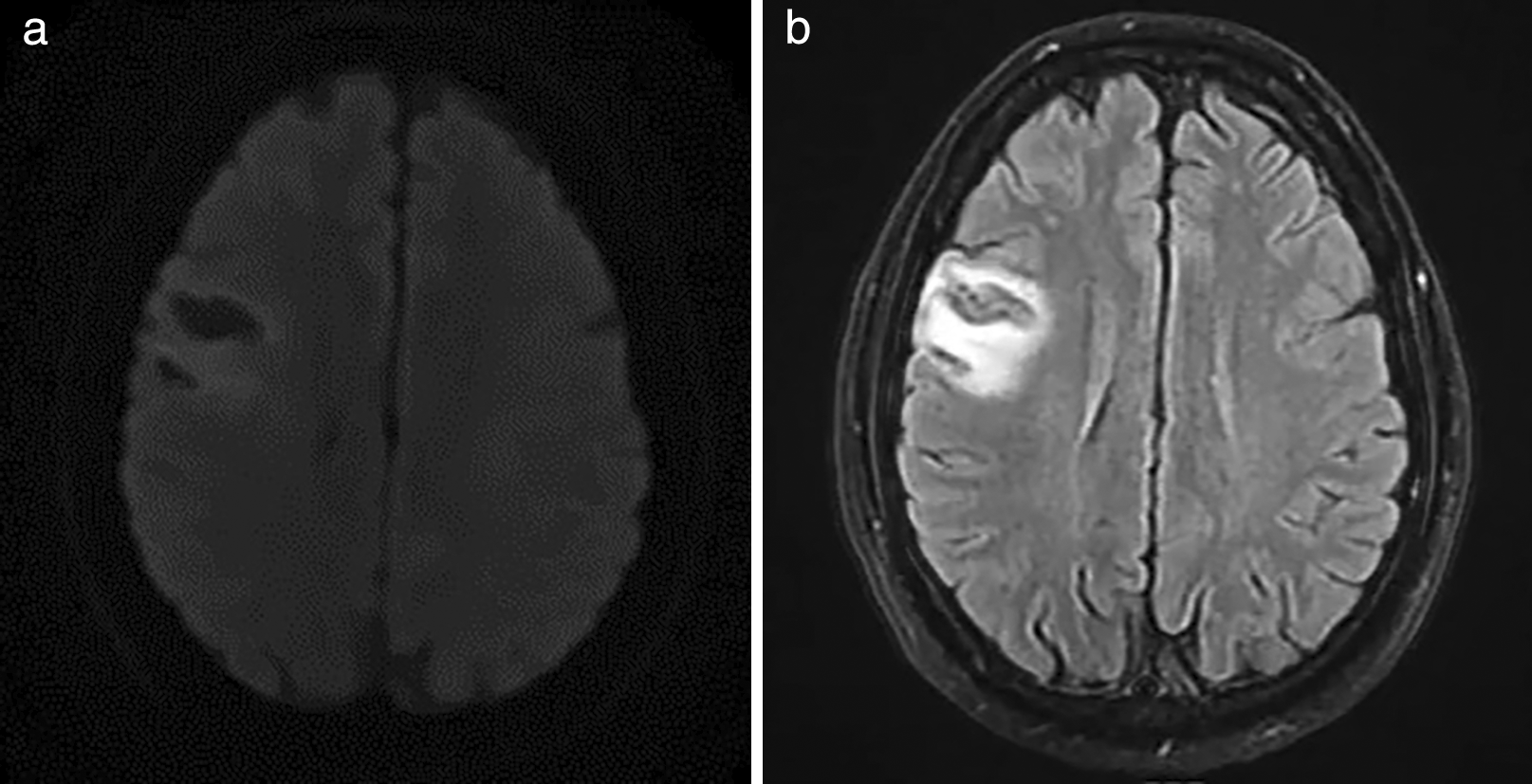
On physical examination, he was found to be tachycardic, with normal oxygen saturation and blood pressure. The auscultation showed arrhythmic heart sounds, and the rest of the general physical examination was without significant alterations. On neurological examination, the patient was alert and dysarthric; however, fluent, understood, named, and repeated, with preserved orientation. There was evidence of left central facial paralysis (Figure 1) and tongue deviation to the left (Figure 2). Left upper limb mono paresis 4/5 on muscle strength scale, with distal predominance at regional evaluation. Reflexes were symmetrical with normal gait and coordination. Complementary studies showed average blood cell count, moderate hypokalemia, persistently elevated blood glucose levels, and urinalysis with glycosuria and preserved renal function. Atrial fibrillation on electrocardiogram. Based on these findings, it was considered an acute stroke of probable cardioembolic etiology, which is confirmed with brain MRI that evidenced right frontal cortico subcortical lacunar image with gliosis in T2 and FLAIR sequence and that restrings in diffusion (Figure 3).

The echocardiogram reported mitral valve prolapse, mild insufficiency, and normal biventricular systolic diastolic function with preserved ejection fraction. Carotid ultrasound reported no alterations and 24-hour holter monitoring with basal atrial fibrillation rhythm with variable ventricular response throughout the study. Diagnosis of ischemic stroke of cardioembolic etiology was made, first-diagnosis atrial fibrillation o with rapid ventricular response CHA2DS2-VASc 1 and HAS-BLED 1 and de novo type 2 diabetes mellitus. At discharge, he continued antiarrhythmic treatment and insulin therapy with oral antidiabetic and direct oral anticoagulant.
The hypoglossal nerve nucleus is located in the tegmentum of the medulla oblongata, located between the midline and the dorsal motor nucleus of the vagus nerve. This thin-structured nucleus receives innervation from the precentral gyrus and is fed information from the inferior frontal cortex and the premotor cortex.6 This information is transmitted through corticobulbar tracts and distributed bilaterally to the hypoglossal nerve nuclei, resulting in unilateral lesions with a clinically insignificant effect.
A study involving 300 patients with unilateral cerebral infarctions showed that 29% of them had tongue deviation towards the side of the motor deficit. Most of these cases were associated with ipsilateral face and arm weakness. It was found that a lesion in the precentral gyrus in the tongue area could cause a contralateral palsy of the hypoglossal nerve, a phenomenon known as “pseudoperipheral tongue palsy.” In this type of situation, the tongue deviated towards the opposite side of the lesion, but atrophy or fasciculations were not observed, as took place in the present case.8
Disruption of the cortical lingual pathway plays a crucial role in developing dysarthria after cerebrovascular events due to the presence of a central mono paresis of the tongue.9,10
Supranuclear paralyzes of the hypoglossal nerve of vascular origin, which cause tongue deviation, tend to be accompanied by other signs such as hemiparesis or, less frequently, weakness in the facial, pharyngeal, or masticatory musculature of the same side. These presentations set up an opercular syndrome on the affected side.11 However, in our case, the presentation was atypical since a tongue deviation and facial weakness were observed, without marked hemiparesis, in the context of a unilateral supranuclear lesion.
We obtained written informed consent for publication of their clinical details and clinical images from the participants.
| Views | Downloads | |
|---|---|---|
| F1000Research | - | - |
|
PubMed Central
Data from PMC are received and updated monthly.
|
- | - |
Is the background of the case’s history and progression described in sufficient detail?
Partly
Are enough details provided of any physical examination and diagnostic tests, treatment given and outcomes?
Partly
Is sufficient discussion included of the importance of the findings and their relevance to future understanding of disease processes, diagnosis or treatment?
Partly
Is the case presented with sufficient detail to be useful for other practitioners?
Partly
Competing Interests: No competing interests were disclosed.
Reviewer Expertise: Paediatrics, General Medicine
Competing Interests: No competing interests were disclosed.
Reviewer Expertise: Stroke, Epilepsy, Headache.
Is the background of the case’s history and progression described in sufficient detail?
Yes
Are enough details provided of any physical examination and diagnostic tests, treatment given and outcomes?
Yes
Is sufficient discussion included of the importance of the findings and their relevance to future understanding of disease processes, diagnosis or treatment?
Yes
Is the case presented with sufficient detail to be useful for other practitioners?
Yes
Competing Interests: No competing interests were disclosed.
Reviewer Expertise: Stroke, Epilepsy, Headache.
Alongside their report, reviewers assign a status to the article:
| Invited Reviewers | ||
|---|---|---|
| 1 | 2 | |
|
Version 2 (revision) 08 Jul 24 |
read | read |
|
Version 1 25 Apr 24 |
read | |
Provide sufficient details of any financial or non-financial competing interests to enable users to assess whether your comments might lead a reasonable person to question your impartiality. Consider the following examples, but note that this is not an exhaustive list:
Sign up for content alerts and receive a weekly or monthly email with all newly published articles
Already registered? Sign in
The email address should be the one you originally registered with F1000.
You registered with F1000 via Google, so we cannot reset your password.
To sign in, please click here.
If you still need help with your Google account password, please click here.
You registered with F1000 via Facebook, so we cannot reset your password.
To sign in, please click here.
If you still need help with your Facebook account password, please click here.
If your email address is registered with us, we will email you instructions to reset your password.
If you think you should have received this email but it has not arrived, please check your spam filters and/or contact for further assistance.
Comments on this article Comments (0)