Keywords
Endometriosis, pelvic pain, etiology, animal model, Epithelial-mesenchymal transition, fibrosis
This article is included in the Manipal Academy of Higher Education gateway.
This article is included in the Endometriosis collection.
Endometriosis is a benign, estrogen-dependent, persistent chronic inflammatory heterogeneous condition that features adhesions caused by estrogen-dependent periodic bleeding. It is characterised by a widely spread fibrotic interstitium that comprising of fibroblasts, myofibroblasts, collagen fibres, extracellular proteins, inflammatory cells, and active angiogenesis found outside the uterus. Thus, fibrosis is recognized as a critical component because of which current treatments, such as hormonal therapy and surgical excision of lesions are largely ineffective with severe side effects, high recurrence rates, and significant morbidity. The symptoms include dysmenorrhea (cyclic or non-cyclic), dyspareunia, abdominal discomfort, and infertility. The significant lack of knowledge regarding the underlying root cause, etiology, and complex pathogenesis of this debilitating condition, makes it challenging to diagnose early and to implement therapeutic approaches with minimal side effects presenting substantial hurdles in endometriosis management. Research on understanding the pathogenesis of endometriosis is still ongoing to find biomarkers and develop non-hormonal therapeutic approaches. Current clinical research indicates a close relationship between endometriosis and fibrosis, which is thought to be tightly linked to pain, a major factor for the decline in the patient’s quality of life but little is known about the underlying pathophysiological cellular and molecular signaling pathways that lead to endometriosis-related fibrosis. The available experimental disease models have tremendous challenges in reproducing the human characteristics of the disease to assess treatment effectiveness. Future translational research on the topic has been hindered by the lack of an adequate fibrotic model of endometriosis emphasizing the necessity of etiological exploration. This review article’s goal is to examine recent developments in the field and pinpoint knowledge gaps that exist with a focus on the development of novel fibrotic mouse models for the early diagnosis and treatment of endometriosis and how this knowledge aids in the development of novel anti-fibrotic treatments which opens fresh avenues for a thorough investigation and extended research in the field of endometriosis.
Endometriosis, pelvic pain, etiology, animal model, Epithelial-mesenchymal transition, fibrosis
Endometriosis is an inflammatory disorder dependent on estrogen and results from the implantation of viable endometrial, epithelial, and stromal cells (a lesion) outside of the uterus, often associated with infertility.1 The condition affects at least 10% (~247 million) of women worldwide, with Asian women reporting the highest prevalence with over ~ 42 million girls and women from India2,3 which can hurt the outcome of IVF treatments.4,5 Endometriosis can result in severe dysmenorrhea, dyspareunia, menorrhagia, excruciating pelvic/abdominal pain, and eventually lead to infertility due to considerable damage to the structure and function of reproductive organs, even compromising entire bodily systems due to the accumulation of fibrotic tissue.6 Even after several decades of research, the etiology is still unclear and dependent on a few key assumptions. The retrograde menstruation theory, embryonic remnants, coelomic metaplasia, immune dysfunction, inflammation and oxidative stress, hormones, dysfunctional apoptosis, microbiome, metabolomics, endocrinology, and genetic expression differences are the main theories of pathophysiology2,7 Currently, the most widely recognized theory explaining how endometriosis begins is “Sampson’s theory”, which holds that the misplaced viable endometrium-like tissue is transferred onto the pelvic peritoneum by retrograde menstruation via the fallopian tubes.8 The diagnosis can take 4 to 11 years due to difficulties in classifying and identifying the disease as well as its peculiar symptoms, as well as a lack of diagnostic indicators.9 The disease’s variable severity can be due to superficial peritoneal, deep peritoneal (DIE), ovarian endometriomas, extra-abdominal endometriosis, and iatrogenic endometriosis.10 According to Maddern et al., endometriosis has a significant impact on a person’s quality of life, their reproductive health, and society at large. Therefore, a thorough understanding of the mechanisms underlying the origin and evolution of endometriosis is crucial for managing and evaluating the risks associated with the condition.11 Although imaging procedures such as transvaginal ultrasonography (TVUS), and magnetic resonance (MRI) imaging are common diagnostic tools, the gold standard diagnosis method for endometriosis remains the histological investigation of lesions obtained after laparoscopic surgery.12 Endometriosis pelvic adhesions have also had a major impact on the American Society of Reproductive Medicine (rASRM) categorization score approach.13,14 It does not, however, account for the pathology-based staging that is based on the normal course of endometriosis and fibrosis aspect which includes epithelial to mesenchymal transition (EMT) or mesenchymal to epithelial transition (FMT), Smooth muscle metaplasia (SMM). This means that patients with fibrotic characteristics and adhesions may fail to get a reliable diagnosis.15
Retrograde menstruation is prevalent in healthy women and only a small population of women develop the condition contributing to the understanding of complex mechanisms that underlie the onset of this challenging condition.16 While 90% of women of reproductive age undergo retrograde menstruation to the pelvic cavity, only 10% of them go on to develop endometriosis. This suggests that the onset and progression of the disease in the peritoneal cavity depend on additional relevant factors.17 This entails understanding how cells from the normal lining of the uterus find atypical locations, multiply excessively, escape immune and apoptotic processes, and acquire the necessary blood supply and nutrients that ultimately result in the formation of aberrant lesions.18 None of the available theories fully captures the intricacies of fibrotic endometriosis, emphasizing the need for additional studies to identify the pathophysiology of endometriosis.19 Therefore, it is imperative to investigate and characterize the molecules involved in the emergence of this crippling disease, including the acquisition of characteristics in these normal endometriotic cells such as increased proliferation, invasion, vascularization, angiogenesis, and migration.20 The formation, invasion, and angiogenesis of fibrotic ectopic lesions have been associated with disrupted immunoregulatory processes and a variety of inflammatory markers, including immune cells, cytokines, chemokines, matrix metalloproteinases, and other substances associated with the immune system.21,22 Thus, a thorough understanding of the mechanisms underlying the origin and evolution of endometriosis is crucial for managing and evaluating the risks associated with the condition. In this review, we describe and comment on existing endometriosis models, research gaps, proposals, or ideas for the most essential and underappreciated aspect of the condition of EMT and fibrosis, and how focused research on it can lead to novel therapeutics.
We conducted an electronic database literature search of PubMed and Google Scholar for published research articles on endometriosis and endometriotic animal models. “Endometriosis”, “endometriosis mice model”, “Primate model of endometriosis”, “endometriotic patients”, and “endometriosis-associated fibrosis” were the search terms that were employed. Articles with thorough experimental data and definitive results were considered for inclusion; those with inconclusive research findings were eliminated. We incorporated clinical trials, surveys of endometriosis-affected women, and observational and experimental studies including animal research as references. Research written in languages other than English was not considered. All the graphics were prepared using Biorender software (BioRender.com).
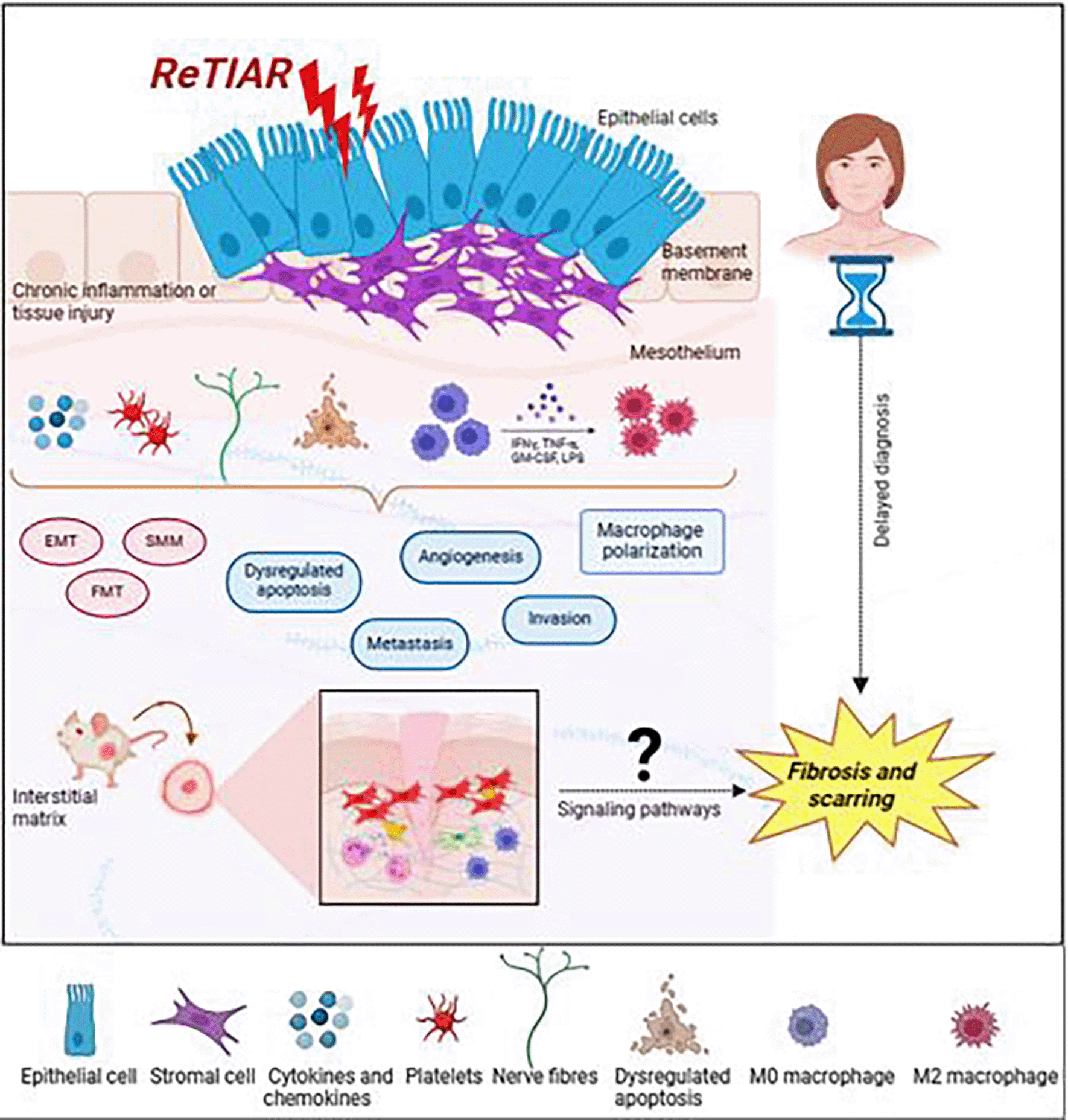
Endometriosis is characterized by the persistent occurrence of fibrosis and myofibroblasts within endometriotic lesions, which play a critical role in the disease’s development, making fibrosis a molecular hallmark of endometriosis.23 Significant scarring is commonly linked to endometriosis.23 Although the initial onset of endometriosis is associated with the existence of endometrial stromal and glands in abnormal locations, often the endometrial components are soon replaced by fibrotic and smooth muscle components.24 Rectovaginal nodules frequently display glandular epithelium embedded deeply within fibromuscular tissue, devoid of any surrounding stroma.25 Additionally, in 40% of ovarian endometriomas, there is no detection of endometrial epithelium, and the interior of the cyst is covered solely by fibrotic tissue.26 Despite being a crucial pathological feature of the disease, pelvic adhesions generally lack any endometrial components.14 These adhesions contribute to the pathology of some common symptoms of endometriosis, including chronic pelvic pain, deep dyspareunia, and infertility, which may be influenced by pelvic adhesions.14 In fibrosis of organs like lungs, liver, and kidney, involvement of TGF-β signaling pathway is well documented.27 TGF-β is an influential growth factor and a chemical that attracts monocytes and is capable of triggering fibrosis and angiogenesis in abnormal growths and promoting the advancement of endometriosis.28 In comparison to normal women, the peritoneal fluid of stage III and IV endometriosis patients exhibits greater levels of TGF-β.29 The process by which endometriosis progresses to a malignant condition remains unknown. However, continuous inflammation, immunological dysregulation, and fibrosis, most likely caused by iron-induced oxidative stress, may lead to genetic changes, which may lead to a malignant feature.30,31 Fibrosis is believed to be linked to pain, which is the disease’s most common symptom and the principal cause of the patient’s poor quality of life.32 If the underlying mechanisms are uncovered, they may explain why the disease’s morphological characteristics do not match the extent and nature of fibrosis-induced pain sensations.33 Fibrotic tissue is defined by excessive development of extracellular matrix (ECM) components inside and around inflamed or damaged tissue, and it is a typical and significant phase of tissue repair in all organs. Fibrosis involves activated platelets, macrophages, and myofibroblasts, which result in increased collagen deposition.34 Furthermore, fibrosis occurs with the transition from epithelial to mesenchymal cells in variety of malignancies, which is associated with poor prognosis.35 Fibrosis and smooth muscle metaplasia are two of the main characteristics of endometriosis in women; fibrosis is found surrounding the endometriotic tissue, and the degree of fibrosis is connected with the degree of smooth muscle metaplasia.36 Based on these data, we postulate that fibrotic-based EMTs’ involvement in chronic inflammatory responses may be a factor in the invasive nature of endometriotic lesions. Also, angiogenesis which stimulates endothelial function, vascular permeability, and the emergence of experimental endometriosis, is commonly associated with this heightened invasive and metastatic potential.37 Endometriotic lesions are thought to be “wounds” that undergo repeated tissue injury and repair (ReTIAR) through the processes of smooth muscle metaplasia (SMM), fibroblast-to-myofibroblast trans-differentiation (FMT), and epithelial-mesenchymal transition (EMT). This process ultimately leads to fibrosis and is a common feature of all endometriotic diseases.31,38 Epithelium-mesenchymal transition (EMT) is characterized by polarised, stationary epithelial cells change into highly motile mesenchymal cells.39 This makes it possible for solitary cells to pass through the basement membrane, grow invasively, and metastasize by both intra- and extravasation. Sampson’s implantation theory states that each of these occurrences is necessary for the development of an endometriotic lesion.8
Regretfully, due to differences in opinion over the etiology of the disease, the EMT route has received less attention in the context of endometriosis than it does in cancer research. In recent times, most research on EMT in endometriosis focuses on tissues; very few examine the specific transcription factors involved in EMT signaling that are present in endometriotic cells.40,41 EMT-related processes in endometriosis have been reported to be much higher in ectopic endometrial lesions than in eutopic endometrium.42 As a result, endometriosis etiology may involve EMT. We speculate that EMT and fibrosis processes may be involved in the evolution of endometriosis, given its chronic nature and the possibility of it leading to fibrotic adenomyosis. Additionally, in their mouse model of endometriosis, Modi et al., discovered significant inflammation but no histological fibrosis and no epithelial-mesenchymal transition concluding EMT and fibrosis are not typical in endometriosis.43 Consequently, studies on the molecular pathways based on EMT or possible targets for therapeutic intervention for EMT and fibrosis in endometriosis were stopped due to the unavailability of a animal model of endometriosis that mimicked the human condition. Endometriosis research is mostly based on non-human primate or rodent models due to the apparent limitations and ethical concerns of human experimentation. The available mice models have aided in the investigation into several aspects of the disorder, such as early disease phases,44 steroid hormone involvement,45 host inflammatory mechanisms,46,47 oxidative stress,48,49 neuro-angiogenesis,50 and infertility51 were also studied in mice. However, there is a paucity of information on the development of pre-clinical models that define clinically effective endpoints such as fibrosis or EMT-induced metastasis. Furthermore, 50-70% of drugs that advance to phase II and III of clinical trials are unable to show efficacy.52 This suggests that there are not enough disease models to investigate crucial biological processes. To conclude, we want to highlight that an ideal animal model developed specifically to study endometriosis should include the same cellular and pathophysiological pathways and clinical behaviours that are observed in endometriotic patients, such as fibrosis leading to scar formation and EMT linked to invasion and metastasis.
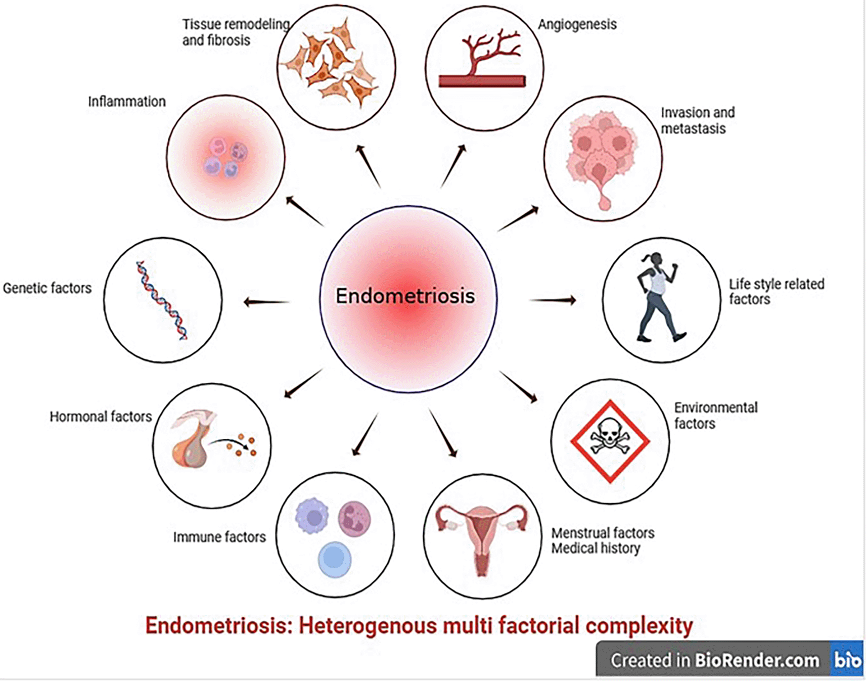
According to Greaves et al., endometriosis is currently being studied using two basic approaches: human-based in vitro samples and experimental in vivo animal models. The first type involves experimental in vitro research using tissue biopsies and fluids obtained from resected lesions or aspiration biopsies, such as endometrial and peritoneal explants, endometriotic cell lineages, primary endometrial stromal cells, endometrial stem cells, and immune cells.53 In vivo animal models are essential for assessing drug candidates and for preclinical trial testing. Our knowledge of the early phases of disease development, including the effects of peritoneal microenvironment, inflammatory responses, and steroid responsiveness has improved because to these models.54 For several reasons, it is challenging to create in vitro or in vivo models that mimic or represent the salient characteristics observed in endometriotic patients, such as EMT or fibrosis. One, the condition is multifactorial, heterogeneous complexity, as none is certain of the condition’s onset or duration. Second, there are many disease characteristics associated with endometriosis, including peritoneal, deep infiltrative lesions, and ovarian endometrioma. Lastly, endometriosis cannot be modelled based on a particular pathophysiological mechanism.55 Furthermore, endometriosis has been connected to genetic,56 immunological,57 environmental,58,59 and hormonal changes such as progesterone resistance60 and estrogen dependency61 further posing challenge in creating a suitable animal model (Figure 1).

One of the most significant obstacles in endometriosis research is the lack of reliable animal models. Ideally, a disease model should mirror human disease while also allowing researchers to investigate the effects of intrinsic (e.g., genes) and extrinsic (e.g., environment) factors on disease progression. Many previous studies linked fibrosis secondary to the development of endometriosis and there has not been much research on the study of fibrosis. Based on research from animal models of the condition, it became clear that a percentage of women receiving hormone therapy in human trials were not responding to these drugs62 requiring surgical lesion removal to alleviate symptoms. Women may have endometriotic lesions that have progressed to a fibrotic state by the time they seek medical attention, rendering treatment ineffective. Significant evidence supporting the process of fibrosis comes from in vitro experiments conducted on humans and from in vivo higher vertebrates such as baboons.
Despite a recent surge in endometriosis research, the underlying pathobiology of the disease remains poorly known, implying that animal models of the disorder are crucial for future studies in this field. Non-human primates and higher vertebrates are regarded to be potential candidates for disease research due to their anatomical resemblance to human reproductive organs.63 Controlled experimental investigations on humans are limited because assessing disease prevalence and development necessitates numerous laparoscopies, which are challenging for a variety of reasons. Though endometriosis occurs spontaneously in humans, human investigations have been limited for ethical and practical reasons which is one of the primary reasons being the difficulty of studying the disease. As a result, understanding the mechanisms that cause this disease requires the use of an appropriate animal model. Endometriosis has long been investigated in both primate and non-primate animals. The spontaneous endometriosis of the baboon64–66 limitation is that baboons have vast and effective mechanisms for clearing and regenerating their peritoneum66 the rhesus monkey67,68 where limitation is the significance of peritoneal cysts in endometriosis pain and discomfort was not investigated. The cynomolgus monkey69,70 has been described, with the limitations that deep lesions were difficult to diagnose and time course changes in the condition were not investigated. Though non-human primates are an excellent model for studying endometriosis, they are expensive to maintain and are extremely sensitive to captivity. Furthermore, spontaneous endometriosis occurs at a low frequency, limiting the use of primates in research.71 However, because research facilities for primates are restricted, non-primate experimental animal species, such as mice or rats, are regarded to provide an ideal first-line technique for investigating the etiology of this mysterious disease.
Every menstrual cycle, endometriosis is characterized by the development of new lesions and the advancement of pre-existing lesions. Therefore, additional research is required to comprehend the endometriosis lesion’s natural course and their gradual development.72 There is evidence of gradual lesion clearing, but only a small number of studies using mouse models of endometriosis have studied disease induction and regression.72,73 It is unethical to perform many laparoscopies on endometriosis patients to monitor disease progression. So, longitudinal studies of lesion formation and progression can considerably increase the translational efficiency of pre-clinical model of endometriosis.72
Mice are the most popular experimental animal models due to their ease of gene manipulation, availability, and handling, tissue similarity in vivo, small size and large litter, which make them cost-effective, and their relatively short gestation, which allows for transgenerational examination.74 Based on the vast majority of already available research publications, two types of mice models have been successfully used to implant endometriotic lesions. The first approach involves suturing, whereby human endometriotic implants are surgically auto-transplanted into the peritoneum of immunocompromised mice.75–77 The second approach involves intraperitoneal or subcutaneous implantation of autologous uterine segments into the peritoneum of recipient mice from a syngeneic donor.50,54,78 Although there are numerous reports describing the spontaneous attachment, growth, and proliferation of endometriotic lesions, these lesions do not accurately reflect human endometriosis because they do not exhibit characteristics like chronic, persistent fibrosis for internal scarring, or invasiveness based on EMT. Moreover, the animal models provide data on the inflammatory processes generated by implanted lesions rather than those caused by endometriosis. Rats can only produce superficial lesions, which are the most fundamental and possibly least clinically significant types of lesions. The inability of any study to recreate fibrotic endometriotic lesions may account for the failure of rat models to yield data relevant to the pathophysiology and treatment of human endometriosis. This situation demonstrates that the preclinical animal studies that have been established are not transferable.79 Many studies using rodents as a model for endometriosis have looked at the gene expression patterns of ectopic tissue deposits in rats in an attempt to correlate them with human endometriotic lesions. Chronic inflammation, angiogenesis, and extracellular matrix remodeling have all been found to be common pathways.80–82 While some aspects of the disease are replicated in the rodent model, all the modifications involve suturing uterine fragments (endometrium plus myometrium) to different sites, which does not accurately represent the formation of lesions from those shed endometrial tissue or the dissemination of menstrual tissue into the peritoneum. It is noteworthy that, particularly in terms of understanding its pathophysiology and treatment options, the current rodent models have not been successful in yielding findings that apply to human endometriosis. Therefore, fibrosis a mostly disregarded component of human endometriosis be taken into consideration.83,84 However, efforts to translate the results into humans were unsuccessful in offering effective endometriosis treatments. Therefore, developing novel animal models that mirror the continuous fibrotic process seen in endometriotic patients is essential in improving our understanding of the disease. An increasing amount of research has recently brought attention to the role that fibrosis plays in clinical-grade endometriosis. On the other hand, little is known about fibrosis treatment strategies. Therefore, it is critical to develop a fibrotic mouse model of endometriosis, elucidate the regulatory processes behind fibrosis in endometriosis, and identify more precise specific biomarkers for the disease. These markers can also be utilized to find effective therapy targets and identify endometriosis in its early phases (Figure 2).

To mimic the fibrotic scarring observed in endometriosis, many endometriotic fibrotic animal models have been developed (Table 1). Furthermore, new in vivo models that use stromal cells generated from menstrual blood have been created to study endometriosis; these models show enhanced endometriotic cell migration and proliferation.60 Many cues, including estrogen stimulation, may trigger the epithelial-mesenchymal transition.85 Furthermore, estrogen-induced EMT of Ishikawa cells promotes adenomyosis.86 However, it remains still unknown how estrogen causes EMT in endometriosis at the molecular level. To prevent fluctuations in the mice’s estradiol levels during the estrous cycle, the majority of established mouse models use ovariectomized mice.87–90 As a result, the steady availability of estradiol in the circulation may help to promote lesion establishment and growth. But this makes it impossible to research how estrogen-induced EMT in endometriosis affects fertility like in women with normal circulating estrogen. Therefore, studies of endometriosis produced in intact mice call for more research on the connection between ectopic tissue and fertility.
| Model | Fibrotic gene | Mechanism | References |
|---|---|---|---|
| Balb/C | CD41, TGF-β1, p-Smad3, CCN2, α-SMA, collagen I and LOX | EMT, FMT, SMM and fibrosis | 87 |
| C57BL/6 | VEGF, PCNA, COX-2, p-p65, collagen I, α-SMA, Fibronectin, FGFR2 and MVD, Platelets | Inflammation and fibrosis | 88 |
| Balb/C | CD41, VEGF, CD31 MVD, PCNA, p-p65, COX-2, TGF-β1, α-SMA, and collagen I | Fibrosis | 89 |
| EESCs from females | Thromboxane B2 (TXB2), thromboxane A2 (TXA2) | Fibrosis | 90 |
| C57BL/6 Klf11−/− | TGF-β and KLF 10 and 11 | Fibrosis | 91,92 |
| C57/BL6 Klf11−/− | COL1A1/Col1a1 | Fibrosis | 93 |
| C57BL/6 | Nrf2 | Fibrogenesis | 94 |
| Balb/C | CD41, PCNA, VEGF, CD31, collagen I, a-SMA, and LOX | FMT and fibrosis | 95 |
| Balb/C | β-catenin | Fibrosis | 96 |
| C57BL/6 | PIM2 | Glycolysis and fibrosis | 97 |
| C57BL/6 | TGF-β/ERK | Fibrosis | 98 |
| KLF11−/− and Smad3−/− | KLF11 and TGF-βR | Fibrosis | 99 |
In endometriotic lesions, it is known that TGF-β family members, Notch receptor, and bioactive sphingolipid sphingosine 1-phosphate (S1P) cause tissue fibrosis and change signaling pathways.91 It has been found that NF-κB is activated in endometriotic lesions and peritoneal macrophages, which are essential for the inflammation associated with endometriosis. It has been demonstrated that inhibiting NF-κB lowers the development and progression of endometriosis in women as well as its associated symptoms.92 Estrogen can promote the formation and dissemination of endometriosis ectopic lesions by upregulating the expression of the transcription factor Slug in ectopic endothelial cells and inducing the epithelial-mesenchymal transition.86 Fibrogenesis in endometriosis may be facilitated by aberrant Wnt/β-catenin pathway activation and reversed by blocking the Wnt/β-catenin pathway.93 TGF-β1 may stimulate the expression of N-cadherin, OCT4, and Snail in ectopic stromal cells, implying that TGF-β1 facilitates cell invasion.94 The AKT and ERK signaling pathways may work synergistically to promote the formation of deep endometriotic lesions by increasing endometriotic stromal cell proliferation in a fibrotic milieu in vitro.95
By inducing EMT and FMT in endometriotic lesions, platelet-derived TGF-β1 stimulates smooth muscle metaplasia (SMM) and fibrosis.96 Evidence suggests that EMT induces fibrogenesis in addition to increasing cellular invasiveness. For instance, TGF-β1/Smad3 signaling pathway, which is driven by platelets, is known to induce EMT and FMT in endometriotic lesions, which eventually results in SMM and fibrosis.96 Targeting TGF-β1 may be an effective strategy to prevent fibrosis and adhesion formation since endometriotic cells release TGF-β1, which induces ECM disorganisation and fibrosis in the tissues of ovarian endometriotic patients.97 Oxidative stress has been linked to the ADAM17/Notch signaling pathway and perhaps fibrosis, according to a study done on endometriosis patients.98 Furthermore, it is known that endometriotic cells of endometriomas express Smad2, Smad3, and Smad4 (as well as their phosphorylated forms), which causes fibrosis and adhesion to ovarian tissues, suggesting a role for TGF- β1/Smad signaling.97 FOXP1 uses Wnt signaling to increase fibrosis in endometriosis.99 Through their effects on tissue repair, senescence, EMT, FMT, and proliferation of fibroblasts/myofibroblasts, mutations in TP53, PTEN, ARID1A, PIK3CA, KRAS, and PPP2R1A appear to hasten the development and fibrogenesis of endometriosis.100 A significant increase was observed in the mRNA levels of α-SMA, vimentin, N-cadherin, fibronectin, PAI-1 (Serpine1), Snail, Slug, and LOX.101 Growth factors such TGF-β1, PDGF, EGF, and CTGF are released by activated platelets in lesions, facilitating fibrogenesis in endometriotic patients with deep endometriosis and ovarian endometrioma.102 NR4A1 is a novel pro-endometriotic transcription factor that accelerates the development of endometriosis.103 HOXC8 stimulates TGF-β signaling, which affects adhesion, cell proliferation, migration, and ovarian endometrioma.104 FAK intracellular non-receptor tyrosine kinase mediates a series of processes in the development of endometriosis, including cell adhesion, inflammatory response, and fibrosis signaling in patients with endometriomas.105 In ectopic ESCs derived from retrograde menstruation, PGE2/thrombin is known to induce modifications such FMT and EMT, which are linked to fibrotic changes in the lesions.41 Through EMT and FMT processes, proinflammatory substances such PGE2 and thrombin in retrograde menstrual fluid have been jointly implicated in generating endometriosis fibrosis in endometriotic patients. This suggests potential targets for treatment to mitigate fibrosis.41 Apart from the fibrotic and EMT markers, numerous processes, such as pyroptosis, NLRP3 inflammasome, and deregulation of the long noncoding RNA MALAT1 are identified to cause fibrosis in endometriotic patients.87 It has been discovered that reducing the number of lesions by targeting inflammatory molecules like IL-8 also known to reduce fibrosis and adhesions, highlighting the potential for disease-modifying therapy.106 While existing research has shed light on the genes involved, there is still a potential to uncover the intricate downstream signaling networks that govern this complex disease. As a result, sophisticated additional approaches, such as knockout models that incorporate high throughput RNA sequencing and omics methodologies should be emphasised to further validate the role and mechanism of fibrotic markers in the development of fibrosis, providing solid proof for the discovery of drugs that hinder, terminate, and reverse fibrosis progression and benefit endometriotic patients.
Endometriosis is a common benign gynaecological disease with a high propensity for migration and invasion. The cell-to-cell or cell-ECM connections allow the cells to migrate, invade, and proliferate in new locations. MMPs are linked to adhesion, invasion, and the severity of endometriosis. This indicates that MMPs have a role in extracellular matrix remodeling, which is necessary for the development of ectopic endometriosis lesions.107 They are also significantly higher in the endometrial and peritoneal fluid of endometriosis patients.108,109 Matrix metalloproteinases (MMPs) are a family of enzymes that are mostly found in the endometrium’s functional layer. They are secreted by the resident immune cells and stromal fibroblasts, which facilitate the remodeling of the extracellular matrix including collagen, elastins, and other glycoproteins and endometrial disintegration during menstruation. Tissue inhibitors of matrix metalloproteinases (TIMP) are endogenous antagonists that reduce MMP overexpression, and ovarian steroid hormones are known to control MMP activity.110 For early clinical studies of EMT, the nude mouse is a suitable model, particularly for the identification of MMP-2 and TIMP-2, proteins that seem to play a significant role in the pathophysiology of EMT. It has been found that estrogen specifically increases MMP-2 expression to encourage ectopic implantation of the endometrium. Progestin, on the other hand, can suppress TIMP-2 expression, increasing the MMP-2/TIMP-2 ratio and increasing the invasiveness of ectopic endometrium to facilitate implantation.111 In ovarian endometriosis, MMP7 facilitated the epithelial-mesenchymal transition; EGF increased MMP7 expression by activating the ERK1-AP1 pathway.112,113 MMP14 affects the development and function of invadopodia, which in turn modulates mesenchymal cells’ capacity for invasion and migration.114 MMP-2 and MMP-9, two important enzymes involved in the destruction of diverse types of ECM, have been linked to the development of endometriosis by regulating endometrial cell invasion.115 MMP-2 and MMP-9 have been shown to operate as both biomarkers of EMT and triggered factors that contribute to the progression of EMT.116 As a result, we hypothesize that MMPs may be crucial in controlling the endometriosis-related EMT process. However, further research is required to fully understand the connection between MMPs and the epithelial-mesenchymal transition in endometriosis, as there are not enough comprehensive studies on the subject. Despite this, it is apparent that MMPs play a crucial role in collagen production, which is necessary for endometriosis fibrosis to develop gradually.107 These findings suggest that there may be a precise equilibrium between collagen synthesis and breakdown, which should be investigated further.
Endometriosis is an underdiagnosed chronic inflammatory disease that affects millions around the world. The primary explanation for endometriosis growth is the transplantation of living endometrial cells that are refluxed after menstruation, thereby attaching to and invading other pelvic organs developing inflammation and fibrosis.2 Despite its broad incidence and importance, endometriosis research has significant limitations.117 The gaps include a lack of understanding of the disease’s etiology, a delay in diagnosis that necessitates invasive treatments, and the difficulties of integrating electronic health records for research, which aids in identifying potential therapeutic tools and reminds us to look beyond endometriotic lesions.118 50 to 70 percent of endometriotic drugs that advance to phase II and III in clinical trials are unable to show efficacy, suggesting an unfulfilled research gap in developing appropriate animal models.119 Endometriosis fibrosis shares characteristics with other fibrotic conditions, including increased myofibroblast and smooth muscle cell activity, high levels of fibrotic-associated growth factor and protein production, epithelial to mesenchymal transition, and collagen deposits.15 Molecular hallmarks of endometriosis include immunological dysregulation, ER expression, progesterone resistance, chronic inflammation, angiogenesis, and epigenetic changes. There is substantial evidence that fibrosis is a molecular characteristic of endometriosis etiology.15 Interestingly, fibrosis as a histologic feature in lesions can progress, most likely due to repeated tissue injury and repair caused by inflammation-induced recurrent menstrual bleeding31,120 Thus, a thorough understanding of the disease process is required for progress in the fields of biomarker identification and nonhormonal therapy. Fibrosis may impair drug administration and efficacy. Rather, a study into the mechanisms that resolve fibrosis will uncover new possibilities by discovering new targets for pharmacologically regulating the condition, notably in the pharmacology of multi-component medications.100,121 Because EMT-induced fibrosis is numerous and diverse and plays vital functions in various human body systems, robust longitudinal studies are required to [1] Confirm biomarkers and underlying mechanisms linked with fibrosis progression, providing insights into disease causes and potential diagnostic or prognostic tools. [2] To investigate temporal dynamics to record the advancement of fibrosis over time, allowing researchers to better comprehend its development from early stages to advanced forms thereby allowing early intervention and personalised treatment methods. [3] To investigate treatment efficacy, or the effectiveness of various interventions for fibrosis, to provide useful data on long-term outcomes and responses. [4] To better understand the natural course of fibrosis, including its variations among individuals, potential triggers, and variables influencing its progression, to create preventive and targeted therapeutics. [5] To determine whether endometriosis’s inflammatory environment participates in fibrosis. The proposed pathways of endometriosis participation in fibrosis require more investigation. Indeed, discovering fibrosis-specific therapies for endometriosis remains a significant issue. As a result, further inquiry and investigation are needed in the future. Finding the underlying etiology of endometriosis is made more difficult by the disease’s missing components, such as EMT and fibrosis, which have yet to be replicated in experimental rodent models that use heterologous or homologous endometriotic tissue. Filling in these gaps may lead to more accurate patient diagnoses, more effective medications, and a better knowledge of how the disorder affects women’s lives. Any treatments that help to reduce the fibrotic aspect of the disease will have far-reaching implications for the individual, the population, and the healthcare system. These thought-provoking articles show our reliance on carefully selected animal models to advance our understanding of endometriosis. They emphasize the multisystem character of pro-inflammatory mechanisms in endometriosis, as well as the need for researchers to think beyond the endometrial lesion. As we have come to expect, no single cause can explain endometriosis. Yet these studies give us optimism that more therapeutic methods to improve the quality of life for affected people are on the way. The breakthrough in the construction of models is promising research that could have substantial beneficial consequences for patients. Translating these research findings into clinical care will undoubtedly aid in shortening the extended delay to diagnosis and understanding the epidemiological underpinnings of the condition.
Endometriosis is a prevalent gynaecological disorder with a significant influence on female patients’ physical and emotional well-being due to its intrusive, and recurring nature. The association between endometriosis and fibrosis imbalance is poorly understood; additionally, EMT may play a role in the etiology of endometriosis through immunological regulation, pro-inflammatory cytokines, and other mechanisms. Clinical trials have shown that targeting EMT-induced fibrosis can help treat endometriosis, establishing a new research direction and theoretical foundation for the diagnosis and treatment of fibrotic endometriotic patients. Thus, it is vital to examine the molecular pathways that drive and sustain fibrosis in endometriosis using a novel fibrotic-based animal model, to discover new pharmacological targets and provide creative therapeutics for patients. Furthermore, the research connecting endometriosis and fibrosis has added a further complicating factor to the shared strategy for dealing with endometriotic patients with infertility, as well as a potentially essential concern in the counselling and management of the condition for those desiring future fertility. Well-designed longitudinal studies are needed to improve clinical decision-making in these contexts. Although gynecologic surgeons are aware of the complex role of fibrosis in the surgical treatment of endometriosis, the molecular pathways that relate fibrosis to endometriosis-associated pain and infertility remain unknown. More research is needed to better understand the clinical implications of fibrosis and identify it as a molecular marker of endometriosis etiology. A potentially important element to consider while counselling and managing endometriotic patients who plan to have children in the future. Well-designed longitudinal studies are required to make more informed clinical decisions in these contexts. Efforts should be focused on building trustworthy disease models that incorporate physiologically relevant cells, such as organoids and microfluidics. The continued creation of animal models to aid in understanding the processes of endometriosis development offers the best chance of creating therapeutic options to prevent or reverse this mysterious disease. This review aims to spark a debate on the need to revise present understandings by focusing on the fibrotic features of endometriosis pathogenesis. We believe that this approach will shed new light on the condition and suggest areas that need to be investigated further.
We would like to thank bioRENDER (biorender.com) for assisting in drawing all the graphics.
| Views | Downloads | |
|---|---|---|
| F1000Research | - | - |
|
PubMed Central
Data from PMC are received and updated monthly.
|
- | - |
Is the topic of the review discussed comprehensively in the context of the current literature?
Partly
Are all factual statements correct and adequately supported by citations?
Partly
Is the review written in accessible language?
Yes
Are the conclusions drawn appropriate in the context of the current research literature?
Partly
Competing Interests: No competing interests were disclosed.
Reviewer Expertise: Endometriosis; Translational study; Preclinical animal model
Alongside their report, reviewers assign a status to the article:
| Invited Reviewers | ||
|---|---|---|
| 1 | 2 | |
|
Version 3 (revision) 03 Dec 24 |
read | |
|
Version 2 (revision) 02 Oct 24 |
read | read |
|
Version 1 01 Jul 24 |
read | |
Provide sufficient details of any financial or non-financial competing interests to enable users to assess whether your comments might lead a reasonable person to question your impartiality. Consider the following examples, but note that this is not an exhaustive list:
Sign up for content alerts and receive a weekly or monthly email with all newly published articles
Already registered? Sign in
The email address should be the one you originally registered with F1000.
You registered with F1000 via Google, so we cannot reset your password.
To sign in, please click here.
If you still need help with your Google account password, please click here.
You registered with F1000 via Facebook, so we cannot reset your password.
To sign in, please click here.
If you still need help with your Facebook account password, please click here.
If your email address is registered with us, we will email you instructions to reset your password.
If you think you should have received this email but it has not arrived, please check your spam filters and/or contact for further assistance.
Comments on this article Comments (0)