Keywords
UniProt ID P21453, S1PR1, sphingosine 1-phosphate receptor 1, EDG1, antibody characterization, antibody validation, western blot, immunoprecipitation, immunofluorescence
This article is included in the YCharOS (Antibody Characterization through Open Science) gateway.
UniProt ID P21453, S1PR1, sphingosine 1-phosphate receptor 1, EDG1, antibody characterization, antibody validation, western blot, immunoprecipitation, immunofluorescence
Sphingosine 1-phosphate receptor 1 (S1PR1), is a G-protein coupled receptor which binds to its abundant ligand, sphingosine 1-phosphate, inducing intracellular signalling pathways in cell growth, differentiation, migration and trafficking.1–4 S1PR1 activation by sphingosine 1-phosphate is essential for neuronal events, and its dysregulation may contribute to the pathogenesis of Alzheimer’s disease.5,6 High-performing S1PR1 antibodies would facilitate S1PR1 research and uncover therapeutic strategies.
This research is part of a broader collaborative initiative in which academics, funders and commercial antibody manufacturers are working together to address antibody reproducibility issues by characterizing commercial antibodies for human proteins using standardized protocols, and openly sharing the data.7–9 Here we evaluated the performance of nine commercial antibodies for S1PR1 for use in western blot, immunoprecipitation, and immunofluorescence, enabling biochemical and cellular assessment of S1PR1 properties and function. The platform for antibody characterization used to carry out this study was endorsed by a committee of industry and academic representatives. It involves identifying appropriate cell lines with adequate target protein expression, developing or contributing equivalent knockout (KO) cell lines and finally, characterizing most commercially available antibodies against the corresponding target protein. The standardized antibody characterization protocols are openly available on Protocol Exchange (DOI: 10.21203/rs.3.pex-2607/v1).10
The authors do not engage in result analysis or offer explicit antibody recommendations. A limitation of this study is the use of universal protocols - any conclusions remain relevant within the confines of the experimental setup and cell line used in this study. Our primary aim is to deliver top-tier data to the scientific community, grounded in Open Science principles. This empowers experts to interpret the characterization data independently, enabling them to make informed choices regarding the most suitable antibodies for their specific experimental needs. Guidelines on how to interpret antibody characterization data found in this study are featured on the YCharOS gateway.11
Our standard protocol involves comparing readouts from WT (wild type) and KO cells.12,13 The first step is to identify a cell line(s) that expresses sufficient levels of S1PR1 to generate a measurable signal using antibodies. To this end, we examined the DepMap transcriptomics database to identify all cell lines that express the target at levels greater than 2.5 log2 (transcripts per million “TPM” + 1), which we have found to be a suitable cut-off (Cancer Dependency Map Portal, RRID:SCR_017655). The SK-HEP-1 cells expresses the S1PR1 transcript at 6.5 log2 (TPM+1) RNA levels, which is above the average range of cancer cells analyzed. Parental and S1PR1 KO SK-HEP-1 cells were obtained from Abcam (Table 1).
| Institution | Catalog number | RRID (Cellosaurus) | Cell line | Genotype |
|---|---|---|---|---|
| ATCC | HTB-52 | CVCL_0525 | SK-HEP-1 | WT |
| Abcam | - | - | SK-HEP-1 | S1PR1 KO |
For western blot experiments, WT and S1PR1 KO protein lysates were rain on SDS-PAGE, transferred onto nitrocellulose membranes, and then probed with nine S1PR1 antibodies in parallel (Table 2, Figure 1).
| Company | Catalog number | Lot number | RRID (Antibody Registry) | Clonality | Clone ID | Host | Concentration (μg/μl) | Vendors recommended applications |
|---|---|---|---|---|---|---|---|---|
| Abcam | ab233386** | GR3404607-2 | AB_2928162 | recombinant-mono | EPR21202 | rabbit | 0.43 | Wb |
| ABclonal | A12935 | 59370101 | AB_2759781 | polyclonal | - | rabbit | 1.10 | Wb |
| Aviva Systems Biology | ARP80838 | QC56393-190325 | AB_3083071 | polyclonal | - | rabbit | 0.50 | Wb |
| Novus Biologicals (a Bio-Techne brand) | NBP2-67129** | HN1019 | AB_3083072 | recombinant-mono | JM10-66 | rabbit | 1.00 | Wb, IF |
| Cell Signaling Technology | 63335** | 1 | AB_3083073 | recombinant-mono | E8U3O | rabbit | 0.20 | Wb, IP, IF |
| Proteintech | 55133-1-AP | 89828 | AB_10793721 | polyclonal | - | rabbit | 1.00 | Wb, IP |
| Thermo Fisher Scientific | MA5-32587** | XC3523726 | AB_2809864 | recombinant-mono | JM10-66 | rabbit | 1.00 | Wb, IF |
| Thermo Fisher Scientific | MA5-35431** | XC3523881 | AB_2849332 | recombinant-mono | ARC0881 | rabbit | 0.29 | Wb |
| Thermo Fisher Scientific | MA5-38484* | XC3523353 | AB_2898397 | monoclonal | 8EAH5 | mouse | 1.00 | Wb |
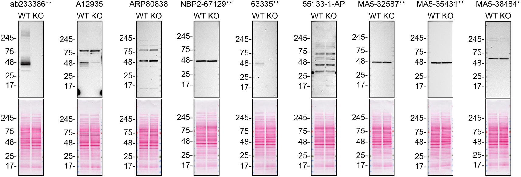
Lysates of SK-HEP-1 (WT and S1PR1 KO) were prepared and 30 μg of protein were processed for western blot with the indicated S1PR1 antibodies. The Ponceau stained transfers of each blot are presented to show equal loading of WT and KO lysates and protein transfer efficiency from precast midi 4-20% Tris-Glycine polyacrylamide gel (Thermo Fisher Scientific, cat number WXP42012BOX) to the nitrocellulose membrane. Antibody dilutions were chosen according to the recommendations of the antibody supplier. Antibody dilution used: ab233386** at 1/1000, A12935 at 1/1000, ARP80838 at 1/500., NBP2-67129** at 1/1000, 63335** at 1/1000, 55133-1-AP at 1/1000, MA5-32587** at 1/1000, MA5-35431** at 1/1000, MA5-38484* at 1/500. Predicted band size: 42.8 kDa. *Monoclonal antibody, **Recombinant antibody.
We then assessed the capability of all nine antibodies to capture S1PR1 from SK-HEP-1 protein extracts using immunoprecipitation techniques, followed by western blot analysis. For the immunoblot step, a specific S1PR1 antibody identified previously (refer to Figure 1) was selected. Equal amounts of the starting material (SM), the unbound fraction (UB), as well as the whole immunoprecipitate (IP) eluates were separated by SDS-PAGE (Figure 2).
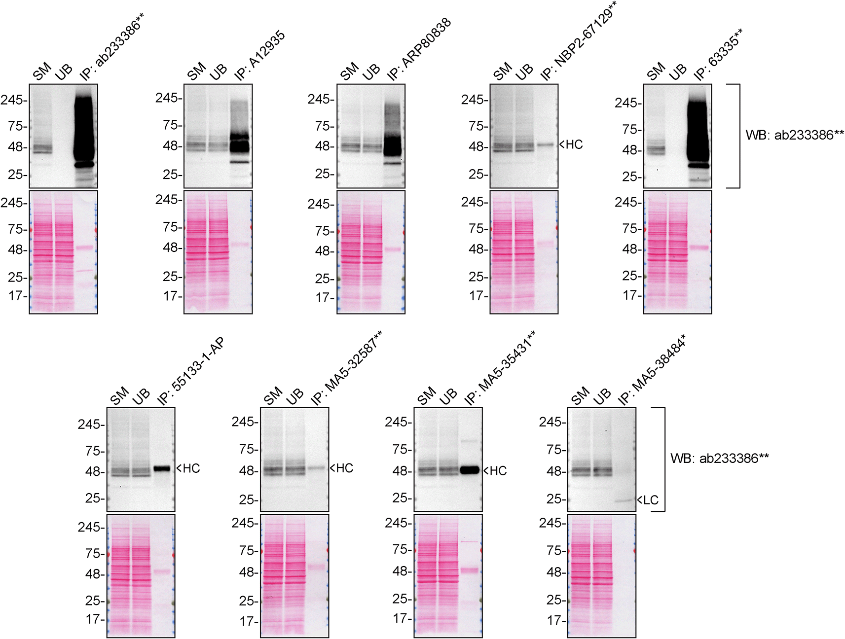
SK-HEP-1 lysates were prepared, and immunoprecipitation was performed using 2.0 μg of the indicated S1PR1 antibodies pre-coupled to Dynabeads protein A or protein G. Samples were washed and processed for western blot on a precast midi 4-20% Tris-Glycine polyacrylamide gel with the indicated S1PR1 antibodies. For western blot, ab233386** was used at 1/1000. The Ponceau stained transfers of each blot are shown. SM = 4% starting material; UB = 4% unbound fraction; IP = immunoprecipitate, HC = antibody heavy chain, LC = antibody light chain. *Monoclonal antibody, **Recombinant antibody.
For immunofluorescence, nine antibodies were screened using a mosaic strategy. First, SK-HEP-1 WT and S1PR1 KO cells were labelled with different fluorescent dyes in order to distinguish the two cell lines, and the S1PR1 antibodies were evaluated. Both WT and KO cells were imaged in the same field of view to reduce staining, imaging and image analysis bias (Figure 3). Quantification of immunofluorescence intensity in hundreds of WT and KO cells was performed for each antibody tested,10 and the images presented in Figure 3 are representative of this analysis.
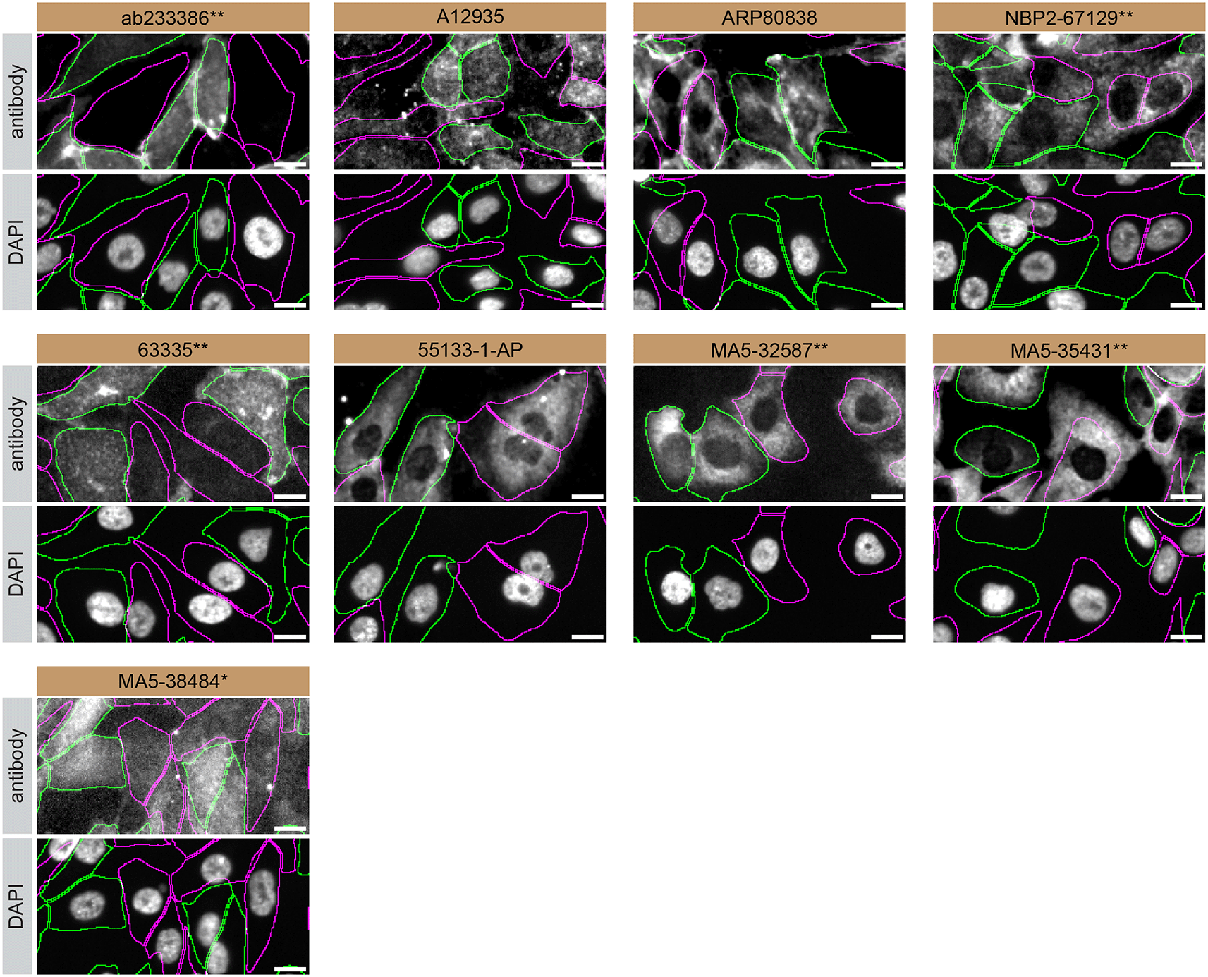
SK-HEP-1 WT and S1PR1 KO cells were labelled with a green or a far-red fluorescent dye, respectively. WT and KO cells were mixed and plated to a 1:1 ratio in a 96-well plate with optically clear flat-bottom. Cells were stained with the indicated S1PR1 antibodies and with the corresponding Alexa-fluor 555 coupled secondary antibody including DAPI. Acquisition of the blue (nucleus-DAPI), green (identification of WT wells), red (antibody staining) and far-red (identification of KO cells) channels was performed. Representative images of the merged blue and red (grayscale) channels are shown. WT and KO cells are outlined with green and magenta dashed line, respectively. When an antibody was recommended for immunofluorescence by the supplier, we tested it at the recommended dilution. The rest of the antibodies were tested at 1 and 2 μg/ml and the final concentration was selected based on the detection range of the microscope used and a quantitative analysis not shown here. Antibody dilution used: ab233386** at 1/400, A12935 at 1/1000, ARP80838 at 1/250., NBP2-67129** at 1/1000, 63335** at 1/2000, 55133-1-AP at 1/500, MA5-32587** at 1/1000, MA5-35431** at 1/300, MA5-38484* at 1/200. Bars = 10 μm. *Monoclonal antibody, **Recombinant antibody.
In conclusion, we have screened nine S1PR1 commercial antibodies by western blot, immunoprecipitation, and immunofluorescence by comparing the signal produced by the antibodies in human SK-HEP-1 WT and S1PR1 KO cells. Several high-quality antibodies that successfully detect S1PR1 under our standardized experimental protocol can be identified. Researchers who wish to study S1PR1 in a different species are encouraged to select high-quality antibodies, based on the results of this study, and investigate the predicted species reactivity of the manufacturer before extending their research.
The underlying data for this study can be found on Zenodo, an open-access repository for which YCharOS has its own collection of antibody characterization reports.14,15
The standardized protocols used to carry out this KO cell line-based antibody characterization platform was established and approved by a collaborative group of academics, industry researchers and antibody manufacturers. The detailed materials and step-by-step protocols used to characterize antibodies in western blot, immunoprecipitation and immunofluorescence are openly available on Protocol Exchange, a preprint server (DOI: 10.21203/rs.3.pex-2607/v1).10
Cell lines used and primary antibodies tested in this study are listed in Tables 1 and 2, respectively. To ensure that the cell lines and antibodies are cited properly and can be easily identified, we have included their corresponding Research Resource Identifiers, or RRID.16,17
| Views | Downloads | |
|---|---|---|
| F1000Research | - | - |
|
PubMed Central
Data from PMC are received and updated monthly.
|
- | - |
Provide sufficient details of any financial or non-financial competing interests to enable users to assess whether your comments might lead a reasonable person to question your impartiality. Consider the following examples, but note that this is not an exhaustive list:
Sign up for content alerts and receive a weekly or monthly email with all newly published articles
Already registered? Sign in
The email address should be the one you originally registered with F1000.
You registered with F1000 via Google, so we cannot reset your password.
To sign in, please click here.
If you still need help with your Google account password, please click here.
You registered with F1000 via Facebook, so we cannot reset your password.
To sign in, please click here.
If you still need help with your Facebook account password, please click here.
If your email address is registered with us, we will email you instructions to reset your password.
If you think you should have received this email but it has not arrived, please check your spam filters and/or contact for further assistance.
Comments on this article Comments (0)