Keywords
O00767, SCD, SCD1, Steroyl-CoA desaturase, antibody characterization, antibody validation, western blot, immunoprecipitation, immunofluorescence
This article is included in the YCharOS (Antibody Characterization through Open Science) gateway.
O00767, SCD, SCD1, Steroyl-CoA desaturase, antibody characterization, antibody validation, western blot, immunoprecipitation, immunofluorescence
This revised version incorporates edits made in response to reviewer comments, which have improved the clarity of the manuscript. A new Table 3 has been added to assist antibody users in interpreting the data presented. Additionally, a limitations section has been included to acknowledge the constraints inherent to this antibody guide.
See the authors' detailed response to the review by Michael L. Garelja
See the authors' detailed response to the review by Cecilia Williams and Matilda Holm
Stearoyl-CoA desaturase (SCD1) is a membrane-bound enzyme which catalyzes the rate-limiting step in the conversion of saturated fatty acids into mono-unsaturated fatty acids.1,2 The regulation of SCD1 is physiologically important as maintaining a proper ratio of saturated to monounsaturated fatty acids is essential for membrane fluidity. Disruption to this ratio can lead to pathological conditions, including cardiovascular disease, obesity, non-insulin dependent diabetes mellitus, hypertension, neurological diseases, immune disorders and cancer.2–7
SCD1 is of particular importance in Parkinson’s disease (PD) research as its inhibition has been found to rescue α-Synuclein cytotoxicity and inclusion formation, both hallmarks of PD progression. The neurotoxic mechanisms underlying PD progression are not yet clearly defined.8–10 Mechanistic studies would be facilitated with the availability of high-quality SCD1 antibodies.
This research is part of a broader collaborative initiative in which academics, funders and commercial antibody manufacturers are working together to address antibody reproducibility issues by characterizing commercial antibodies for human proteins using standardized protocols,11 and openly sharing the data.12–14 Here we evaluated the performance of nine commercial antibodies for SCD1 for use in western blot, immunoprecipitation, and immunofluorescence (also referred to as immunocytochemistry), enabling biochemical and cellular assessment of SCD1 properties and function. The platform for antibody characterization used to carry out this study was endorsed by a committee of industry academic representatives. It consists of identifying human cell lines with adequate target protein expression and the development/contribution of equivalent knockout (KO) cell lines, followed by antibody characterization procedures using most commercially available renewable antibodies against the corresponding protein. The standardized consensus antibody characterization protocols are openly available on Protocol Exchange (DOI: 10.21203/rs.3.pex-2607/v1).15
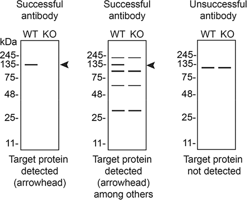
The authors do not engage in result analysis or offer explicit antibody recommendations. A limitation of this study is the use of universal protocols – any conclusions remain relevant within the confines of the experimental setup and cell line used in this study. Our primary aim is to deliver top-tier data to the scientific community, grounded in Open Science principles. This empowers experts to interpret the characterization data independently, enabling them to make informed choices regarding the most suitable antibodies for their specific experimental needs Guidelines on how to interpret antibody characterization data found in this study are featured on the YCharOS gateway16 and in Table 3 of this data note.
Our standard protocol involves comparing readouts from WT (wild type) and KO cells.17,18 The first step was to identify a cell line(s) that expresses sufficient levels of a given protein to generate a measurable signal using antibodies. To this end, we examined the DepMap transcriptomics database to identify all cell lines that express the target at levels greater than 2.5 log2 (transcripts per million “TPM” + 1), which we have found to be a suitable cut-off (Cancer Dependency Map Portal, RRID:SCR_017655). The HeLa cell line expresses the SCD1 transcript at 6.7 log2 (TPM+1) RNA levels, which is above the average range of cancer cells analyzed, and does not carry mutations in the SCD gene that could affect antibody–epitope binding, as seen on DepMap. A SCD KO HeLa cells were obtained from Abcam ( Table 1).
Inherent limitations are associated with the antibody characterization platform used in this study. Firstly, the YCharOS project focuses on renewable (recombinant and monoclonal) antibodies and does not test all commercially available SCD1 antibodies. YCharOS partners provide approximately 80% of all renewable antibodies, but some top-cited polyclonal antibodies may not be available through these partners.
Secondly, the YCharOS effort employs a non-biased approach that is agnostic to the protein for which antibodies have been characterized. The aim is to provide objective data on antibody performance without preconceived notions about how antibodies should perform or the molecular weight that should be observed in western blot. As the authors are not experts in SCD1, only a brief overview of the protein’s function and its relevance in disease is provided. SCD1 experts are invited to analyze and interpret observed banding patterns in western blots and subcellular localization in immunofluorescence.
Thirdly, YCharOS experiments are not performed in replicates primarily due to the use of multiple antibodies targeting various epitopes. Once a specific antibody is identified, it validates the protein expression of the intended target in the selected cell line, confirms the lack of protein expression in the KO cell line and supports conclusions regarding the specificity of the other antibodies. Moreover, the same antibody clones are often donated by 2–3 manufacturers—such as the SCD1 antibodies ab19862 and MA5-27542 (clone CD.E10, cross-licensed between Abcam and Thermo Fisher)—effectively serving as replicates and enabling validation of test reproducibility. All experiments are performed using master mixes, and meticulous attention is paid to sample preparation and experimental execution. In IF, the use of two different concentrations serves to evaluate antibody specificity and can aid in assessing assay reliability. In instances where antibodies yield no signal, a repeat experiment is conducted following titration. Additionally, our independent data is performed subsequently to the antibody manufacturers internal validation process, therefore making our characterization process a repeat.
Lastly, as comprehensive and standardized procedures are respected, any conclusions remain confined to the experimental conditions and cell line used for this study. The use of a single cell type for evaluating antibody performance poses as a limitation, as factors such as target protein abundance significantly impact results. Additionally, the use of cancer cell lines containing gene mutations poses a potential challenge, as these mutations may be within the epitope coding sequence or other regions of the gene responsible for the intended target. Such alterations can impact the binding affinity of antibodies. This represents an inherent limitation of any approach that employs cancer cell lines.
For western blot experiments, WT and SCD KO protein lysates were ran on SDS-PAGE, transferred onto nitrocellulose membranes, and then probed with nine antibodies in parallel ( Table 2, Figure 1).
| Company | Catalog number | Lot number | RRID (Antibody Registry) | Clonality | Clone ID | Host | Concentration (μg/μl) | Vendors recommended applications |
|---|---|---|---|---|---|---|---|---|
| Abcam | ab19862* | 1057200-1 | AB_445179 | monoclonal | CD.E10 | mouse | 1.00 | Wb, IP, IF |
| Abcam | ab236868** | 1007366-14 | AB_2928123 | recombinant mono | EPR21963 | rabbit | 0.61 | Wb, IP, IF |
| Abcam | ab39969 | 1036585-6 | AB_945374 | polyclonal | rabbit | 0.90 | Wb | |
| Aviva Systems Biology | ARP32797_T100 | QC2226-43641 | AB_841676 | polyclonal | rabbit | 1.00 | Wb | |
| Bio-Techne | AF7550 | CGOP0121061 | AB_3107036 | polyclonal | sheep | 0.20 | Wb | |
| Proteintech | 28678-1-AP | 00103543 | AB_2923581 | polyclonal | rabbit | 0.40 | Wb, IF | |
| Thermo Fisher Scientific | MA5-27542* | YH4004441A | AB_2723611 | monoclonal | CD.E10 | mouse | 1.00 | Wb, IP, IF |
| Thermo Fisher Scientific | PA5-75757 | YJ4089139 | AB_2719485 | polyclonal | rabbit | 1.00 | Wb, IF | |
| Thermo Fisher Scientific | PA5-95762 | YJ4090059A | AB_2807564 | polyclonal | rabbit | 1.35 | Wb, IF |
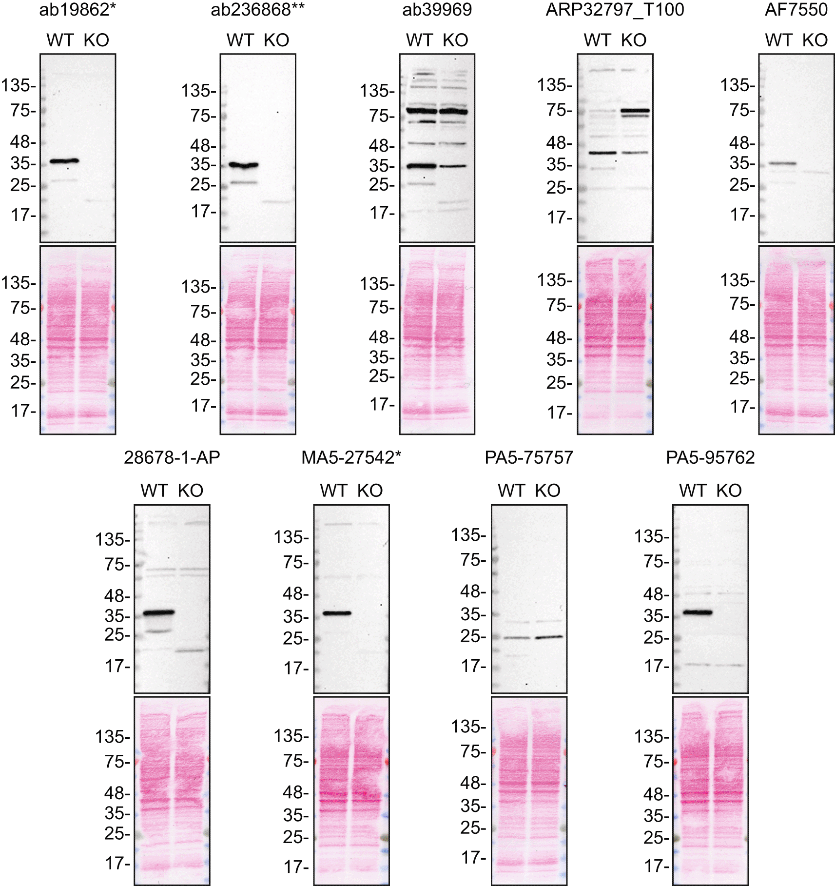
Lysates of HeLa WT and SCD KO were prepared, and 35 μg of protein were processed for western blot with the indicated SCD1 antibodies. The Ponceau stained transfers of each blot are presented to show equal loading of WT and KO lysates and protein transfer efficiency from the acrylamide gels to the nitrocellulose membrane. Tris-Glycine 4-20% gels were used. Antibody dilutions were chosen according to the recommendations of the antibody supplier. An exception was given for antibody AF7550 which was titrated because the signal was too weak when following the supplier’s recommendations. Antibody dilution used: ab19862* at 1/1000, ab236868** at 1/1000, ab39969 at 1/1000, ARP32797_T100 at 1/1000, AF7550 at 1/200, 28678-1-AP at 1/1000, MA5-27542* at 1/1000, PA5-75757 at 1/200 and PA5-95762 at 1/1000. Predicted band size: 41.5 kDa *Monoclonal antibody, **Recombinant antibody.
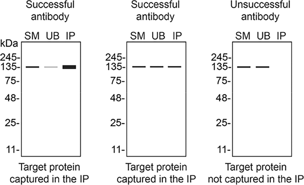
We then assessed the capability of all nine antibodies to capture SCD1 from HeLa protein extracts using immunoprecipitation techniques, followed by western blot analysis. For the immunoblot step, a specific SCD1 antibody identified previously (refer to Figure 1) was selected. Equal amounts of the starting material (SM), the unbound fraction (UB), as well as the whole immunoprecipitate (IP) eluates were separated by SDS-PAGE ( Figure 2).
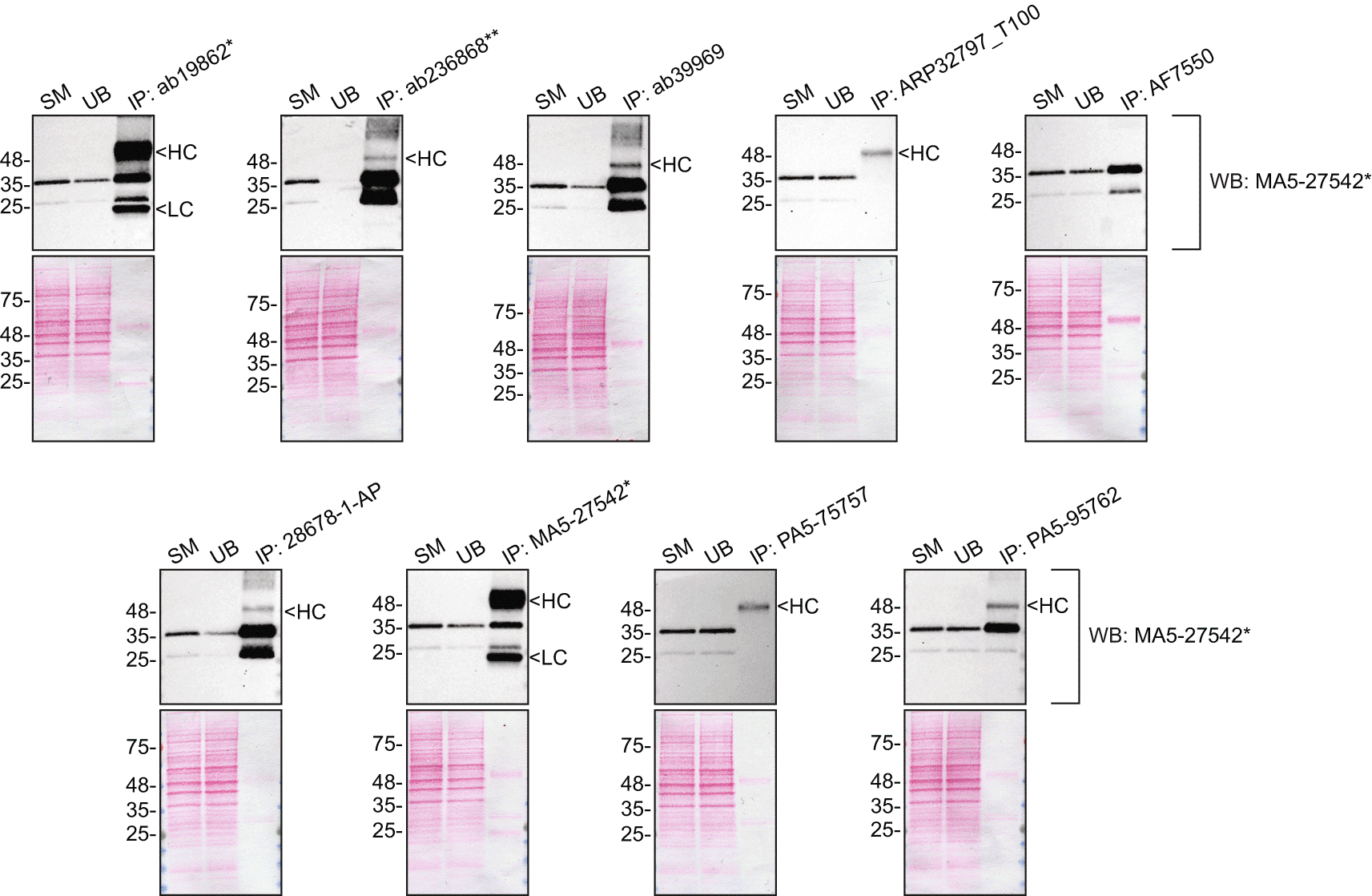
HeLa lysates were prepared, and immunoprecipitation was performed using 1 mg of lysate and 2.0 μg of the indicated SCD1 antibodies pre-coupled to Dynabeads protein A or protein G. Samples were washed and processed for western blot with the indicated SCD1 antibody. For western blot, MA5-27542* was used at 1/1000. Tris-Glycine 4-20% gels were used. The Ponceau stained transfers of each blot are shown. Predicted band size: 41.5 kDa. SM=4% starting material; UB=4% unbound fraction; IP=immunoprecipitate, HC= antibody heavy chain, LC= antibody light chain. *Monoclonal antibody, **Recombinant antibody.
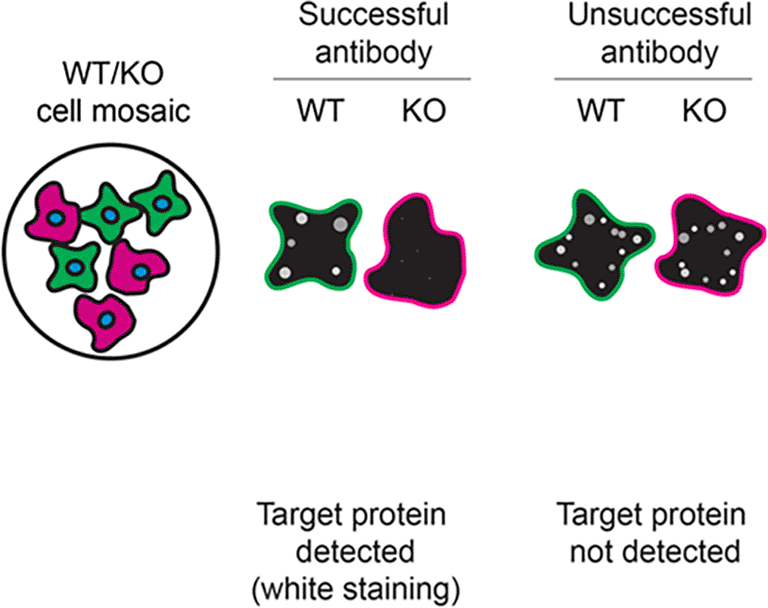
For immunofluorescence, nine antibodies were screened using a mosaic strategy. First, HeLa WT and SCD KO cells were labelled with different fluorescent dyes in order to distinguish the two cell lines, and the SCD1 antibodies were evaluated. Both WT and KO lines imaged in the same field of view to reduce staining, imaging and image analysis bias ( Figure 3). Quantification of immunofluorescence intensity in hundreds of WT and KO cells was performed for each antibody tested, and the images presented in Figure 3 are representative of this analysis.15
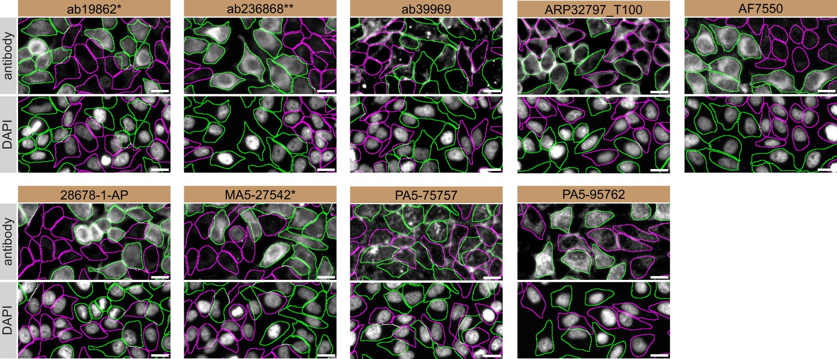
HeLa WT and SCD KO cells were labelled with a green or a far-red fluorescent dye, respectively. WT and KO cells were mixed and plated to a 1:1 ratio on coverslips. Cells were stained with the indicated SCD1 antibodies and with the corresponding Alexa-fluor 555 coupled secondary antibody including DAPI. Acquisition of the blue (nucleus-DAPI), green (WT), red (antibody staining) and far-red (KO) channels was performed. Representative images of the blue and red (grayscale) channels are shown. WT and KO cells are outlined with green and magenta dashed line, respectively. When an antibody was recommended for immunofluorescence by the supplier, we tested it at the recommended dilution. The rest of the antibodies were tested at 1 and 2 μg/mL and the final concentration was selected based on the detection range of the microscope used and a quantitative analysis not shown here. Antibody dilution used: ab19862* at 1/1000, ab236868** at 1/600, ab39969 at 1/150, ARP32797_T100 at 1/500, AF7550 at 1/200, 28678-1-AP at 1/400, MA5-27542* at 1/1000, PA5-75757 at 1/1000 and PA5-95762 at 1/1300. Bars = 10 μm. *Monoclonal antibody, **Recombinant antibody.
| Western blot | Immunoprecipitation | Immunofluorescence |
|---|---|---|
| 
| 
|
In conclusion, we have screened nine SCD1 commercial antibodies by western blot, immunoprecipitation, and immunofluorescence by comparing the signal produced by the antibodies in human HeLa WT and SCD KO cells. High-quality and renewable antibodies that successfully detect SCD1 were identified in all applications. Researchers who wish to study SCD1 in a different species are encouraged to select high-quality antibodies based on the results presented and investigate the predicted species reactivity of the manufacturer before extending their research.
The standardized protocols used to carry out this KO cell line-based antibody characterization platform was established and approved by a collaborative group of academics, industry researchers and antibody manufacturers. The detailed materials and step-by-step protocols used to characterize antibodies in western blot, immunoprecipitation and immunofluorescence are openly available on Protocol Exchange (DOI: 10.21203/rs.3.pex-2607/v1).15
Cell lines used and primary antibodies tested in this study are listed in Tables 1 and 2, respectively. To ensure that the cell lines and antibodies are cited properly and can be easily identified, we have included their corresponding Research Resource Identifiers, or RRID.19,20 All cell lines used in this study were regularly tested for mycoplasma contamination and were confirmed to be mycoplasma-free.
| Views | Downloads | |
|---|---|---|
| F1000Research | - | - |
|
PubMed Central
Data from PMC are received and updated monthly.
|
- | - |
Provide sufficient details of any financial or non-financial competing interests to enable users to assess whether your comments might lead a reasonable person to question your impartiality. Consider the following examples, but note that this is not an exhaustive list:
Sign up for content alerts and receive a weekly or monthly email with all newly published articles
Already registered? Sign in
The email address should be the one you originally registered with F1000.
You registered with F1000 via Google, so we cannot reset your password.
To sign in, please click here.
If you still need help with your Google account password, please click here.
You registered with F1000 via Facebook, so we cannot reset your password.
To sign in, please click here.
If you still need help with your Facebook account password, please click here.
If your email address is registered with us, we will email you instructions to reset your password.
If you think you should have received this email but it has not arrived, please check your spam filters and/or contact for further assistance.
Comments on this article Comments (0)