Keywords
Peripheral nerve regeneration, Digital nerve injury, Transcutaneous electrical stimulation, TENS, Randomized controlled trial, Digital nerve
Peripheral nerve regeneration, Digital nerve injury, Transcutaneous electrical stimulation, TENS, Randomized controlled trial, Digital nerve
This version includes several revisions to improve methodological clarity and interpretation of the findings. We clarified the anatomical targeting of the stimulation protocol, specifying that electrode placement was individualized according to the injured digit and surgical repair site, while the median nerve motor threshold was used only as an operational reference for stimulation intensity. We also added numerical between-group estimates, including unadjusted mean differences and 95% confidence intervals for SWM, s2PD, CSS, and PDI at the 3-month assessment, to support interpretation of the null findings beyond p-values alone. The Discussion and Study limitations sections were expanded to address the sensitivity of SWM and s2PD, the possibility of ceiling effects, the limited generalizability of isolated digital nerve injuries to more complex nerve lesions, the absence of a dose–response framework, the potential masking effect of postoperative rehabilitation, and the limitations of a 3-month follow-up. We also clarified that the findings should be interpreted as showing no detectable clinical advantage of this specific surface PES protocol, rather than definitive evidence against electrical stimulation as a broader strategy for peripheral nerve regeneration.
See the authors' detailed response to the review by Johannes Heinzel
See the authors' detailed response to the review by Nish Mohith Kurukuti
The human hand is a rich sensory and motor multifunctional tool with dexterous control to perform essential manipulation tasks.1 Peripheral nerve injuries, especially of the upper limb, can result in severe disability and reduced quality of life.2–4 Several strategies5–8 including the use of neurotrophic factors, stem cell therapy,9,10 and electrical stimulation,11 have been investigated to promote peripheral nerve regeneration as well as functional recovery after these traumas.12 Electrical stimulation has also been considered as an ancillary to surgical repair, and its effects on nerve recovery has been the focus of several studies.13–25
It is to be noted that the characteristics and regenerative potential of peripheral nerves differ markedly depending on the location and type of lesion.26 Differences in digital nerve lesions compared with more proximal and mixed lesions are described.26,27 Digital nerves are almost exclusively sensory, and injuries to these nerves, properly repaired, generally have shorter regeneration distances and can serve as a model for evaluating the effects of transcutaneous peripheral electrical stimulation (PES), delivered through surface electrodes (commonly referred to as TENS).27 By contrast, proximal nerve injuries, or nerve injuries with larger gaps to overcome, may be more difficult to completely regenerate, given the increased length for axonal growth and the complexity of motor and sensory functional recovery.28
There are different ways to deliver the PES such as implanted electrodes,15,16 percutaneous electrostimulation17,18 (acupuncture needles inserted into the skin and connected to an electric current generator), intraoperative electrostimulation,19–23 thin-film wireless implantable nerve stimulators,24 and surface electrodes.25 The use of transcutaneous surface electrodes is a non-invasive, practical, and simple option, avoiding the reactions provoked by implant surgery or percutaneous stimulation.29
Based on these considerations, we hypothesized that a brief, early postoperative session of transcutaneous PES could modulate biological mechanisms associated with nerve regeneration following digital neurorrhaphy. This hypothesis was grounded on evidence from experimental and clinical studies showing that short-duration electrical stimulation applied soon after nerve repair may enhance activity-dependent regenerative processes.23,30 In the present study, we intentionally adapted these principles to a non-invasive and clinically feasible protocol, consisting of a single 1-hour session of surface stimulation delivered at motor threshold within 24 hours after surgery. The stimulation protocol (20 Hz, 1 hour) was selected based on the findings of Al-Majed et al. (2000),19 which identified this window as optimal for the upregulation of BDNF and TrkB mRNA. Although the digital nerve is sensory, the intensity was calibrated to the motor threshold of the median nerve to ensuring supramaximal recruitment of large-diameter A-beta sensory fibers, which share similar electrical thresholds with A-alpha motor fibers. Although this approach aimed to reproduce, in a transcutaneous manner, stimulation parameters previously associated with regenerative benefits, its biological effectiveness in purely sensory digital nerves remains unclear. Therefore, we conducted a randomized clinical trial to investigate whether this specific surface PES protocol could influence sensory recovery, cold sensitivity, and pain-related disability in patients undergoing digital nerve repair.
This double-blind, randomized, controlled clinical trial was conducted at a general hospital in Bahia, Brazil, from December 19, 2020, to June 10, 2022. The study was prospectively registered in the Brazilian Clinical Trials Registry (ReBEC) on December 18, 2020 (registration number: U1111-1259-1998; available at: https://ensaiosclinicos.gov.br/rg/RBR-8xn3qq5). Ethical approval was obtained from the Research Ethics Committee of the Faculty of Medicine of Bahia, and the study protocol was published31 in advance to ensure methodological transparency and compliance with the Declaration of Helsinki.32
Adult patients aged 18 to 60 years with an acute, non-segmental digital nerve injury of the hand were eligible for inclusion if surgical repair was successfully performed within two weeks of injury. Exclusion criteria comprised the presence of metal implants at the surgical site, history of seizures, use of a cardiac pacemaker, local infection or skin lesions at the intervention site, associated bone or tendon injuries, and any pre-existing neuropathies.
All patients underwent standardized microsurgical neurorrhaphy under ultrasound-guided axillary block, with epineural approximation using 2 to 4 nylon 8-0 sutures to align nerve fascicles and minimize trauma. Within 24 hours after surgery, participants were randomly allocated to one of two groups.
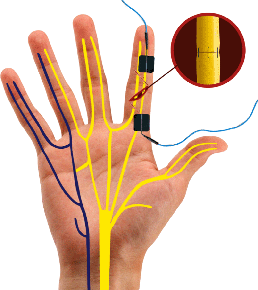
The stimulation parameters were chosen based on previous studies related to nerve regeneration and patient safety.11,14,15,19 Group A (Surgery + PES) received one hour of transcutaneous electrical stimulation using the Neurodyn II device (Ibramed, Brazil), delivering a square-pulsed, biphasic, symmetrical current at 20 Hz with a 0.4 ms pulse width. Stimulation intensity was standardized using the median nerve motor threshold as an operational reference, rather than as the anatomical stimulation target. Electrode placement was individualized for the injured digit and the surgical repair site. In all participants, two 1 × 1 cm silicone-carbon gel electrodes were positioned proximal and distal to the neurorrhaphy, along the presumed course of the repaired digital nerve. Therefore, the intervention was anatomically directed to the injured digital nerve branch, although selective and equivalent recruitment of the repaired nerve across all digits cannot be fully confirmed. Group B (Surgery + sham PES) underwent an identical setup with the same device, electrode positioning, and duration, but after an initial perceptible activation, the device output was reduced to zero for the remainder of the session ( Figure 2). A certified physiotherapist, blinded to the group allocation, supervised the rehabilitation protocol. Sessions were remotely monitored via electronic platforms such as WhatsApp or Skype. Patients underwent a hand sensory re-education program based on the approach proposed by Dellon & Jabaley (1982),33 focused on hand sensory re-education over 3-month period. Participants were also encouraged to perform complementary exercises in a home-based program.
*Intention-to-treat (ITT) analysis.
Patients were randomly assigned in a 1:1 ratio to Group A (surgery + PES) or Group B (surgery + sham) using an electronic randomization sequence generated with the website randomization.com (available at the time of study planning). Allocation concealment was ensured through centralized management by an independent researcher who was the only person with access to the randomization list. A physiotherapist, blinded to group assignment, administered all stimulation sessions using identical devices with the same electrode placement and duration. For sham sessions, the device was initially activated to produce perceptible stimulation cues before being set to zero output. Although real PES could induce subtle muscle contractions, the identical device design and protocol helped maintain blinding for both participants and the administering physiotherapist.
All patients were evaluated in person by the same surgeon responsible for both the surgical procedure and postoperative follow-up. Assessments were scheduled at four time points: (1) pre-intervention; (2) one-week post-intervention; (3) one-month post-intervention (including ongoing rehabilitation sessions); (4) three months post-intervention (upon completion of all 20 rehabilitation sessions). The three-month follow-up period was selected based on the expected timeframe for peripheral nerve regeneration over short distances (2 to 6 cm), assuming an average axonal growth rate of 1 to 3 mm per day.34
The primary outcome was sensory recovery of digital nerves following microsurgical neurorrhaphy, assessed using quantitative sensory tests. Specifically, the Semmes-Weinstein Monofilament (SWM) test and the static two-point discrimination (s2PD) test were applied during four scheduled in-person evaluations. Outcome differences between the two groups (intervention vs. sham) were analyzed post-randomization.
The SWM test, a crucial marker of functional recovery, assesses perception of pressure thresholds related to peripheral reinnervation.35 During the test, participants rested their hands on a table and closed their eyes. In three trials, we applied scored probes perpendicularly to the pulp side of the affected finger for 1 to 1.5 seconds. A positive response in at least two of three trials indicated the sensory threshold.23
The secondary outcome included self-reported measures of cold sensitivity and pain-related functional disability. These were evaluated using the Cold Sensitivity Severity Scale (CSS)36 and the Pain Disability Index (PDI),37 both validated tools for assessing postoperative sensory complaints and pain impact on daily life, aimed to measure improvements in terms of cold sensitivity and pain disability in social functions for individuals who underwent neurorrhaphy of digital nerves in the hand. We used two patient-reported outcome questionnaires: the Cold Sensitivity Severity Scale (CSS)36 and the Pain Disability Index (PDI).37 These patient-reported outcomes were assessed only at the 3-month follow-up, after completion of the rehabilitation protocol.
The s2PD test serves as an established assessment tool for evaluating tactile gnosis.2,38 It measures the ability to distinguish between two nearby points touching the skin, ensuring they are truly distinct rather than perceived as a single point. The test estimates the minimum distance necessary for the patient to perceive the two pressure points as separate contacts.39 It reflects the degree of innervation in a specific skin area. The Medical Research Council classification, modified by Mackinnon & Dellon, allows grouping based on different value ranges related to the sensitive recovery threshold35,40 ( Table 2).
The CSS offers a reliable way to assess cold sensitivity. In cases like amputation or nerve damage, hypersensitivity can occur and lead to significant disability. The CSS consists of four questions related to cold-induced symptoms. The total score provides the cold-sensitivity severity score.
The PDI comprises a seven-item questionnaire evaluating how pain affects various aspects of daily life. Each item is rated from 0 (no disability) to 10 (total disability), and the final score (ranging from 0 to 70) reflects the level of disability due to pain. The PDI has demonstrated consistency, validity, and reliability in studies related to nerve damage.37
The sample size was estimated based on effect size data reported by Gordon et al. (2010).23 We calculated the sample size considering a repeated measures analysis of variance (ANOVA) test, accounting for interactions between and within factors. The effect size, as reported by Gordon et al. (2010),23 was 0.26. Additionally, we set an alpha-type error of 5%, a statistical power of 80%, and worked with two groups and three measures.
Adjustments were applied to account for correlations among repeated measures (correlation coefficient = 0.5) and a non-sphericity correction factor of 1.0 (assuming compound symmetry). Based on these assumptions, the minimum required sample was calculated to be 26 participants.
To account for potential losses, we increased the sample size by 20%, resulting in a final sample of 32 patients (16 per group).
The data were evaluated in a paired and non-paired way through within and between-group comparisons. For within-group evaluations, repeated measures ANOVA or the Friedman test was applied, followed by the Student–Newman–Keuls post hoc test. For between-group comparisons, one-way ANOVA or the Kruskal-Wallis test was used, followed by the Student-Newman-Keuls post hoc test. Specifically, repeated measures ANOVA was used for normally distributed SWM and s2PD data across time points, while the Friedman test was applied when normality assumptions were not met. Between-group comparisons at each time point were performed using one-way ANOVA or Kruskal–Wallis tests accordingly. Independent samples t-tests were used for CSS and PDI comparisons at 3 months.
The choice of statistical tests was based on the distribution characteristics of the data, and normality was assessed using the Shapiro-Wilk test or the nature of the data. A 95% confidence interval was considered for statistical analysis, with statistical significance set ai p < 0.05 an alpha of 5% (P < 0.05) and a power of 80%.
Descriptive analysis was conducted using means and standard errors or medians and interquartile range (25th/75th percentiles), as appropriate to data distribution. Both measurements of the variables and the statistical analysis were performed under blinded conditions by assessors unaware of group allocation. The independent variable for both groups was the use of electric current. The dependent variables were derived from the pre- and post-treatment assessments (SWM, s2PD, CSS, and PDI). All statistical tests were performed using JASP (V0.18.3).
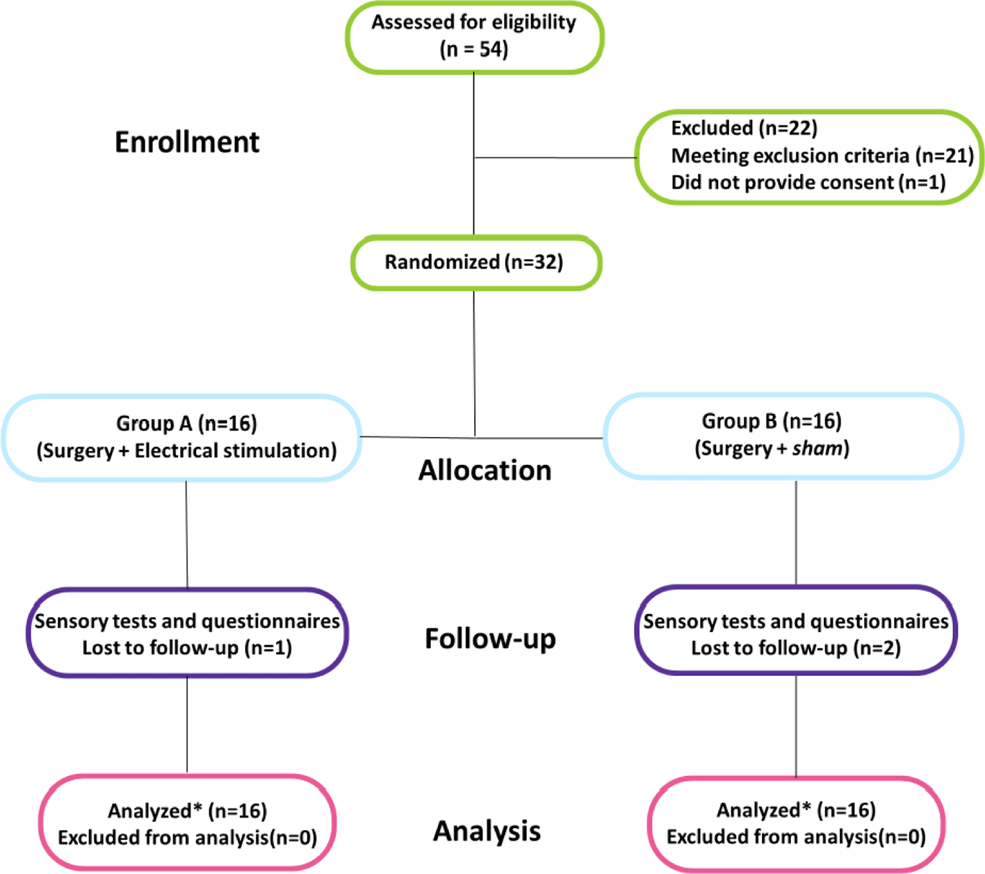
Eligibility was evaluated in a total of 54 patients. Of these, 21 did not meet the inclusion criteria, and one declined to participate. Thus, 32 patients were randomized, with 16 allocated to the PES group and 16 to the sham group ( Figure 1). Baseline characteristics were comparable between groups, and all participants presented with severe sensory impairment on preoperative evaluation, assessed by MSW and s2PD tests ( Table 1). No significant differences between groups were observed during the immediate postoperative period (up to one week; p > 0.05), so subsequent statistical analyses focused on the 1- and 3-month follow-up data.
Sensory outcomes improved over time in both groups, with significant changes observed between 1 and 3 months postoperatively. Repeated measures ANOVA demonstrated a significant effect of time for both SWM (p = 0.012) and s2PD (p = 0.002), indicating progressive sensory recovery in both groups. No significant group effect or group–time interaction was observed for either outcome (p > 0.05). Age showed a marginal influence on SWM outcomes (p = 0.082), becoming significant when scores were converted to needle size (p = 0.014) ( Figure 3).
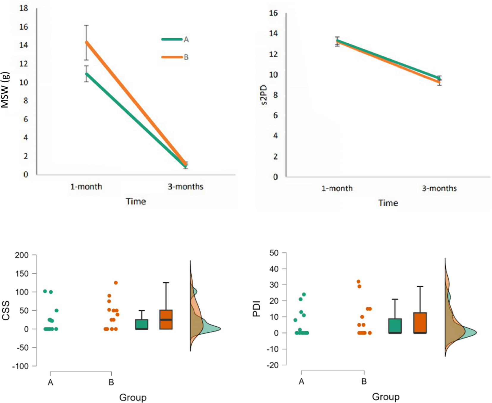
Top panels show mean Semmes-Weinstein monofilament thresholds (SWM, grams) and static two-point discrimination (s2PD, millimeters) at 1 and 3 months postoperatively. Both groups demonstrated progressive sensory recovery over time, with no significant differences between PES and sham. Bottom panels show Cold Sensitivity Severity Scale (CSS) and Pain Disability Index (PDI, 0–70) scores at 3 months. Error bars represent standard error of the mean.
As CSS and PDI were assessed only at the 3-month follow-up, comparisons between groups were performed using an independent samples t-test at this single time point. No statistically significant differences were found for CSS (p = 0.305) or PDI (p = 0.477). However, the two scales were highly correlated (r = 0.819, p < 0.001), suggesting consistent subjective perception of disability and cold sensitivity among patients. Age was initially included as a covariate due to its known influence on nerve regeneration. To assess the robustness of the results, sensitivity analyses were performed by (1) excluding age as a covariate and (2) removing an outlier, a 57-year-old participant from the PES group with discrepant SWM values (z-score ≈ 5).
In summary, patients in both groups gradually recovered sensitivity as measured by SWM and s2PD, reaching satisfactory levels at the final assessment. However, no significant treatment effect was found across time points. At 3 months, the unadjusted mean between-group difference between PES and sham was 0.10 g for SWM (95% CI, -0.94 to 1.15) and 0.79 mm for s2PD (95% CI, -0.26 to 1.83). For patient-reported outcomes, the mean between-group difference was -13.71 points for CSS (95% CI, -40.55 to 13.13) and -2.46 points for PDI (95% CI, -9.46 to 4.54) ( Figure 3). Importantly, these sensitivity analyses did not alter the primary conclusions of the study.
This randomized controlled trial investigated the effectiveness of surface PES in promoting sensory recovery following digital nerve neurorrhaphy. Both groups exhibited a gradual recovery of sensory function over the 3-month follow-up, as measured by SWM and s2PD tests. From a theoretical perspective, electrical stimulation is thought to enhance nerve regeneration through activity-dependent mechanisms that require effective recruitment of regenerating axons and sufficient current density at the nerve site. In the present study, the transcutaneous delivery of stimulation, applied at motor threshold in a purely sensory nerve injury, may not have provided the selectivity or intensity of fiber activation necessary to trigger these mechanisms.41 Although age showed a near-significant effect in some models, its overall influence appears limited. Cold sensitivity and pain-related disability were assessed only at the final follow-up and showed high inter-individual variability, limiting the ability to draw definitive conclusions regarding these secondary outcomes. Transcutaneous electrical stimulation holds promise in nerve regeneration, offering a non-invasive approach with potential practical benefits.42 It can be utilized to circumvent the complications of surgical implantation or percutaneous stimulation.43,44
Some research indicates that it may take up to 8 weeks for the regenerating axons to cover a distance of 25mm, and the use of PES may reduce this period.20,26 Previous results demonstrated that subjects who received stimulation exhibited earlier and better outcomes around 3 months post-surgery.26 Gordon at al. (2010)23 conducted an innovative randomized controlled trial (RCT) of 21 patients undergoing carpal tunnel decompression surgery. Postoperative direct nerve stimulation using implanted wires (20 Hz, 4-6 volts 0.1-0.8 ms) for one hour led to earlier improvements in electrophysiological parameters when compared to controls. Simillarly, Wong et al. (2015)26 conducted a double-blind RCT involving 31 patients with transected digital nerves and observed significantly improved sensory outcomes with PES (20 Hz, <30 V, 0.1–0.4 ms), although no differences in overall functional recovery were found. In another trial, Power et al. (2020)45 evaluated PES following cubital tunnel decompression in 31 patients. The intervention group received a single 1-hour session of PES (20 Hz, <30 V, 0.1 ms) and demonstrated greater improvements in Motor Unit Number Estimation (MUNE) over time compared to the control group. Importantly, these studies employed direct, implanted, or intraoperative stimulation, ensuring higher current density and selective activation of nerve fibers at the repair site, conditions that differ substantially from transcutaneous stimulation and may explain the discrepancy in findings. The present findings should also be interpreted in light of the absence of a dose–response framework. This trial tested a single 1-hour session of surface PES, selected to mirror parameters previously associated with regenerative effects in experimental and clinical studies. However, neuromodulatory effects may depend on stimulation dose, timing, repetition, current density, and cumulative exposure. Therefore, the absence of benefit with this single-session protocol does not exclude the possibility that repeated sessions, different intensities, alternative timing, or longer stimulation schedules could produce different outcomes.
Some studies reported adverse findings that contradict prior research that has highlighted the advantageous impact of direct current electric fields on the regeneration of peripheral nerves.46,47 In the present trial, neither the s2PD nor the SWM tests demonstrated a significant enhancement in tactile receptor reinnervation in the digital pulp among patients who received PES. Given these results, the effectiveness of transcutaneous applied electrical fields in promoting in vivo peripheral nerve regeneration remains uncertain.
Cold intolerance48 and pain49 are commonly reported sources of substantial morbidity following nerve injuries in the hand. Previous studies have associated the severity of these symptoms with the degree of sensory recovery, with poorer reinnervation correlating with more pronounced functional impairment and discomfort.49,50 In the present sample, isolated digital nerve injuries were not associated with worse outcomes in terms of pain or cold intolerance, as measured by the CSS and PDI. These symptoms appear to be more commonly linked to complex cases involving finger replantation, severe vascular compromise, or proximal nerve injuries of the median or ulnar nerves.
Postoperative rehabilitation following hand neurorrhaphy is considered standard of care51 and was prescribed for all participants in this study. Withholding rehabilitation would be ethically inappropriate, as hand therapy is routinely recommended after surgery in real-world clinical settings. Omission of such care could also compromise the study’s external validity. Nevertheless, the use of structured rehabilitation in both groups may have reduced the ability to isolate the specific contribution of PES. Because sensory re-education and hand therapy are expected to promote functional recovery after neurorrhaphy, improvements driven by rehabilitation in both groups may have masked subtle additive effects of the stimulation protocol.52
The use of a digital nerve injury model in this study presents inherent limitations regarding the generalizability of the findings. Digital nerve injuries differ substantially from proximal or mixed nerve lesions, particularly due to the shorter axonal regeneration distances and the absence of motor components. Because isolated digital nerve injuries typically show rapid and favorable recovery after repair, the short regeneration distance may have produced a ceiling effect by three months. In addition, although SWM and s2PD are widely used and clinically meaningful tools after digital nerve repair, they may have limited sensitivity to detect subtle differences in axonal regeneration, receptor reinnervation, or sensory processing.53–55 Therefore, small biological effects of PES may have remained below the detection threshold of these clinical measures. Consequently, although our findings provide insight into the effects of surface PES in isolated digital sensory nerve injuries, they may not be directly applicable to more clinically challenging scenarios, such as mixed motor-sensory nerve injuries, delayed repairs, segmental defects, nerve gaps, or proximal lesions requiring longer regenerative distances. Future studies should consider the use of more complex clinical models, such as proximal or mixed nerve injuries, to better assess the potential of electrical stimulation in promoting meaningful functional recovery. Therefore, the present findings should be interpreted as evidence regarding the limitations of this specific stimulation paradigm in this clinical model, rather than as evidence against the potential usefulness of electrical stimulation as a broader therapeutic strategy for peripheral nerve regeneration.
One limitation of PES is the potential interference of the anesthetic technique with its effectiveness. Ideally, surgery should be performed under general anesthesia. A recent study in rats56 demonstrated that the perioperative use of lidocaine significantly reduced the beneficial effects of electrical stimulation on nerve regeneration. In our study, an axillary block (far from the stimulation site) was used, and PES was applied after post-anesthetic recovery. However, a potential attenuating effect on nerve due to anesthesia cannot be completely ruled out. Current trends in hand surgery increasingly favor local or regional anesthesia, given their advantages of lower perioperative risk, faster recovery, and superior postoperative analgesia. Therefore, proposing PES as an adjunct treatment in digital nerve repair under general anesthesia (without first evaluating its use under standard anesthetic conditions) may limit its clinical applicability.
Skin impedance may have resulted in insufficient current density reaching the nerve when compared to the levels achieved with implanted electrodes.23 In addition, the short regeneration distance in isolated digital nerve injuries may have contributed to a ceiling effect, while SWM and s2PD may not have been sensitive enough to detect subtle treatment-related differences.
The 3-month follow-up also limits the temporal resolution of the findings. This period captures early sensory recovery after short-distance digital nerve repair, but later outcomes at 6 to 12 months may better reflect sensory refinement, cortical reorganization, functional integration, and patient adaptation during daily hand use.
Another limitation of this study is that part of the functional evaluation relied on subjective, patient-reported data. Although neurophysiological assessments can serve as sensitive tools for evaluating the severity and progression of nerve injuries in adults, their application was limited in this study. All patients had isolated digital nerve lacerations, which complicates electrophysiological interpretation due to signal contamination through volume conduction from the intact digital nerve branch on the opposite side of the finger.26 Nevertheless, recent outcome research in the field of peripheral nerve injury has increasingly emphasized the importance of combining functional assessments with patient-reported outcomes.57
The specific protocol of surface PES tested in this study may not confer additional clinical benefit in isolated digital nerve injuries under standard rehabilitation conditions. No significant differences were observed in cold sensitivity or pain-related disability outcomes between the intervention and control groups. These findings suggest that, within the context of isolated digital nerve injuries, surface PES may not confer additional clinical benefit beyond standard surgical repair and rehabilitation. Further research is warranted to explore the efficacy and safety of electrical stimulation protocols in more complex nerve injury models and under varying clinical conditions.
This study was approved by the Research Ethics Committee of the Faculty of Medicine of Bahia (Federal University of Bahia), under protocol number 4.430.230. All procedures were conducted in accordance with the Declaration of Helsinki. The study was prospectively registered in the Brazilian Clinical Trials Registry (ReBEC) under registration number U1111-1259-1998.
| Views | Downloads | |
|---|---|---|
| F1000Research | - | - |
|
PubMed Central
Data from PMC are received and updated monthly.
|
- | - |
Provide sufficient details of any financial or non-financial competing interests to enable users to assess whether your comments might lead a reasonable person to question your impartiality. Consider the following examples, but note that this is not an exhaustive list:
Sign up for content alerts and receive a weekly or monthly email with all newly published articles
Already registered? Sign in
The email address should be the one you originally registered with F1000.
You registered with F1000 via Google, so we cannot reset your password.
To sign in, please click here.
If you still need help with your Google account password, please click here.
You registered with F1000 via Facebook, so we cannot reset your password.
To sign in, please click here.
If you still need help with your Facebook account password, please click here.
If your email address is registered with us, we will email you instructions to reset your password.
If you think you should have received this email but it has not arrived, please check your spam filters and/or contact for further assistance.
Comments on this article Comments (0)