Keywords
alcohol use disorder, viral infection, biomarkers, ROC analysis, liver enzymes, popula-tion-specific reference values
This article is included in the Addiction and Related Behaviors gateway.
alcohol use disorder, viral infection, biomarkers, ROC analysis, liver enzymes, popula-tion-specific reference values
This revised version was prepared in response to the published peer review report. The manuscript was revised to improve terminology consistency, methodological transparency, and interpretive accuracy. We clarified the definition of alcohol use disorder (AUD) according to ICD-10 F10.2 and replaced stigmatizing or inconsistent terminology with person-first terminology. We also clarified that the viral comorbidities assessed in this study refer to chronic viral infections, including hepatitis B, hepatitis C, and HIV.
The Methods section was expanded to specify the exact recruitment period, inpatient recruitment setting, use of baseline/admission laboratory values before treatment initiation, control group selection, sources of clinical and anthropometric data, withdrawal severity classification, and glucose sampling conditions. Additional clarification was added regarding the De Ritis ratio, fasting versus non-fasting glucose measurements, and documentation of delirium, hallucinations, and delusional symptoms during withdrawal.
The Results and Discussion sections were revised to avoid overinterpretation and to align the conclusions more closely with the available data. In particular, we clarified the interpretation of segmented neutrophils, granulocytopenia/neutropenia, platelet count findings, neuropsychiatric manifestations, creatinine and MELD-related interpretation, bilirubin terminology, and ROC analysis/class coding. The diagnostic interpretation was refined to specify that ROC analyses evaluated discrimination between patients with AUD and alcohol-free controls, and between AUD cohorts with and without chronic viral infection.
The supplementary materials were clarified by adding abbreviation definitions, reference intervals, and glucose sampling details. The Zenodo dataset file was additionally anonymized without changes to study groups, statistical analyses, numerical results, or reported conclusions. References were checked and corrected where needed.
See the authors' detailed response to the review by Julia Sinclair and Julia Morris
Alcohol use disorder (AUD) is a serious public health problem with significant social, economic, and medical consequences, as it primarily affects individuals of working and reproductive age. Globally, 5.3% of deaths are attributable to alcohol consumption, and more than 2.4 million people suffer from alcohol-related cirrhosis.1
Alcohol abuse damages multiple organs and systems, including the liver, pancreas, heart, kidneys, nervous system, and hematopoietic cells.2 Reliable diagnostic and prognostic biomarkers provide a minimally invasive approach, most commonly blood-based, for clinical assessment and management. The integration of biomarker screening with instrumental and clinical data forms the basis of modern diagnostics.
Biomarker variability is strongly influenced by sex, age, ethnicity, and geography,3 reflecting environmental and cultural factors such as diet, lifestyle, and alcohol consumption habits. Even closely related ethnic groups with distinct dietary patterns and attitudes toward psychoactive substances demonstrate significant biochemical differences.4 These phenotypic traits are reinforced at the genetic level through diverse functional combinations of single nucleotide polymorphisms (SNPs), which regulate physiological and pathological processes.5 This underscores the importance of population- and ethnicity-specific research.
The present cross-sectional study assessed hematological and biochemical parameters in patients with AUD, with and without chronic viral infections, in Uzbekistan, compared with alcohol-free controls.
A cross-sectional study was conducted at the Republican Specialized Scientific and Practical Medical Center for Mental Health (RSSPMCMH, Tashkent, Uzbekistan). Clinical and laboratory data were collected from 19 March to 22 June 2025. Recruitment was limited to this period because repeated admissions of the same patients became increasingly frequent in the clinical flow.
A total of 292 male inpatients with alcohol use disorder (AUD; ICD-10 code F10.2) were included, together with 49 alcohol-free male controls. Female patients (n = 8) were excluded because their small number was insufficient for meaningful sex-stratified analysis. Individuals with mild alcohol-related conditions (ICD-10 F10.1) or clinically mild withdrawal presentations (n = 2) were also excluded because of their very small number and because their short alcohol-use history and clinical profile were not comparable with patients with established long-term AUD.
The final study population consisted of three groups: Cohort 1, patients with AUD without chronic viral infection (n = 251); Cohort 2, patients with AUD and chronic viral infection, including hepatitis B, hepatitis C, or HIV (n = 41); and Cohort 3, alcohol-free controls (n = 49). Patients were recruited from inpatient units of the center. Laboratory measurements were obtained at admission, before treatment initiation, and were considered baseline values. Only admission values were included in the analysis; follow-up measurements during treatment were excluded.
The control group consisted of male volunteers of approximately similar age, including employees of the same institution and relatives of employees. Controls had no history of alcohol use disorder or psychoactive substance use, as confirmed by questionnaire-based screening and clinical assessment. One initially screened control participant was excluded because of a self-reported history of chronic hepatitis B, as the control group was defined to include individuals without known chronic viral infections.
Inclusion criteria for patients were male sex, age 20–65 years, inpatient status, and a clinical diagnosis of AUD (ICD-10 F10.2), established by an addiction psychiatrist and supported by AUDIT. Exclusion criteria included female sex, mild alcohol-related conditions or mild withdrawal symptoms, acute infections, incomplete clinical or laboratory data, and clinical profiles inconsistent with established long-term AUD.
Alcohol withdrawal severity was assessed clinically by the treating addiction psychiatrist and classified as mild, moderate, or severe according to the intensity of autonomic, somatic, and neuropsychiatric symptoms, including tremor, agitation, anxiety, sleep disturbance, nausea/vomiting, disorientation, hallucinations, delusional symptoms, and the need for inpatient monitoring or pharmacological management. Mild withdrawal was defined as limited autonomic or anxiety-related symptoms without clinically significant neuropsychiatric manifestations or somatic instability. Moderate withdrawal included more pronounced symptoms requiring inpatient observation and treatment, whereas severe withdrawal was characterized by delirium, hallucinations, delusional symptoms, marked agitation, or clinically significant somatic instability.
Controls were age-matched alcohol-free males without alcohol or substance use disorders who provided informed consent. Individuals with known or self-reported chronic viral infections were not included in the control group.
The study was conducted in accordance with the Declaration of Helsinki. Ethical approval was obtained from the Local Ethics Committee of the Republican Specialized Scientific and Practical Medical Center for Mental Health (RSSPMCMH), Tashkent, Uzbekistan (Approval No. 1/2025, 18 March 2025). Written informed consent was obtained from all participants. All data included in the dataset were anonymized prior to analysis and public sharing.
Somatic status was assessed by clinical examination and instrumental diagnostics. Data on age, ethnicity, comorbidities, alcohol-use history, alcohol withdrawal symptoms, and clinical status were obtained from medical records, physician assessments, patient interviews, available collateral history, ultrasound reports, and documented diagnoses. Anthropometric data, including height and weight, were collected from medical records and questionnaires. Body mass index (BMI) was calculated as weight in kilograms divided by height in meters squared (kg/m2).
Hematological and biochemical parameters were measured using automated analyzers (Mindray, China) with standardized enzymatic and colorimetric methods, as detailed in Supplementary File S1. Blood samples from patients were collected at admission regardless of fasting status, because patients were admitted at different times of the day. Therefore, glucose levels in the patient group represent random/non-fasting measurements. In contrast, blood samples from control participants were obtained under fasting conditions.
The De Ritis ratio was calculated as the ratio of aspartate aminotransferase (AST) to alanine aminotransferase (ALT) and was used to characterize patterns of liver injury. Composite indices included FIB-4, De Ritis ratio, mean arterial pressure (MAP), and BMI, calculated using standard formulas.
Delirium, hallucinations, and delusional symptoms were recorded only when documented by the treating addiction psychiatrist during the withdrawal episode. These data were obtained from clinical examination, patient interview, medical history, available collateral history, and medical record review. Symptoms were considered withdrawal-related when they occurred in the context of alcohol withdrawal and were not documented as part of a primary psychiatric disorder. According to medical records, these patients had no previous registration or documented history of primary psychotic disorders.
Diagnostic accuracy of biomarkers was evaluated by ROC analysis. AUROC values with 95% confidence intervals were estimated using bootstrap resampling (n = 1000). Optimal cut-offs were determined by Youden’s Index, with corresponding sensitivity and specificity reported.
Analyses were performed using Python 3.11 (scikit-learn, numpy, pandas, matplotlib). Nonparametric tests (Mann-Whitney U, Kruskal-Wallis H, χ2 with Bonferroni correction) were applied; significance was set at p < 0.05. A retrospective power analysis confirmed ≥85% power for detecting medium-to-large effect sizes despite the smaller control group.
Among the 292 male patients with AUD examined, 14% (n = 41) were identified as carriers of viral infections, including hepatitis C (n = 18), hepatitis B (n = 18), combined hepatitis B and C (n = 1), triple hepatitis B/C/D (n = 1), HIV (n = 2), and HIV with hepatitis B (n = 1). Despite variation within groups, average age did not differ significantly across cohorts (see Table 1). Patients over 50 years old typically had alcohol use histories exceeding 10 years.
Severe withdrawal symptoms were recorded in 26 of 292 patients with AUD (8.9%). Of these, 23 cases occurred in patients without chronic viral infection (Cohort 1; 7.9% of the total AUD sample and 9.2% within Cohort 1), and 3 cases occurred in patients with chronic viral infection (Cohort 2; 1.0% of the total AUD sample and 7.3% within Cohort 2). Most patients (85.5%) exhibited moderate withdrawal symptoms.
The study population was predominantly Turkic-speaking (94.5%, n = 276), mainly of Uzbek ethnicity. Both patients with AUD (n = 292) and alcohol-free controls (n = 49) demonstrated comparable ethnic composition, ensuring cohort homogeneity. Notably, neuropsychiatric symptoms (delirium and/or hallucinations) were observed in 5.5% (n = 16) of patients with AUD. Among these, 43.8% (n = 7) were of Slavic origin and 18.8% (n = 3) were Tatar, despite these groups comprising only 6.1% and 2.7% of the total AUD cohort, respectively. This disproportion may reflect possible population-specific susceptibility to alcohol-related neuropsychiatric complications.
FIB-4 values were more frequently elevated in patients with AUD and chronic viral infection, with markedly high scores (>4) observed in this subgroup, suggesting a greater likelihood of advanced fibrosis. MAP values were also higher among virus-positive patients, indicating possible vascular dysregulation.
The prevalence of somatic comorbidities varied across study groups (see Figure 1).
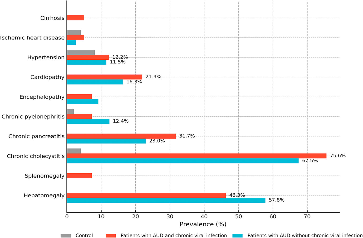
Hypertension was more frequent in patients with AUD (11.5% without viral infection; 12.2% with chronic viral infection) compared to alcohol-free controls (8.2%) (p < 0.05 for both patient groups vs. controls). Cardiopathy occurred in 21.9% of virus-positive patients and 16.3% of virus-negative patients versus 0% in controls (C vs. V, **p < 0.001; C vs. N, **p < 0.001). Chronic pancreatitis was observed in 31.7% of virus-positive and 23.0% of virus-negative patients compared to 0% in controls (C vs. V, **p < 0.001; C vs. N, **p < 0.001). Chronic cholecystitis was the most prevalent condition, affecting 75.6% of virus-positive and 67.5% of virus-negative patients versus 4.1% in controls (C vs. both patient groups, **p < 0.001). Encephalopathy occurred in 7.3% of virus-positive and 9.2% of virus-negative patients, while it was absent in controls (C vs. V, *p < 0.05; not significant for C vs. N or V vs. N). Splenomegaly was found only in virus-positive patients (7.3%, p < 0.01). Cirrhosis was also observed exclusively in this group (4.9%), although differences did not reach statistical significance. No significant variation was identified for ischemic heart disease, hepatomegaly, or chronic pyelonephritis (all p > 0.05).
A comparative analysis of hematological parameters was conducted among virus-positive (n = 41) and virus-negative (n = 251) patients with AUD, and an alcohol-free control group (n = 49). Only values outside physiological reference ranges were considered. Hematological parameters are summarized in Supplementary Table S1.
Hemoglobin (Hb) levels were significantly reduced in both AUD cohorts, falling below normal in 78% of virus-positive and 45% of virus-negative patients. Macrocytosis, defined as MCV > 100 fL, was observed in 17–29% of patients with AUD versus 6.1% of controls (p < 0.001), supporting its association with chronic alcohol exposure.
Erythrocyte sedimentation rate (ESR) was elevated in 63.4% of virus-positive and 57.4% of virus-negative patients (p < 0.001), compared to 10.2% of controls, indicating systemic inflammation, especially with viral comorbidity.
Leukocytosis was more common in virus-positive patients (36.6%) than in virus-negative patients (15.1%). An increased proportion of segmented neutrophils was observed in 7.6-9.8% of patients; however, because direct morphological assessment of neutrophil hypersegmentation was not performed, this finding should be interpreted cautiously and cannot be used to infer megaloblastic anemia or hepatic/renal impairment. Lymphocytosis, likely reflecting antiviral immune activation, was present in 9.2% of virus-positive and 5.0% of virus-negative individuals, while lymphocytopenia occurred in 16.3% of controls. Unexpectedly, monocytosis was more frequent in controls (44.9%) than in patients with AUD (20.0–26.8%), possibly reflecting transient immune responses.
Thrombocytopenia was identified in 13.5–17.1% of patients with AUD versus 4% of controls (p = 0.029). Mean platelet volume (MPV), a marker of platelet activation and turnover, was elevated in 41.5% of virus-positive patients, compared to 16.7–22.4% in the other groups (p < 0.001).
Biochemical parameters were evaluated in virus-positive (n = 41) and virus-negative (n = 251) patients with AUD, as well as in an alcohol-free control group (n = 49). Detailed biochemical characteristics across the three study cohorts are presented in Supplementary Table S2.
As shown in Supplementary Table S2, glucose levels were slightly lower in AUD groups compared to controls. However, within AUD cohorts, virus-positive patients demonstrated 1.2-fold higher glucose levels than virus-negative individuals (p = 0.002), possibly reflecting altered gluconeogenesis and insulin sensitivity. Creatinine concentrations were reduced in both AUD groups by ~1.5-fold vs. controls, likely indicating reduced muscle mass or altered renal tubular function. α-Amylase (AAMY) levels were significantly higher in patients with AUD, with mean values 1.7 times those of controls (p < 0.001; Dunn’s test; r = 0.01). However, elevated enzyme activity was found in only 2.4–8.2% of individuals. This likely reflects total α-amylase activity, not limited to pancreatic isoforms, which is common in chronic alcohol users.
Liver enzymes showed marked alterations. ALT levels were 2.1 times higher in both AUD cohorts versus controls (p < 0.001), and 1.2 times higher in virus-positive vs. virus-negative patients—suggesting additive liver injury from viral and alcohol-related mechanisms. AST levels were approximately threefold higher in AUD groups, with slightly greater elevation in the virus-negative cohort, possibly due to dominant alcohol-related damage.
Bilirubin profiles varied: direct (conjugated) bilirubin was elevated in 72% of virus-negative and 65.9% of virus-positive patients. However, virus-positive individuals also demonstrated elevated indirect (unconjugated) bilirubin, implying compromised hepatic conjugation capacity.
Thus, according to the obtained data, statistically significant differences (p < 0.001) in blood biomarker profiles between patients with AUD and individuals without alcohol abuse (control cohort) were observed among biochemical markers, specifically liver enzymes (ALT, AST), bilirubin fractions, creatinine, glucose, and urea, while among hematological indicators, the most pronounced distinctions were noted for hemoglobin, erythrocyte sedimentation rate, and mean corpuscular volume of erythrocytes. Detailed distributions of biochemical biomarkers across the three cohorts are shown in Supplementary Figure S1.
To evaluate the diagnostic potential of clinical and laboratory markers, Receiver Operating Characteristic (ROC) analysis was performed (see Table 2).
Class coding: (a) positive class = Cohorts 1+2, negative class = Cohort 3; (b) positive class = Cohort 2, negative class = Cohort 1.
Two diagnostic comparisons were assessed. The first comparison evaluated the ability of biomarkers to distinguish all patients with AUD, including both patients without chronic viral infection (Cohort 1) and patients with chronic viral infection (Cohort 2), from alcohol-free controls (Cohort 3). The second comparison evaluated the ability of biomarkers to differentiate patients with AUD and chronic viral infection (Cohort 2) from patients with AUD without chronic viral infection (Cohort 1).
The area under the ROC curve (AUROC) was used to quantify discriminatory performance, with higher values indicating stronger classification ability. Sensitivity and specificity reflected the correct identification of true positive and true negative cases, respectively.
In the comparison of all patients with AUD (Cohorts 1+2) versus alcohol-free controls (Cohort 3), the strongest discriminatory performance was observed for AST (AUROC = 0.951; 95% CI: 0.915–0.987; sensitivity 94%, specificity 89% at 73 U/L), FIB-4 (AUROC = 0.877; 95% CI: 0.820–0.935; sensitivity 76%, specificity 96% at 1.16), and MAP (AUROC = 0.817; sensitivity 96%, specificity 59% at 97 mmHg). ALT (AUROC = 0.808), α-amylase/AAMY (AUROC = 0.802), and direct bilirubin (AUROC = 0.799) also demonstrated good discriminatory performance. In contrast, BMI (AUROC = 0.276) and creatinine (AUROC = 0.060) showed inverse discrimination according to the predefined class coding, reflecting lower values in patients with AUD rather than poor discriminatory performance.
In the comparison between AUD subgroups, markers of immune-inflammatory status were more informative for differentiating patients with AUD and chronic viral infection from those without chronic viral infection. WBC showed the highest discriminatory performance (AUROC = 0.790; 95% CI: 0.705–0.876; sensitivity 95%, specificity 59%), followed by lymphocytes (AUROC = 0.735; sensitivity 90%, specificity 64%), creatinine (AUROC = 0.711; sensitivity 88%, specificity 46%), and monocytes (AUROC = 0.682; sensitivity 98%, specificity 37%). These findings suggest that hepatic and systemic biomarkers are most informative for distinguishing AUD from alcohol-free controls, whereas immune-inflammatory markers better reflect chronic viral comorbidity within AUD cohorts.
This study investigated biochemical, hematological, and somatic characteristics in two AUD cohorts, with and without chronic viral infections, compared to controls. Statistically significant differences were observed across multiple parameters, underscoring the impact of chronic alcohol use and chronic viral comorbidity on systemic physiology. Although the relatively small control group is a limitation, post hoc power analysis for key markers such as ALT indicated sufficient statistical power (≥85%), supporting the robustness of the findings.
Non-invasive indices including BMI, MAP, and the FIB-4 score were employed to assess metabolic and hepatic status. The FIB-4 index identified fibrotic changes in 42% of patients with chronic viral infections and in 27% of virus-negative patients with AUD, consistent with long-term alcohol exposure and previously reported fibrosis rates.6
Our results demonstrate a substantial burden of somatic complications in men with AUD, with notable differences between those with and without chronic viral comorbidity. The high prevalence of chronic cholecystitis and pancreatitis highlights cumulative hepatopancreatic injury, amplified in virus-positive patients. Cardiopathy was disproportionately common in virus-positive patients, supporting the notion that viral infection may aggravate cardiovascular vulnerability. Splenomegaly was observed exclusively in virus-positive individuals (7.3%), suggesting a greater likelihood of portal hypertension and advanced hepatic involvement. Cirrhosis was also detected only in virus-positive patients, though without statistical significance, likely reflecting sample size limitations rather than absence of association. Encephalopathy occurred in both patient cohorts but was absent in controls, suggesting it is primarily linked to alcohol exposure rather than viral status. The lack of difference between patient cohorts suggests that AUD may be the principal driver of neurotoxic complications.7 In contrast, ischemic heart disease, hepatomegaly, and chronic pyelonephritis showed no significant differences, implying closer relation to lifestyle or cumulative alcohol exposure than to viral comorbidity.
Significant reductions in hemoglobin (Hb) were detected in both AUD cohorts, with subnormal levels in 75% of virus-positive and 50% of virus-negative individuals. Ethanol and acetaldehyde exert cytotoxic effects on erythroid precursors and promote oxidative stress.8 Individuals with reduced aldehyde dehydrogenase (ALDH2) activity, common in Asian populations, accumulate more acetaldehyde, leading to enhanced hemoglobin-acetaldehyde adduct (HbAA) formation.9 This mechanism may partly explain higher susceptibility to liver injury and anemia in Asian alcohol users.
Our data are consistent with Russian findings showing reduced hemoglobin in individuals with AUD, although baseline values in the Russian cohort were higher, potentially due to ethnic-genetic or environmental differences.10 Inflammatory markers, including ESR, leukocyte count, and lymphocytes abnormalities, were more pronounced in patients with AUD, particularly in those with chronic viral infection, suggesting ongoing systemic immune activation. This pattern may reflect not only viral-associated inflammatory burden, but also alcohol-related disruption of gut–liver immune signaling. Zeng et al. showed that chronic alcohol exposure is associated with intestinal fungal dysbiosis and an increased Candida albicans-specific Th17 response, with these cells detected in the circulation and liver of patients with alcohol-associated liver disease. In experimental models, ethanol exposure promoted the migration of Candida albicans-specific Th17 cells from the intestine to the liver, where IL-17-mediated signaling contributed to hepatic inflammation and injury. These findings support the concept that alcohol-related immune activation may involve gut-derived microbial and fungal immune pathways, which could amplify systemic and hepatic inflammation in patients with AUD and chronic viral comorbidity.11
Unexpectedly, monocytosis was more frequent in controls, possibly reflecting immune responses to non-viral stimuli or transient inflammatory variation unrelated to AUD. Chronic alcohol use can induce both immune suppression and hyperinflammation through epigenetic alterations of monocyte/macrophage function.8 In addition, alcohol-related disruption of granulopoiesis has been described as a mechanism contributing to impaired innate immune defense and increased susceptibility to infection.12 In the present AUD cohorts, however, neutropenia was rare and comparable between virus-positive and virus-negative patients, suggesting that suppression of the neutrophilic granulocyte compartment was not a prominent hematological feature in these patients.
Thrombocytopenia was also observed in patients with AUD. As shown in Supplementary Table S1, median PLT values were lower in both AUD cohorts than in controls, with the lowest values in patients with chronic viral infection (Cohort 1: 240.0 [190.5–295.5] ×109/L; Cohort 2: 198.0 [163.0–261.0] ×109/L; Cohort 3: 224.0 [204.0–275.0] ×109/L; p = 0.029). PLT values below the reference range were recorded in 13.55% of patients without viral infection and 17.07% of patients with chronic viral infection, compared with 4.1% of controls. Although platelet counts below 119,000/μL have been associated with increased risk of withdrawal seizures and delirium tremens,13 clinically documented delirium and hallucinations during withdrawal were recorded in only 16 of 292 patients with AUD; among them, only one had PLT below the reference range, one had PLT at the lower reference limit, and 14 had values within the reference range. Thus, reduced PLT was an important hematological feature of AUD, but showed no clear correspondence with recorded withdrawal-related delirium or hallucinations in this cohort.
The mean platelet volume (MPV), an indicator of platelet activation, was elevated in 41.5% of virus-positive patients, reflecting increased turnover or stress-induced megakaryopoiesis, potentially mediated by acetaldehyde toxicity and ALDH2 variants.14,15
Our findings on macrocytosis are consistent with prior studies. MCV was elevated in up to 29% of patients with AUD, supporting its role as a biomarker of chronic alcohol exposure.16–18 Mechanistically, macrocytosis may reflect direct erythrocyte toxicity or deficiencies in folate, vitamin B12, and liver function.19
Serum α-amylase was significantly higher in patients with AUD than in controls, despite overall lower activity than reported in Brazilian studies.20 Elevated amylase may reflect pancreatic/hepatic inflammation21 or sympathetic activation, as suggested by King and Nater.22,23
The AST/ALT ratio (De Ritis index) followed expected patterns, being higher in patients with AUD and slightly more elevated in those without chronic viral infections, consistent with alcoholic liver disease and corroborated by Iluz-Freundlich et al.24 Elevated direct (conjugated) bilirubin further supported cholestasis. In contrast, indirect (unconjugated) hyperbilirubinemia was more frequent in virus-positive individuals, suggesting impaired conjugation and hepatocellular function.
Elevated bilirubin in a subset of controls may reflect dietary, metabolic, or lifestyle-related factors reported in other populations. Similar trends were documented in Indian and Chinese populations.25,26
Creatinine was significantly lower in patients with AUD, consistent with prior reports,27 possibly reflecting lower skeletal muscle mass, malnutrition, or altered protein metabolism. Creatinine remains an important component of liver disease severity assessment, including the MELD score, where it is considered together with bilirubin and INR to support risk stratification in advanced liver disease.28 In the present cohorts, lower creatinine should therefore be interpreted as part of the broader metabolic and somatic profile of patients with AUD, rather than as an isolated renal marker.
Glucose and total protein showed paradoxical trends: hyperglycemia and hypoproteinemia were more common in controls, possibly due to obesity. In contrast, elevated globulin fractions in patients with AUD may reflect chronic inflammation.29,30
Hypoglycemia was present in 27.1% of virus-negative patients with AUD, consistent with alcoholic ketoacidosis. This finding warrants expanded biochemical assessment in future studies, including ketone body and osmolality measurements.31–34
Neuropsychiatric symptoms (e.g., delirium, hallucinations) were present in 5.5% of patients with AUD. A disproportionately high frequency of neuropsychiatric symptoms was observed among participants of Slavic origin; however, these findings should be interpreted cautiously because of the small subgroup size. These observations may reflect population-specific differences in enzymatic systems involved in alcohol metabolism.10
Finally, ROC analysis highlights the effectiveness of liver-specific biomarkers (AST, ALT, FIB-4) and systemic parameters (MAP, AAMY) in distinguishing patients with AUD from alcohol-free controls. Elevated AUROC values for AST and FIB-4 align with prior research indicating hepatic injury and fibrotic transformation.35,36 MAP elevation is consistent with alcohol-induced sympathetic activation.37 In subgroup analysis, immune-inflammatory markers (WBC, lymphocytes, monocytes) were more relevant for differentiating patients with AUD and chronic viral infection from those without chronic viral infection, suggesting that viral comorbidity contributes additional immunologic changes.38,39 These findings indicate that combined biomarker panels may be more informative than isolated markers for distinguishing AUD from alcohol-free controls and for identifying viral comorbidity within AUD cohorts. This is clinically relevant because chronic alcohol exposure and viral infections can produce overlapping hepatic, inflammatory, and metabolic abnormalities, making interpretation of single laboratory parameters difficult. However, the limited discriminatory performance of some markers, including platelets, hemoglobin, and BMI, suggests that they should be interpreted as supportive clinical features rather than standalone diagnostic indicators.
This study has several limitations. First, the analysis was restricted to male patients because only eight female patients met the initial screening criteria, which was insufficient for meaningful sex-stratified analysis. Given known sex-related differences in alcohol metabolism, body composition, and biochemical and hematological profiles, women were therefore excluded from the final analytical dataset. Accordingly, the findings should be interpreted as primarily applicable to male patients with AUD, and future studies should include adequately powered female cohorts. Second, the recruitment period was restricted to three months to minimize patient recirculation and ensure the dataset reflected primary screening; while methodologically justified, this limited the overall sample size. Third, the control group was relatively small compared with the patient cohorts. Despite these limitations, the study provides novel population-specific insights into biomarker profiles in AUD with and without chronic viral comorbidity. Importantly, this work represents the first systematic investigation of such biomarkers in a poorly studied population of patients with AUD in Uzbekistan, highlighting the need for further research in this unique setting.
ROC analysis confirmed the strong diagnostic value of liver enzymes (AST, ALT), α-amylase, MCV, direct (conjugated) bilirubin, the De Ritis ratio, FIB-4 index, and mean arterial pressure (MAP) in alcohol use disorder (AUD). Leukocyte, lymphocyte, and creatinine levels were the most informative for distinguishing patients with AUD with and without chronic viral comorbidity. These findings highlight the clinical utility of multimarker panels for differentiation and risk stratification in AUD.
Importantly, deviations between Uzbek control values and international reference ranges underline the need for population- and ethnicity-specific interpretation of biochemical and hematological markers in clinical practice. Establishing regionally validated diagnostic thresholds could improve screening accuracy and support personalized management of patients with AUD in Central Asia.
| Views | Downloads | |
|---|---|---|
| F1000Research | - | - |
|
PubMed Central
Data from PMC are received and updated monthly.
|
- | - |
Provide sufficient details of any financial or non-financial competing interests to enable users to assess whether your comments might lead a reasonable person to question your impartiality. Consider the following examples, but note that this is not an exhaustive list:
Sign up for content alerts and receive a weekly or monthly email with all newly published articles
Already registered? Sign in
The email address should be the one you originally registered with F1000.
You registered with F1000 via Google, so we cannot reset your password.
To sign in, please click here.
If you still need help with your Google account password, please click here.
You registered with F1000 via Facebook, so we cannot reset your password.
To sign in, please click here.
If you still need help with your Facebook account password, please click here.
If your email address is registered with us, we will email you instructions to reset your password.
If you think you should have received this email but it has not arrived, please check your spam filters and/or contact for further assistance.
Comments on this article Comments (0)