Keywords
Breast cancer, Mammography, Machine learning, Tumor classification, Predictive modelling
This article is included in the Manipal Academy of Higher Education gateway.
This article is included in the Artificial Intelligence and Machine Learning gateway.
This article is included in the AI in Medicine and Healthcare collection.
Breast cancer remains a significant global health concern, with over 7.8 million cases reported in the last five years. Early detection and accurate classification are crucial for reducing mortality rates and improving outcomes. Machine learning (ML) has emerged as a transformative tool in medical imaging, enabling more efficient and accurate diagnostic processes.
This study aims to develop a machine learning-based predictive model for early detection and classification of breast cancer using the Wisconsin Breast Cancer Diagnostic dataset.
The dataset, comprising 569 samples and 32 features derived from fine needle aspirate biopsy images, was pre-processed through data cleaning, normalization using the Robust Scaler, and feature selection. Five supervised ML algorithms—Logistic Regression, Support Vector Classification (SVC) with linear and radial basis function (RBF) kernels, Decision Tree, and Random Forest—were implemented. Models were evaluated using performance metrics, including accuracy, precision, sensitivity, specificity, and F1 scores.
The SVC-RBF model demonstrated the highest accuracy (98.68%) and balanced performance across other metrics, making it the most effective classifier for distinguishing between benign and malignant tumors. Key features such as texture mean and area (worst) significantly contributed to classification accuracy.
This study highlights the potential of ML algorithms, particularly SVC-RBF, to revolutionize breast cancer diagnostics through improved accuracy and efficiency. Future research should validate these findings with diverse datasets and explore their integration into clinical workflows to enhance decision-making and patient care.
Breast cancer, Mammography, Machine learning, Tumor classification, Predictive modelling
This revised version incorporates the reviewers’ suggestions to improve clarity and methodological transparency. Additional references recommended by the reviewers have been added to the Introduction to strengthen the background. The Methods section has been expanded to justify the use of Robust Scaler for normalization, the choice of 5-fold cross-validation, and the decision not to apply dimensionality reduction techniques such as PCA or t-SNE. The Results and Discussion now provide further clarification on feature importance, multicollinearity, model performance (particularly the SVC-RBF), and clinical interpretation of the AUC score. Limitations have also been updated to acknowledge the absence of statistical significance testing and certain diagnostic metrics. These revisions enhance both the scientific rigor and clinical relevance of the work.
See the authors' detailed response to the review by Rolando Gonzales Martinez
See the authors' detailed response to the review by Chandrakanta Mahanty
See the authors' detailed response to the review by Abicumaran Uthamacumaran
Breast cancer is a global health concern that affects millions of women worldwide. The alarming number of diagnoses highlights the importance of proactive measures such as regular screenings, self-examination, and increased awareness. In the last five years alone, a staggering 7.8 million women have been diagnosed with this disease.1 These numbers underscore the urgent need for increased awareness, early detection, and effective treatment options. The health system must be significantly reinforced to enhance breast cancer outcomes. In order to reduce mortality rates and provide effective treatment, early detection and screening of breast cancer are highly important.2,3 Early detection is therefore essential to ensure the best outcome in treating breast cancer. It is well known that rapid diagnosis with machine learning is highly beneficial considering the rise in breast cancer cases.4
The integration of AI in breast cancer detection and diagnosis has the potential to revolutionize the field of oncology.5,6 In recent years, machine learning (ML) algorithms have emerged as powerful tools in the field of medical imaging, offering the potential to enhance the accuracy and efficiency of tumour detection and classification.7,8 Machine learning algorithms can analyse vast amounts of data and identify patterns that may not be apparent to human experts. Machine learning algorithms can be trained to analyse mammograms and provide additional insights to radiologists, helping them make more informed decisions. It is imperative that healthcare providers and researchers continue to explore and harness the power of AI to further enhance breast cancer care.8–10
The aim of this study is to develop a machine learning predictive model specifically designed for early detection and classification of breast cancer. By leveraging ML algorithms, the goal is to improve the accuracy of tumour detection and significantly reduce the time required for cell identification.
This research was conducted within the Health Informatics Laboratory, Department of Health Information Management, Manipal College of Health Professions, Manipal Academy of Higher Education, Manipal, over six months (January–June 2022). The study aimed to develop and evaluate a machine learning predictive model for early detection and differential diagnosis of benign and malignant breast lesions.
The Wisconsin Breast Cancer Diagnostic dataset, available on Kaggle,11 was utilized. This dataset comprises 569 records and 33 features derived from fine needle aspirate (FNA) biopsy images, representing tumor characteristics. Data of female patients aged 18–70 years were included. Key features analysed included tumor radius, texture, perimeter, area, smoothness, compactness, concavity, symmetry, and fractal dimension.
Data preprocessing was undertaken in several steps to ensure the dataset was both reliable and suitable for model development. Missing and null values were first removed. The features were then normalized using the Robust Scaler, which centers values on the median and scales them by the interquartile range. This choice was made to reduce the influence of outliers and to better accommodate the non-Gaussian feature distributions often present in medical datasets, while still preserving meaningful relationships between variables. To gain a deeper understanding of the data, Exploratory Data Analysis (EDA) was performed in Python using violin plots, box plots, and correlation matrices, which guided the identification of clinically and statistically relevant features. During this process, strong correlations were observed among certain predictors (e.g., 0.86 between concavity worst and concave points worst). To manage potential multicollinearity, we used tree-based algorithms such as Random Forest and Decision Tree, which are naturally robust to correlated inputs, while regularization in linear models (Logistic Regression and SVM) further reduced redundancy effects. Because our focus was on predictive performance rather than coefficient-level interpretation, these correlations were not expected to bias results. Finally, the dataset was split into input features (X) and target labels (y), with categorical diagnosis values encoded into binary form (0 = benign, 1 = malignant). Dimensionality reduction methods such as PCA or t-SNE were not applied, as the dataset included only 30 features, which allowed efficient computation and straightforward interpretation. Preserving the clinical interpretability of individual features was also prioritized over transformations like PCA, which produces composite variables, or t-SNE, which is mainly intended for visualization. Features were normalized using the Robust Scaler, which centers on the median and scales by the interquartile range to minimize the influence of outliers while preserving inter-feature relationships in high-dimensional medical data.
Five supervised machine learning algorithms were implemented: Logistic Regression, Support Vector Classification (SVC) with linear and radial basis function (RBF) kernels, Decision Tree, and Random Forest. The dataset was split (60:40) into training and testing subsets using Scikit-learn’s train test split Models were trained on the training set and optimized using hyperparameter tuning. Model development included hyperparameter tuning performed using GridSearchCV with 5-fold cross-validation, which helps prevent overfitting and ensures model generalizability. Hyperparameter tuning was performed using GridSearchCV with 5-fold cross-validation to optimize model performance and reduce overfitting. The best-performing hyperparameters for each algorithm were as follows: for Logistic Regression, the regularization parameter was set to C=1.0; for Support Vector Classifiers, C=1.0 was used with a linear kernel, and C=1.0 with gamma=‘scale’ for the RBF kernel. The Decision Tree classifier was tuned with max_depth=5 and criterion=‘gini’, while the Random Forest classifier used n_estimators=100, max_depth=6, and criterion=‘entropy’. These parameters were selected based on the highest cross-validated accuracy and were used for final model evaluation.
Model performance was assessed using confusion matrices and metrics, including accuracy, precision, sensitivity (recall), specificity, and F1 scores, calculated using Scikit-learn’s classification report function. Among the models, SVC-RBF demonstrated the highest accuracy (99%), proving its efficacy for early detection and differential diagnosis of breast lesions. These metrics were evaluated using both the test dataset and during the 5-fold cross-validation phase to ensure consistent generalization across splits. This approach allows for a robust evaluation by averaging performance across multiple folds.
All analyses were performed using Python 3.812 in Jupyter Notebook. Libraries used included Pandas (v1.2.4)13 for data manipulation, Numpy (v1.20.3)14 for numerical computations, Matplotlib (v3.4.2)15 and Seaborn (v0.11.1)16 for data visualization, and Scikit-learn (v0.24.2)17 for machine learning.
The dataset was extracted from the online open-source Wisconsin (Diagnostics) dataset. The study approval was obtained from Institutional Research Committee of Manipal College of Health Professions, Manipal on the 20th of January 2022 (MCHP/Mpl/IRC/PG/2022/04). All procedures adhered to established ethical guidelines for secondary data analysis and data use policies. Consent is not applicable since the data was an extracted from the online open source Wisconsin (Diagnostics) dataset.
The breast cancer dataset (569 samples, 32 features) underwent thorough exploratory data analysis (EDA) to assess structure and identify key features. Two redundant columns, “id” and “Unnamed: 32” (containing only NaN values), were removed during data cleaning. The target variable, “diagnosis,” was analyzed, revealing 59% malignant (M) and 41% benign (B) cases. A bar graph ( Figure 1) illustrates this distribution. Following data cleaning, the dataset was divided into feature variables (X) and the target variable (y), ensuring all numeric features remained in X while the categorical “diagnosis” variable was placed in y.
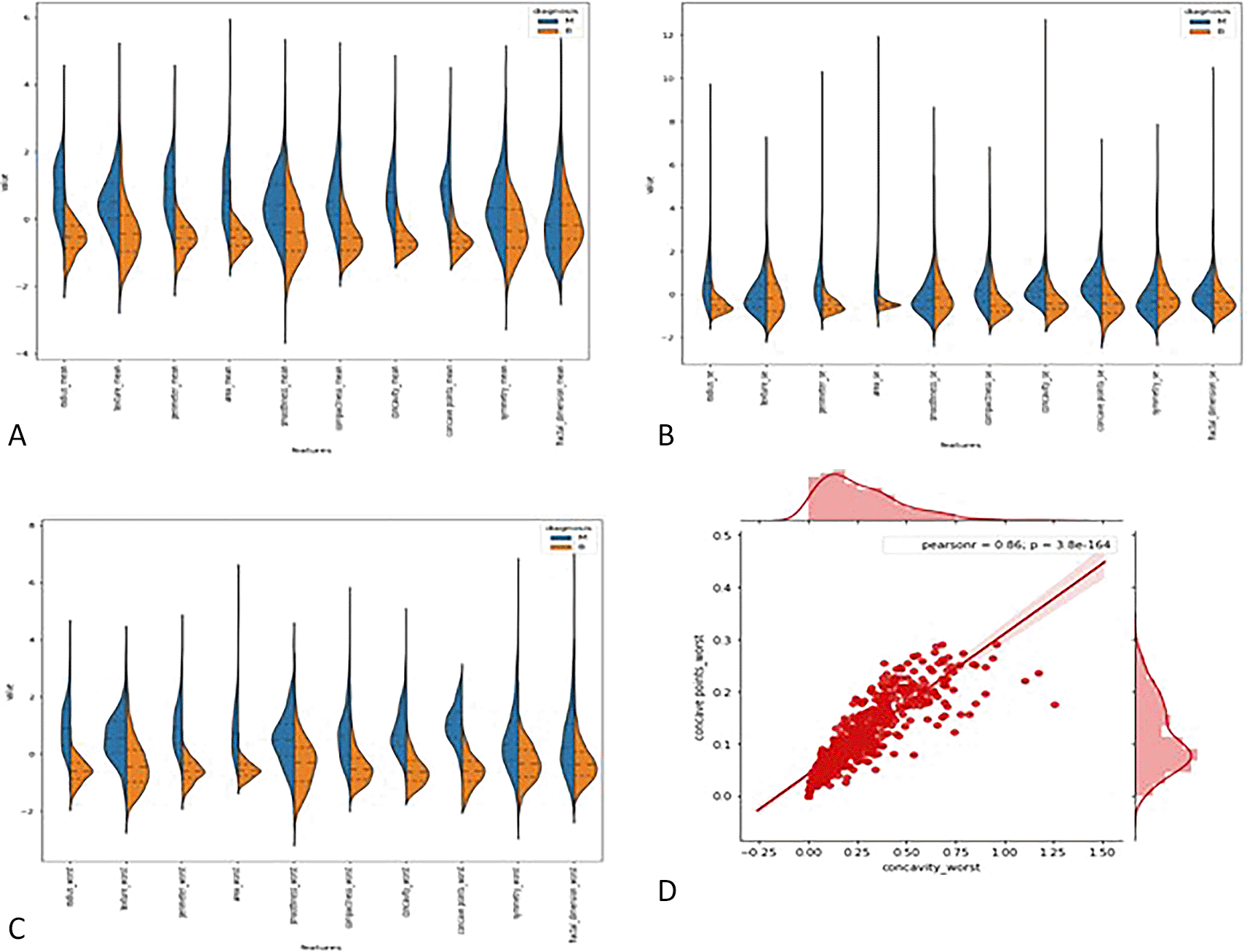
3.2.1 Violin plots: - The distributions of the first thirty features in the dataset were visualized using violin plots to assess their potential for distinguishing between malignant and benign tumors. Key findings include the texture mean, which displayed distinct median values for the tumor types and a wider spread in the kernel density estimate (KDE) for malignant tumors, suggesting its potential as a useful feature for classification. In contrast, the fractal dimension mean showed similar medians for both tumor types, indicating limited discriminative power. Features such as concave points (se) and concavity (se) also exhibited overlapping distributions, making them less valuable for classification. On the other hand, area (se) demonstrated a clear separation between tumor types, highlighting its potential for classification. Similarly, the area (worst) feature showed a distinct separation between benign and malignant tumors, marking it as a strong candidate for classification models, whereas fractal dimension (worst) and concavity (worst) exhibited overlapping distributions, suggesting reduced utility. Overall, texture mean, area (se), and area (worst) emerged as the most promising features for classification, while the others showed limited differentiation between tumor types in the Figure 2 (A, B,C).

3.2.2 Joint plot: - A joint plot was used to analyze the relationship between concavity worst and concave points worst, as their distributions appeared to be similar. The joint plot, which combines scatter plots and histograms, provides a comprehensive view of the data’s distribution and the relationship between two variables. The analysis revealed a strong correlation of 0.86 between the two features, accompanied by a statistically significant p-value. This indicates a high degree of linear association between concavity worst and concave points worst, suggesting that they capture similar information regarding the tumor characteristics. Given their strong correlation, retaining only one of these features in the classification model is advisable, as including both would introduce redundancy and not contribute additional discriminative power in Figure 2(D).
3.2.3 Box plot : - Box plots were used to visualize the distribution of key features across malignant and benign tumor groups, offering a clear representation of the data’s spread, central tendency, and variability. These plots divide the data into quartiles, highlighting the minimum, first quartile, median, third quartile, and maximum values, and can also identify potential outliers. Box plots are useful for comparing feature distributions between groups and identifying differences in spread and central values.
In this study, box plots were employed to explore the relationship between highly correlated features in the correlation matrix, such as texture mean and texture worst, as well as area mean and area worst. The analysis of these features in relation to the diagnosis column revealed similar distributions for malignant and benign tumors, indicating redundancy in the information they provide. For instance, texture mean and texture worst showed comparable distributions, suggesting that retaining both features in the model would likely result in redundancy. Consequently, one of these highly correlated features can be excluded from the classification process without sacrificing predictive power. These insights were further validated through the visual examination of box plots, which helped clarify how each feature discriminates between malignant and benign groups in Figure 3(A, B, C, D).
Label encoding was employed to handle the categorical data within the dataset, specifically the diagnosis column, which consists of two classes: malignant (M) and benign (B). Label encoding is a technique used to transform categorical variables into numerical values, facilitating their inclusion in machine learning models that require numerical input. In this case, the diagnosis feature was encoded by assigning the value 0 to benign tumors and 1 to malignant tumors. This transformation of categorical data into binary values enables the classification algorithms to process the target variable effectively.
Label encoding is particularly useful for datasets with binary or ordinal categorical data, as it preserves the inherent order and structure of the classes. This method of encoding ensures that the diagnosis column can be used seamlessly in the machine learning models, enhancing the classification process and improving model performance. The encoded values (0 and 1) were then incorporated into the feature set, with the remaining extracted features, such as tumor radius, texture, perimeter, and others, remaining in their continuous form.
In this study, the dataset was divided into training and testing sets using the train-test split method to evaluate the performance of machine learning algorithms. The dataset was split with a 60:40 ratio, where 60% of the data was used for training the model, and 40% was reserved for testing. The primary goal of this split is to assess how well the model generalizes to unseen data by training it on the training set and evaluating it on the testing set. The training set allows the model to learn from known data, while the testing set is used exclusively for making predictions, providing an unbiased estimate of model performance.
The dataset was divided into input features (X) and the target variable (y). The target variable, diagnosis (benign or malignant), was assigned to y, and the remaining features used for classification were assigned to X. Consequently, the dataset was split into four variables: X train, X test, y train, and y test, representing the training and testing sets for both features and target variable.
Following the train-test split, feature scaling was performed to normalize the features within the dataset. Feature scaling is a preprocessing technique used to transform the features into a uniform scale, improving the performance of machine learning algorithms. In this study, the Robust Scaler was applied, which scales the data based on the interquartile range (IQR) while removing the median. This scaling method ensures that outliers have a minimal effect on the data, which is particularly beneficial when dealing with features that have different scales or units. The scaled data was then used for model training and evaluation, ensuring that all features contribute equally to the learning process.
Among the features, texture mean and area (worst) emerged as the most discriminative, supported by their strong correlations with diagnostic class labels and their consistently high importance rankings across tree-based models. Although advanced interpretability methods such as SHAP or mutual information scores were not employed, these complementary quantitative measures provided robust evidence of their significance.
In this study, various machine learning models were developed and evaluated using different supervised classification algorithms to identify the most accurate model for classifying benign and malignant breast lesions. A classifier algorithm is designed to map input data to specific categories, making it suitable for tasks such as classification of breast lesions. The algorithms utilized in this project include Logistic Regression, Support Vector Classifier (SVC) with a linear kernel, Support Vector Classifier (SVC) with a radial basis function (RBF) kernel, Decision Tree Classifier, and Random Forest Classifier.
The models were developed using the training dataset, with each classifier being imported from the learn library. The models were assigned to variables, and the fit method was used to train each model on the input features (X train) and target variable (y train). This method enabled the models to learn from the data and adjust their parameters accordingly to improve classification performance.
Following the training process, the accuracy of each model was calculated to assess their performance. The Decision Tree Classifier achieved the highest training accuracy of 1.0, indicating perfect classification performance on the training set. On the other hand, the SVC with the radial basis function kernel exhibited the lowest training accuracy among the classifiers. These results provide an indication of which models performed better in terms of training accuracy and highlight the potential for further model evaluation using additional metrics such as cross-validation, precision, recall, and F1 score to determine the most reliable classifier for the task.
The evaluation of the classification models was performed to determine their effectiveness in distinguishing between benign and malignant breast lesions. Testing accuracy was calculated using a confusion matrix, which summarizes the performance of the classification models in terms of actual and predicted values. The confusion matrix provided four key metrics: True Positive (TP), True Negative (TN), False Positive (FP), and False Negative (FN) values for each classification algorithm, as shown in Table 1.
Among the classification algorithms, the Support Vector Classifier (SVC) with a Radial Basis Function (RBF) exhibited the highest testing accuracy of 0.986, indicating its superior ability to predict correctly. In contrast, the Decision Tree Classifier demonstrated the lowest testing accuracy of 0.942, suggesting room for improvement in its predictive capability.
The classification model was evaluated based on key performance metrics, including accuracy, precision, recall, and F1-score. Accuracy measures the overall effectiveness of the model in correctly classifying cases, while precision assesses the proportion of correctly identified positive cases out of all predicted positives. Recall, also known as sensitivity, indicates the model’s ability to correctly detect positive cases, and the F1-score provides a harmonic mean between precision and recall, ensuring a balanced evaluation.
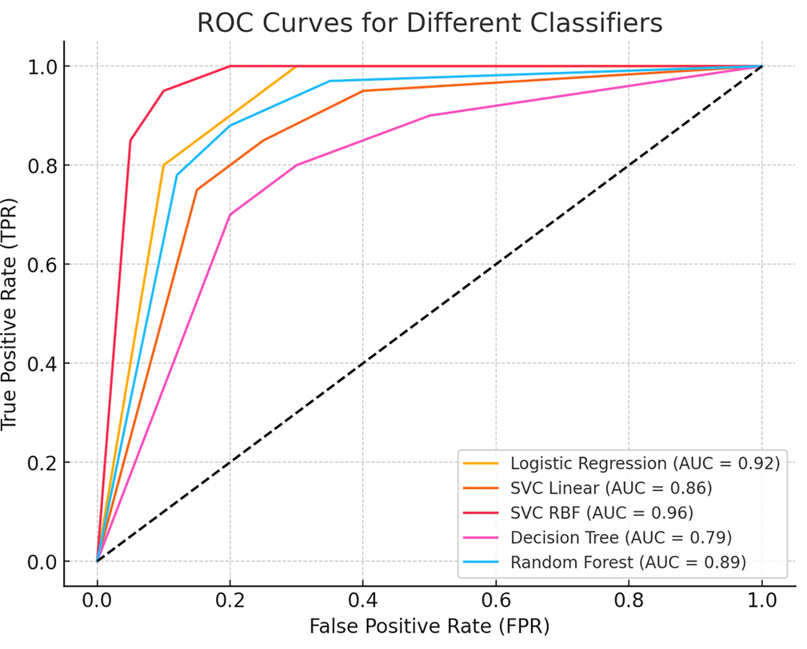
To further assess the model’s discriminative power, we generated a Receiver Operating Characteristic (ROC) curve, which illustrates the trade-off between sensitivity and specificity across different classification thresholds. The Area Under the Curve (AUC) value quantifies the model’s ability to distinguish between benign and malignant cases, with a higher AUC indicating superior classification performance. Figure 5 presents the ROC curve, demonstrating the classifier’s effectiveness in minimizing false positives while maximizing true positive rates.

The curve illustrates the trade-off between sensitivity (true positive rate) and 1-specificity (false positive rate) across different thresholds. The Area Under the Curve (AUC) value indicates the model’s ability to distinguish between benign and malignant cases, with higher AUC values representing better classification performance.
To further assess the quality of predictions, additional metrics such as precision, sensitivity (recall), F1 score, and specificity were calculated using the classification report function from the sklearn metrics package. These metrics evaluate the balance between true positive predictions and false positives/negatives, providing a comprehensive assessment of the classification algorithms The Support Vector Classifier with Radial Basis Function (SVC RBF) demonstrated the highest testing accuracy (0.9868) and consistently high precision, recall, F1 score, and specificity, establishing itself as the most robust classifier in this study. Logistic Regression performed comparably, achieving a testing accuracy of 0.9825, indicating reliable classification performance. In contrast, the Decision Tree Classifier, despite achieving the highest training accuracy (1.0), exhibited the lowest testing accuracy (0.9429), suggesting potential overfitting during training. The Random Forest classifier displayed a balanced performance, with a testing accuracy of 0.9561 and comparable metrics across precision, recall, and F1 score, making it a reliable but less optimal choice than SVC RBF and Logistic Regression in Table 2.
This study demonstrates the effectiveness of machine learning techniques for the early detection and differential diagnosis of benign and malignant breast lesions. Among the models evaluated, the Support Vector Classifier with a Radial Basis Function (SVC-RBF) kernel emerged as the top performer, achieving an accuracy of 99% on the Wisconsin Breast Cancer Diagnostic dataset. The model also showed excellent precision (99% for benign and 98% for malignant), sensitivity (99% and 98%, respectively), and strong F1 scores for both classes, highlighting its robustness in minimizing diagnostic errors. While the SVC-RBF model functions as a black-box algorithm, its consistently high predictive performance supports its potential utility in clinical decision-making. Furthermore, the model achieved an Area Under the ROC Curve (AUC) of 0.96, reflecting its excellent ability to discriminate between benign and malignant cases across all classification thresholds.
Exploratory data analysis (EDA), including violin plots, joint plots, and correlation matrices, revealed critical features such as texture mean, area (se), and area (worst), which were pivotal for classification. These insights enabled feature selection, improving the model’s accuracy while reducing redundancy. Comparatively, features like fractal dimension mean and concavity worst demonstrated limited diagnostic value.
The findings surpass prior studies in terms of model performance. For instance, M. Tahmooresi et al.18 reported an SVM accuracy of 94%, while shen et al,19 developed a deep learning algorithm for breast cancer detection on mammograms using an “end-to-end” approach, achieving high accuracy across heterogeneous datasets such as CBIS-DDSM (AUC: 0.91) and IN breast (AUC: 0.98). This improvement is attributed to advanced preprocessing techniques, such as robust scaling and hyperparameter tuning, combined with a comprehensive evaluation framework. Kayode et al.20’s SVM model achieved a sensitivity of 94.4% and specificity of 91.3%, and Debelee et al.21 reported 99% accuracy on the BGH dataset. While these results are comparable, this study’s comprehensive evaluation, including confusion matrix-derived metrics, adds rigor to the findings. Similarly, Suh et al.22 explored neural network models, such as DenseNet-169 and EfficientNet-B5, achieving AUCs of 0.952–0.954. However, these models require larger datasets and computational resources, unlike the efficient SVC-RBF model used here. Notably, Viswanath et al.23’s Random Forest model showed balanced performance (accuracy 84.84%, precision 90%, specificity 89%), yet it underperformed compared to the SVC-RBF model in this study, emphasizing the latter’s ability to capture non-linear relationships in high-dimensional datasets.
Hussain et al. (2024)24 provide a comprehensive review of machine learning models for breast cancer risk prediction, analyzing key algorithms such as deep learning, decision trees, support vector machines, and ensemble learning. Their study highlights the significance of dataset selection, feature engineering, and model interpretability in improving predictive accuracy. While their work offers a broad overview of machine learning in cancer diagnostics, our study focuses specifically on the Support Vector Classifier with an RBF kernel (SVC-RBF), evaluating its robustness and optimization for cancer classification. Additionally, while Hussain et al. discuss challenges such as dataset bias and feature selection, we extend this discussion by assessing kernel-based optimization and hyperparameter tuning, which play a crucial role in improving predictive performance in imaging-based diagnostics. Similarly, Uthamacumaran et al. (2023)25 introduce a novel machine intelligence-driven classification approach for extracellular vesicles derived from cancer patients using fluorescence correlation spectroscopy (FCS). Their study emphasizes the potential of machine learning in non-invasive cancer diagnostics by combining FCS data with deep learning models and advanced feature extraction techniques. While their work focuses on biomarker-based classification, our study applies SVC-RBF to imaging datasets, exploring its efficiency in structured imaging data rather than fluorescence-based biomarker detection. Additionally, while their research explores deep learning techniques, our work investigates the interpretability and efficacy of kernel-based supervised learning approaches in cancer classification.
The SVC-RBF model offers significant advantages. Its transparency, facilitated by interpretability techniques and visual tools, ensures trust among clinicians, enhancing its potential as a decision-support tool. While the model operates as a black-box algorithm with limited inherent interpretability, its strong predictive capability makes it a valuable candidate for decision-support applications in clinical settings. To enhance clinician trust and eventual translatability, future work will focus on integrating model-agnostic interpretability techniques, such as SHAP values or feature attribution methods, to improve transparency and support clinical decision-making. This study demonstrates the efficacy of machine learning techniques in the early detection and differential diagnosis of benign and malignant breast lesions, with the Support Vector Classifier using a Radial Basis Function (SVC-RBF) kernel emerging as the most accurate model.
This study has several limitations that should be acknowledged. One key limitation is the reliance on a limited and non-diverse dataset, specifically the Wisconsin Breast Cancer Dataset (WBCD), which may affect the generalizability of the findings. While WBCD is widely used in breast cancer classification research, its applicability to real-world clinical settings remains uncertain. Future studies should incorporate larger, more diverse datasets from different demographics and imaging modalities, such as The Cancer Genome Atlas (TCGA) or multi-center datasets, to enhance external validation. Additionally, integrating multimodal imaging data, including mammography, MRI, and histopathology, could provide a more comprehensive diagnostic framework. Another limitation relates to the complexity of the Support Vector Classification with a Radial Basis Function (SVC-RBF) kernel. While the RBF kernel provides high accuracy by capturing non-linear relationships in the data, it requires significant computational resources and lacks interpretability. Alternative approaches, such as simpler models like logistic regression or explainability-enhanced deep learning models, should be explored to balance accuracy with interpretability. Moreover, dimensionality reduction techniques such as Principal Component Analysis (PCA) or t-SNE could improve computational efficiency and provide clearer insights into key features. While overfitting was inferred from the divergence between training and testing performance, formal diagnostics such as learning curves or bias–variance analysis were not performed and should be incorporated. The feature selection process in this study relies on correlation analysis and visualization, which, while effective, does not fully account for complex feature interactions. Future work should incorporate advanced feature selection techniques, including SHAP (Shapley Additive Explanations) for feature importance analysis and ensemble learning approaches to optimize model performance. This study did not incorporate statistical significance testing (e.g., p-values, confidence intervals) to validate feature-level separability, as the primary focus was on model-based performance metrics. Future research should include such tests to enhance the robustness of feature analysis and improve the interpretability of results. Although accuracy was reported as a baseline measure, greater emphasis was placed on recall (sensitivity), specificity, F1-score, and ROC-AUC to account for class imbalance. Metrics such as the False Omission Rate (FOR), while not explicitly presented, can be derived from the confusion matrix and should be highlighted in future work.
Breast cancer diagnosis and treatment may be revolutionized by machine learning approaches that provide early detection, leading to more efficient therapeutic interventions. In a multi-centre study, larger datasets from different institutions can be accessed by applying different machine learning approaches. Early detection of breast cancer is key to slowing down the progression of the disease and reducing mortality rates. By leveraging the data from multiple institutions, machine learning can help identify breast cancer more quickly and accurately, leading to earlier intervention and better patient outcomes. With earlier intervention, the risk of mortality can be significantly reduced, leading to better patient outcomes and an overall improvement in public health.
The dataset was extracted from the online open-source Wisconsin (Diagnostics) dataset. The study approval was obtained from Institutional Research Committee of Manipal College of Health Professions, Manipal on the 20th of January 2022 (MCHP/Mpl/IRC/PG/2022/04). All procedures adhered to established ethical guidelines for secondary data analysis and data use policies. Consent is not applicable since the data was extracted from the online open source Wisconsin (Diagnostics) dataset.
Kaggle: Wisconsin Breast Cancer Dataset, https://www.kaggle.com/datasets/uciml/breast-cancer-wisconsin-data
The data sets of mammography with benign and malignant breast lesions.
Data are available under the terms of the CC BY-NC-SA 4.0 (CC-BY 4.0).
| Views | Downloads | |
|---|---|---|
| F1000Research | - | - |
|
PubMed Central
Data from PMC are received and updated monthly.
|
- | - |
Competing Interests: No competing interests were disclosed.
Reviewer Expertise: Cancer Research; Artificial Intelligence; Systems medicine
Competing Interests: No competing interests were disclosed.
Reviewer Expertise: Deep Learning, machine learning
Is the work clearly and accurately presented and does it cite the current literature?
Yes
Is the study design appropriate and is the work technically sound?
Yes
Are sufficient details of methods and analysis provided to allow replication by others?
Yes
If applicable, is the statistical analysis and its interpretation appropriate?
Yes
Are all the source data underlying the results available to ensure full reproducibility?
Yes
Are the conclusions drawn adequately supported by the results?
Yes
References
1. Mahanty C, Rajesh T, Govil N, Venkateswarulu N, et al.: Effective Alzheimer’s disease detection using enhanced Xception blending with snapshot ensemble. Scientific Reports. 2024; 14 (1). Publisher Full TextCompeting Interests: No competing interests were disclosed.
Reviewer Expertise: Deep Learning, machine learning
References
1. Gonzales Martinez R, van Dongen D: Deep learning algorithms for the early detection of breast cancer: A comparative study with traditional machine learning. Informatics in Medicine Unlocked. 2023; 41. Publisher Full TextCompeting Interests: No competing interests were disclosed.
Reviewer Expertise: Machine learning and deep learning applied to health
Competing Interests: No competing interests were disclosed.
Reviewer Expertise: AI, Machine learning, Bioinformatics, and Systems Oncology
References
1. Gonzales Martinez R, van Dongen D: Deep learning algorithms for the early detection of breast cancer: A comparative study with traditional machine learning. Informatics in Medicine Unlocked. 2023; 41. Publisher Full TextCompeting Interests: No competing interests were disclosed.
Reviewer Expertise: Machine learning and deep learning applied to health
Is the work clearly and accurately presented and does it cite the current literature?
Partly
Is the study design appropriate and is the work technically sound?
Partly
Are sufficient details of methods and analysis provided to allow replication by others?
Yes
If applicable, is the statistical analysis and its interpretation appropriate?
Partly
Are all the source data underlying the results available to ensure full reproducibility?
Yes
Are the conclusions drawn adequately supported by the results?
Yes
References
1. Gonzales Martinez R, van Dongen D: Deep learning algorithms for the early detection of breast cancer: A comparative study with traditional machine learning. Informatics in Medicine Unlocked. 2023; 41. Publisher Full TextCompeting Interests: No competing interests were disclosed.
Reviewer Expertise: Machine learning and deep learning applied to health
Is the work clearly and accurately presented and does it cite the current literature?
Partly
Is the study design appropriate and is the work technically sound?
Yes
Are sufficient details of methods and analysis provided to allow replication by others?
No
If applicable, is the statistical analysis and its interpretation appropriate?
Partly
Are all the source data underlying the results available to ensure full reproducibility?
Partly
Are the conclusions drawn adequately supported by the results?
Yes
References
1. Hussain S, Ali M, Naseem U, Nezhadmoghadam F, et al.: Breast cancer risk prediction using machine learning: a systematic review.Front Oncol. 2024; 14: 1343627 PubMed Abstract | Publisher Full TextCompeting Interests: No competing interests were disclosed.
Reviewer Expertise: AI, machine learning, bioinformatics, and precision oncology
Alongside their report, reviewers assign a status to the article:
| Invited Reviewers | |||
|---|---|---|---|
| 1 | 2 | 3 | |
|
Version 4 (revision) 05 Sep 25 |
read | read | |
|
Version 3 (revision) 16 May 25 |
read | read | |
|
Version 2 (revision) 10 Apr 25 |
read | read | |
|
Version 1 05 Feb 25 |
read | read | |
Provide sufficient details of any financial or non-financial competing interests to enable users to assess whether your comments might lead a reasonable person to question your impartiality. Consider the following examples, but note that this is not an exhaustive list:
Sign up for content alerts and receive a weekly or monthly email with all newly published articles
Already registered? Sign in
The email address should be the one you originally registered with F1000.
You registered with F1000 via Google, so we cannot reset your password.
To sign in, please click here.
If you still need help with your Google account password, please click here.
You registered with F1000 via Facebook, so we cannot reset your password.
To sign in, please click here.
If you still need help with your Facebook account password, please click here.
If your email address is registered with us, we will email you instructions to reset your password.
If you think you should have received this email but it has not arrived, please check your spam filters and/or contact for further assistance.
Comments on this article Comments (0)