Keywords
Solitary fibrous tumour, Eyelid, Children, Pediatric, eye tumor
This article is included in the Oncology gateway.
This article is included in the Eye Health gateway.
Solitary fibrous tumour, Eyelid, Children, Pediatric, eye tumor
Solitary fibrous tumors (SFTs) are a special group of spindle cell tumors of mesenchymal origin. They were first reported in the pleura by Klemperer and Rabin in 1931.1 It seldom involves extra-pleural sites such as the lung, pericardium, liver, kidney, parotid, thyroid gland, peritoneum, nasal cavity, and orbit. In 1994 Dorfman et al. and Westra et al. first elucidated SFT of the orbit.1 They have also been reported in the lacrimal drainage system with their specific characteristics and tailored surgical managements.2 Eyelid involvement is extremely rare, and to the best of author’s knowledge, only 10 cases exist in the English literature out of which only one was reported in the paediatric age group.3,4 Cellularity of SFT varies, with frequent hypocellular areas rich in collagen, and the stroma is myxoid. SFT has a prominent vasculature with dilated blood vessels similar to hemangiopericytoma. Characteristically, the tumour cells exhibit immunoreactivity for CD34 (human hematopoietic progenitor cell antigen) and vimentin (a marker of mesenchymal elements).5 The highly vascular nature of the tumour and tendency for bleeding at surgery suggest special precautions and care in their surgical removal. Complete excision with blunt dissection is recommended whereever possible. Careful and long-term follow-up is important and necessary because the tumour may recur locally several years after excision of the primary tumor.5 Herein, we report successful outcome in a rare case of eyelid SFT in a 5-years-old-child managed with surgical excision in toto along with a review of all the paediatric cases of eyelid and orbit SFT reported in literature. Patient consent has been obtained for publishing identifiable photographs. The case report adhered to the ethical principles outlined in the Declaration of Helsinki as amended in 2013 and also HIPAA compliant.
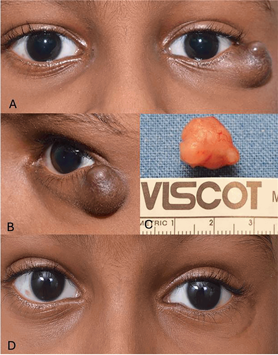
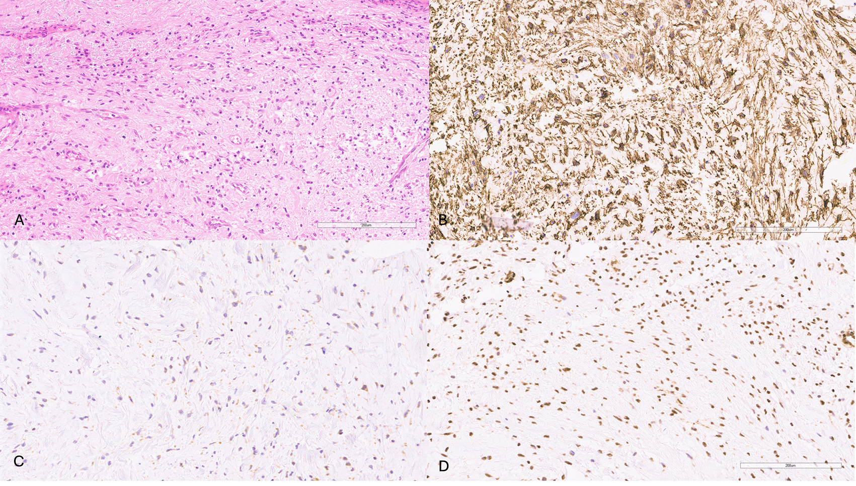
A 5-years-old boy presented with a gradually progressive painless nodule over the temporal aspect of the left lower eyelid of three months duration. External examination revealed a temporal, firm, bilobed subcutaneous well-circumscribed marginal nodule measuring 11×12×6 mm in lower eyelid ( Figures 1A, 1B). There was mild hyperpigmentation of the overlying skin with no associated lash loss or posterior lamellar involvement. The nodule was excised in toto through a sub ciliary skin incision. Gross examination showed it to be a well-capsulated globular white mass ( Figure 1C). Histopathology showed fragments of tissue had spindle cells with indistinct cell border and few of the spindle cells also showed long cytoplasmic processes. These spindle cells were arranged in loose clusters and ill-defined fascicles in the background of entrapped small blood vessels, myxoid degeneration and thick hyalinized collagen bundles. Few admixed mast cells, multinucleated and scattered chronic inflammatory cells were also noted. These spindle cells showed intense positive immunoreactivity for CD34, STAT-6 and negative immunoreactivity for smooth muscle actin (SMA) compatible with diagnosis of solitary fibrous tumour eyelid ( Figures 2A to 2D). The patient is tumour free at 2 year follow up ( Figure 1D).
Solitary fibrous tumor (SFTs) is amongst a group of rare tumors composed of proliferating CD34- positive specialized fibroblasts. Because of overlapping morphologic and immunohistochemical features the term solitary fibrous tumor is used as an encompassing terminology for the entities like SFT, hemangiopericytoma, giant cell angiofibroma and fibrous histiocytoma.6 They most often arise from the pleura but may also occur in multiple locations as well, such as the lungs, peritoneum, pericardium, kidneys, liver, thyroid, parotid, adrenal gland, breast, genito-urinary tract, and meninges. Although the superior orbit is the most frequently reported location for periocular SFTs, it may also occur in the intraconal space, sclera, lacrimal sac, conjunctiva, lacrimal gland fossa, and eyelids.1–3 Herein, we describe an isolated case of SFT of the eyelid, which has rarely been reported in the literature.
SFTs are predominantly benign tumors, but there are reported cases of residual tumors, recurrence, and malignant transformation. The treatment of choice is complete resection, as incomplete excision significantly increases the risk of recurrence and malignant conversion. However, achieving clear margins can be challenging due to the tumor’s fragile capsule, high vascularity, which poses a significant risk of intraoperative hemorrhage, and poor cohesiveness. On histopathological examination, the eyelid mass in this patient was compatible with SFT. Nevertheless, to corroborate the diagnosis, CD34 immunotyping of the neoplastic cells was carried out in light of the peculiar location. The tumor in this case was immunoreactive for vimentin and STAT-6 in addition to CD34, as was previously observed in tumors of a similar nature. Before the widespread use of immunohistochemical research and CD34 labelling, the diagnosis of orbital and periorbital SFT was frequently confounded with other benign tumors such as meningioma, schwannoma, and fibromatosis.7 A hypercellular and hypocellular zone of spindle cells with no specific pattern can be seen in a solitary fibrous tumor against a backdrop of dense collagen bands. Mesenchymal markers like Vimentin are positive in solitary fibrous tumors; however, desmin, cytokeratin, factor VIII-related antigen (a vascular marker), S-100 protein (a neural marker), smooth muscle actin, and muscle-specific actin are negative. In 79–100% of instances, there is immunoreactivity to the marker CD34. Vimentin and CD34-uniform positivity support the diagnosis of SFT. Solitary fibrous tumors generally behave in a benign manner and do not metastasize. Malignancy is defined by having more than four mitoses per high-power field or having abnormal mitotic figures, as well as having cellular pleomorphism or tumor giant cells. Recurrent SFT is more aggressive and has higher mitotic activity than the original tumor.6–10
Solitary fibrous tumors (SFT) of the eyelids are exceedingly rare. To date, only ten additional cases within the HPC-SFT-GCA spectrum have been reported in the literature ( Table 1).8–14 Among these, only one case involved a pediatric patient. Our case involves a 5-year-old patient with an isolated eyelid SFT, making him the youngest documented patient with this condition. The mean age of reported cases is 38 years (median: 35; range: 5 to 78 years), and the mean duration of symptoms is 21 months (median: 12; range: 3-96 months). The majority of these patients are male (6 out of 8, 75%) where gender was specified. The upper and lower eyelids were equally affected in reported cases. Most isolated eyelid SFTs presented as slowly progressive, well-defined subcutaneous eyelid nodules. All patients demonstrated immunoreactivity to CD34. At an average follow-up of 30 months (median: 24; range: 6-96 months), 77% (7 out of 9) of patients were disease-free, while 2 lost to follow-up. One patient with tuberous sclerosis had all four eyelids involved, presenting with pedunculated eyelid lesions of giant cell angiofibroma and experienced multiple recurrences.
| S.No | Author | Year | Age (years)/Sex | Duration (Months) | Morphology & Location | Management | Histopathology | Follow up |
|---|---|---|---|---|---|---|---|---|
| 1 | Sekundo W et al.8 | 1996 | 31/F | 3 | Left medial canthus | Tumour was excised | Mesenchymal tumour consisting of densely packed oval-shaped and spindle-shaped cells growing haphazardly around sinusoidal vascular channels. The latter were lined by a single endothelial layer and showed in part a typical staghorn pattern | 8 months follow-up with no recurrence |
| 2 | Hayashi N et al.9 | 1999 | 78/F | 96 | Nodular lesion at the medial aspect of the left upper lid | At the patient’s insistence, only an incision biopsy was performed | Pattern less, spindle-cell proliferation with a moderate to high degree of cellularity with numerous blood vessels CD34 and vimentin positive | Lost to follow up |
| 3 | Mawn L et al.10 | 1999 | 30/M | 24-36 | Large pedunculated mass involving all four eyelid in a patient with tuberous sclerosis | Resected twice | Fibromyxoid lesion involving the dermis and subcutaneous tissue, scattered floret cellsCD34+, Bcl-2+ (HP- Giant cell angiofibroma). SMA and S-100 negative | Follow up for 8 months, multiple recurrences |
| 4 | Song A et al.11 | 2005 | 24/M | 12 | Right painless subcutaneous nodule medial lower eyelid with predominant lobulated inferior forniceal conjunctival component | Excision biopsy | Tumor demonstrated numerous capillary-sized blood vessels with several tumor giant cells, and loosely arranged tumor cells corresponding to the angiectoid, or pseudovascular spaces. Focal areas containing wavy bundles resembling neural tissue (S100+) seen within the tumor (Giant cell angiofibroma) CD34+, Vimentin +, Bcl-2+ | 8 months follow-up with no recurrence |
| 5 | Kim HJ et al.12 | 2008 | 40/M | 6 | Right lower eyelid nodular well defined mass CT- Isodense homogeneous contrast enhancement | Complete excision of the tumour | Pattern less arrangement of alternating hypercellular and hypocellular regions of spindle cells against a collagenous background of variable vascularity CD34+ | 24 months follow-up with no recurrence |
| 6 | Kakizaki H et al.13 | 2010 | 35/M | 12 | Slow growing painless hard immobile right lower eyelid mass with ectropion and telangiectasia and ulceration, bleed and crusting (history of recent rapid increase+) | Total tumour excision and lateral tarsal strip | Spindle cells arranged in short, ill-defined fascicles, set against a rich reticulin mesh. CD34 and Vimentin positive (HPC- SFT intermediate) | 6 months follow-up with no recurrence |
| 7 | Irene Pecorella et al.14 | 2014 | 78/M | 12 | Slow growing painless hard elastic left lower eyelid subcutaneous | Total tumour excision. | Moderately cellular tumour, composed of pattern less uniform, bland-looking bipolar spindle or oval cells with indistinct eosinophilic cytoplasm, in a heavily collagenized stroma. CD34+, Bcl-2+ | 24 month follow-up with no recurrence |
| 8 | Blandamura S et al.4 | 2014 | 50 | NS | Left upper eyelid | Excisional biopsy was performed | Alternating hypercellular and hypocellular areas. They were composed of an intimate mixture of well-formed, thick-walled, often branching vessels of variable size, with intervening proliferation of spindle to round cells, showing oval nuclei with finely dispersed chromatin CD34, CD99 and Bcl2 were diffusely positive | 6 years follow-up with no recurrence |
| 9 | Blandamura S et al.4 | 2014 | 37 | NS | Right upper eyelid | Excisional biopsy and an year later primary excision was performed | Alternating hypercellular and hypocellular areas. They were composed of an intimate mixture of well-formed, thick-walled, often branching vessels of variable size, with intervening proliferation of spindle to round cells, showing oval nuclei with finely dispersed chromatin CD34, CD99 and Bcl2 were diffusely positive | In spite of the residual disease, the patient is alive and well after 8 years |
| 10 | Blandamura S et al.4 | 2014 | 5 | NS | Left upper eyelid | Excisional biopsy was performed after which patient lost follow-up | Alternating hypercellular and hypocellular areas. They were composed of an intimate mixture of well-formed, thick-walled, often branching vessels of variable size, with intervening proliferation of spindle to round cells, showing oval nuclei with finely dispersed chromatin The giant cells were numerous and scattered throughout the lesions | Lost to follow-up |
| 11 | Present case | 2020 | 5/M | 3 | Left lower lid temporal firm, bilobed subcutaneous well-circumscribed marginal nodule with overlying mild hyperpigmentation of the skin | Nodule was excised in toto through a sub ciliary skin incision | Fragments of tissue with pseudo cystic spaces and surrounding stroma showed spindle cells having long cytoplasmic processes arranged in loose clusters. Background of spindle cells had fibro myxoid substance and hyalinized collagen bundles. CD34, vimentin and STAT-6 positive | 24 months follow up with no recurrence |
To conclude, eyelid SFTs are rare neoplasms that typically present as slow-growing, well-defined subcutaneous nodules on the eyelid. They predominantly affect individuals in their third or fourth decade of life, with a higher incidence in males. The treatment of choice is complete surgical excision. Accurate diagnosis relies on histopathology combined with immunohistochemistry. It is crucial to be aware of this entity involving eyelids and consider in the differential diagnosis of vascular spindle-cell eyelid tumors in children. No data are associated with this article.
| Views | Downloads | |
|---|---|---|
| F1000Research | - | - |
|
PubMed Central
Data from PMC are received and updated monthly.
|
- | - |
Provide sufficient details of any financial or non-financial competing interests to enable users to assess whether your comments might lead a reasonable person to question your impartiality. Consider the following examples, but note that this is not an exhaustive list:
Sign up for content alerts and receive a weekly or monthly email with all newly published articles
Already registered? Sign in
The email address should be the one you originally registered with F1000.
You registered with F1000 via Google, so we cannot reset your password.
To sign in, please click here.
If you still need help with your Google account password, please click here.
You registered with F1000 via Facebook, so we cannot reset your password.
To sign in, please click here.
If you still need help with your Facebook account password, please click here.
If your email address is registered with us, we will email you instructions to reset your password.
If you think you should have received this email but it has not arrived, please check your spam filters and/or contact for further assistance.
Comments on this article Comments (0)