Keywords
α-guaiene, patchouli oil, antifungal activity, Minimum Inhibitory Concentration, Minimum Fungicidal Concentration
This article is included in the Plant Science gateway.
This article is included in the Pathogens gateway.
α-guaiene, patchouli oil, antifungal activity, Minimum Inhibitory Concentration, Minimum Fungicidal Concentration
The revised version of our article has undergone substantial improvements compared to the earlier submission. First, we have clarified methodological inconsistencies, particularly regarding the minimum inhibitory concentration (MIC) values, fungal strains (including ATCC numbers), and the precise plant species used (Pogostemon cablin). These revisions ensure greater scientific accuracy and reproducibility.
Second, we refined the presentation of results by addressing ambiguities in retention times, compound identification, and concentration reporting. For example, the discussion of guaiene concentration, bulnesene, and inhibition zones against Aspergillus niger has been corrected to provide clearer interpretation and scientific justification. In addition, formatting issues such as italicization of Latin names and consistency of terminology have been standardized throughout the text.
Third, we improved the rationale for analytical methods. The purpose of using mass spectrometry (MS) alongside retention indices has been explicitly stated, and additional references have been incorporated to support the use of MIC and MFC data. Sentences related to export statistics and contextual background were revised and combined for better readability and logical flow.
Finally, the results and discussion section has been reorganized to strengthen coherence. Ambiguous expressions (e.g., “155%,” “60%”) were clarified as concentrations or relative values, and supporting references were provided. These adjustments enhance both the technical precision and readability of the manuscript.
Overall, the revised manuscript addresses all reviewer comments, corrects technical inaccuracies, and improves clarity in both scientific content and presentation. We believe these modifications significantly strengthen the article and make it suitable for peer review.
See the authors' detailed response to the review by Irmanida Batubara
See the authors' detailed response to the review by Harlinda Kuspradini
Patchouli oil is an essential oil extracted from various plant parts such as flowers, leaves, stems, and roots (Pandey et al., 2022; van Beek & Joulain, 2018). Patchouli (Pogostemon cablin Benth) is a crucial aromatic plant in the perfume industry (Jain et al., 2022) and used in perfume, soap, pharmaceutical, cosmetic, and other industries. Patchouli oil is also renowned for its active constituents and therapeutic benefits (Leong et al., 2019). Furthermore, patchouli oil accounts for approximately 85% of Indonesia’s essential oil exports, with a current annual value of 1,200-1,500 tons. Indonesia exports 90% of the world’s patchouli oil (Rahmayanti et al., 2018). Notably, patchouli oil’s constituent components, such as patchouli alcohol, α-guaiene, δ-guaiene, α-patchoullene, and seychellene, have several benefits (Pandey et al., 2021). Pressure, temperature, reflux ratio, and fractionation column all play crucial roles in the fractionating process used to separate these constituents (Almeida et al., 2018; Nurjanah et al., 2020a).
Essential oils have antiviral, antiparasitic, antifungal, bactericidal, insecticidal, and nematocidal properties (Khaledi & Zahani, 2018). They can be used as bactericides and fungicides against various human-infecting fungus and bacterium types. A pathogenic fungus Candida albicans causes candidiasis, whereas Microsporum gypseum and Trichophyton mentagrophytes cause dermatophytosis (Moskaluk & VandeWoude, 2022). Aspergillus niger is another fungal pathogen that affects the respiratory system by causing different diseases such as aspergillosis (Fiema et al., 2022). Fortunately, essential oils can be developed as antifungal agents for preventing these. Most compounds derived from essential oils are terpenes and their metabolites derivatives (D’agostino et al., 2019), such as α-guaiene, a sesquiterpene comprising an average of 11% of the total mass (Orf et al., 2021). Patchouli oil’s main constituents are patchouli alcohol (27.0%-35%), bulnesene (13.0%-21.0%), and α-guaiene (11.0%-16.0%) (Górski et al., 2021). Terpenes can inhibit protein and DNA synthesis and promote cell rupture in antibiotic-susceptible and antibiotic-resistant bacteria (Masyita et al., 2022).
The antimicrobial activity of essential oils, notably patchouli oil, has been reported. Patchouli essential oil has been shown to inhibit Malassezia furfur and to be effective as an antimicrobial and anti-inflammatory agents (Srivastava et al., 2022). Treatment of C. albicans and T. mentagrophytes with patchouli oil fraction 8 containing 55.59% patchouli alcohol content yielded inhabitation zone of 9.24 mm and 7.70 mm in both fungi, respectively (Setyaningrum et al., 2017).
Patchouli alcohol is the primary antifungal component of patchouli oil and can be used as a fixative in perfume and related industries. After patchouli alcohol extraction, other components such as α-guaiene can increase patchouli oil’s utilization. Studies on α-guaiene antifungal activity are few, necessitating more investigations on this compound. While patchouli alcohol (PA) exhibited antimicrobial properties against pathogenic bacteria (Escherichia coli, Pseudomonas aeruginosa, Bacillus proteus, Shigella dysenteriae, Typhoid bacillus, Staphylococcus aureus) (Yang et al., 2013). In addition, PA proved effective in treating some germs that were resistant to antibiotics, such as methicillin-resistant Staphylococcus aureus (MRSA) (Wan et al., 2021). Therefore, this study explores the antifungal activity of fraction 1 patchouli oil containing 38.8% α-guaiene against C. albicans, A. niger, M. gypseum, and T. mentagrophytes.
Several studies shown that α-guaiene can be isolated using molecular distillation and spinning band vacuum distillation. However, to obtain pure α-guaiene is very still difficult. Molecular distillation can only isolate α-guaiene with a putity level 0f 18.80% (Widyasanti et al., 2021) while using spinning band distillation can produce α-guaiene with higher purity of 31.05% (Nurjanah et al., 2020b). For this reason, this study used α-guaiene with purity 38.8%, which was obtained from modifying the previous process conditions (Nurjanah et al., 2020b).
Patchouli oil was obtained from distiller in Subang, West Java, Indonesia. The nutrient agar culture used was Potato Dextrose Agar (PDA), whereas the liquid medium was Potato Dextrose Broth (PDB). Other chemicals such as 1% BaCl2, 1% H2SO4, sterile distilled water, 70% alcohol, lactophenol cotton blue (LPCB), 0.85% NaCl, ketoconazole, fluconazole, and n-hexane were also utilized. Chemical compound were obtained from Bratachem, Indonesia while drugs were obtained from Kimia Farma, Bandung, Indonesia. The fungi used were A. niger, C. albicans ATCC 7102, M. gypseum ATCC 14683, and T. mentagrophytes ATCC 16404.
The instruments used included Spinning Band Distillation System Model 36-100 from B/R Instrument USA, autoclave, erlenmeyer flask, beaker glass, scotch bottle, bunsen, ose needle, petri dish, cuvette, spectrophotometer, micropipette, microplate, fin pipet, microscope, laminar airflow, oven, spatula, and vortex. The study was conducted using a laboratory experimental method with descriptive analysis, with ketoconazole serving as the positive control for all fungi tests, fluconazole for M. gypseum and T. mentagrophytes, and n-hexane serving the negative control for the four fungi.
To obtain the dominant α-guaiene, the sample was fractionated with a pressure of 10 mmHg, column length of 90 cm, and reflux ratio of 20:1. Patchouli oil was divided into five fractions: 1 at 249°C-254°C, 2 at 254°C-259°C, 3 at 259°C-264°C, 4 at 264°C-269°C and 5 at 269°C-274°C. Fraction 4 was suspected to contain α-guaiene as the most predominant content.
GCMS analysis of the sample was carried out on a Agilent® 6890 GC-MS, equipped with a split-spitless injector, attached to an Agilent HP-5MS capillary column (30 m x 250 μm, 0.25 μm film thickness). The carries gas was helium at a flowrate of 1.0 mL/min, split ratio 400:1, injector temperature was 280°C, pressure was 10.48 psi. The transfer line was heated to 280°C.
The modified microdilution technique (Eloff, 1998) was used to determine the antifungal activity of the sample.
Conidia were wasted from the surfaces of the agar slant with sterile 0.85% saline containing 0.1% Tween 20 (vol/vol). The conidia suspension was adjusted with sterile 0.85% saline to a concentration of roughly 1.0 × 10.5 in a final amount of 100 μL per well. The inocula were stored at -20°C for the firther use. Dilutions of the inocula were cultured on the solid MEA to verify the absence of contamination and to check the validity of the inocula.
Determination of the inhibited fungal growth (MIC) was performed by a serial dilution technique using 96-well microtitre plates. Different volume of investigated sample was dissolved in malt extract broth (MEB) with fungal inoculums (10 μL). The microplates were incubated for 72 hours at 28°C. The lowest concentration without visible growth were defined as the concentration that completely MIC. The minimal fungicidal concentration (MFC) was determined by serial subcultivation of 2 μL into microtitre plates containing 100 μL of MEB. The lowest concentration with no discernible growth concentration that totally inhibited fungal growth (MFC) was the lowest concentration at which no growth was defined as the MFC, indicating 99.5% killing of the origin inoculum.
To obtain the dominant α-guaiene, the patchouli oil was fractionated using a BR Instrument Spinning Band Distillation System Model 36-100 and divided into five fractions. Based on the GC-MS test results, the compound was higher in fraction 1 than in the others. After fractionation, the fractions were tested for antifungal activity.
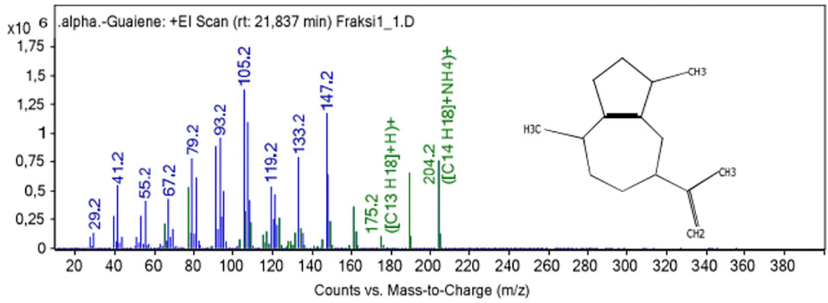
Figure 1 show fragmentation pattern of α-guaiene.
Macroscopic identification involved observation with the eyes in order to directly identify the physical appearance ( Figure 2). Figure 2 (Line 1) show the physical appearance of C. albicans and A. niger grown on PDA medium. C. albicans cultures were white and formed round colonies, corroborating Lee et al’s finding (Le et al., 2022), that C. albicans has a round shape. Allen et al. (2018) reported that A. niger culture exhibited white mycelium and brown-black conidia heads. Despite its higher colony size, A. niger had less dense colonies than C. albicans, which tend to form a firm line based on the groove of the ose needle stroke. In addition, C. albicans colonies were spherical, and had a relatively uniform size without hyphae due to their unicellularity, whereas A. niger colonies were irregularly spherical, had a nonuniform size, and formed hyphae owing to their multicellularity.
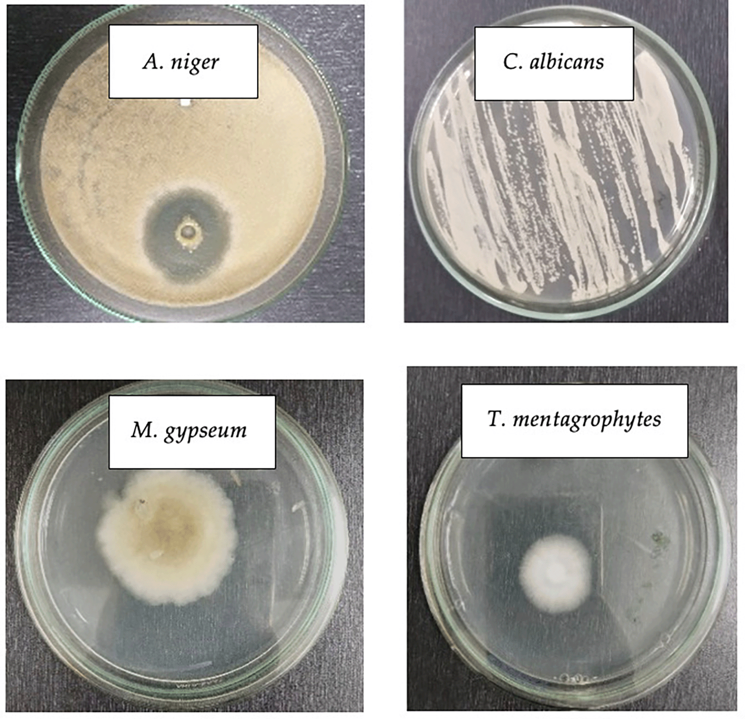
The colony of the fungus suspected to be M. gypseum resembled a pile of fine white cotton on top with a little brown powder scattered on the hyphae, corroborating a previous finding (Putriningsih & Arjentinia, 2018) that M. gypseum colonies grew rapidly, were slightly powdery with a blackish-red brown color, and were scattered with a flat surface containing macroconidia.
Macroscopically, although M. gypseum and T. mentagrophytes did not differ significantly, having white hyphae, T. mentagrophytes colonies appeared denser and slightly whiter with a protruding rough or powdery surface corroborating Frías-De-León et al’s findings (Frías-De-León et al., 2020) that T. mentagrophytes colonies were often white to slightly yellowish-white and could sometimes turn violet-red, brown, or pale yellowish with a surface resembling cotton, wax wovwn or granules.
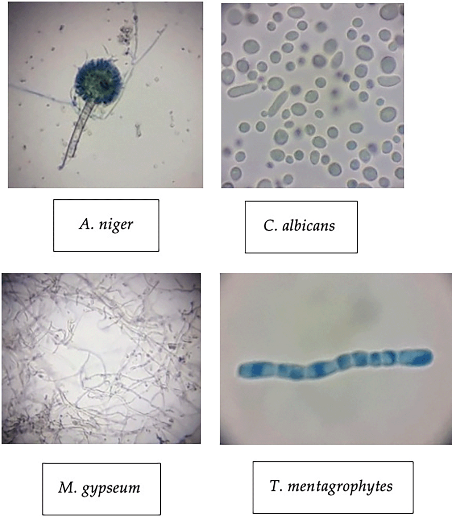
The four test fungi were also identified microscopically (1000x) to corroborate their characteristics. A. niger, C. albicans, M. gypseum and T. mentagrophytes shared similar characteristics with the same fungi theoretically. Figure 3 showed the identification results of the test. The A. niger shows that this fungus has an elongated shape with visible fungal part such as conidia, vesicles, and conidiophores that were obvious by the staining process. Meanwhile, Figure 3 depicting the C. albicans compared to A. niger. C. albicans has a variable round shape with a bluish color and nonuniform cell size. Each cell shows a different individual because this fungus is a unicellular microorganism.
MIC result on A. niger
The MIC test results for A. niger showed that well with a clear appearance only occurred at 60% concentration treatment, whereas those at 30%, 15%, 7.5% and 3.75% were cloudy with fungal colonies on the surface medium. These observations are relatively weak compared to other studies, such as Yanti et al. (2017) which examined the essential antifungal test on kaffir lime against five type of Aspergillus fungi, revealing an inhibitory mechanism as delayed spore germination and the formation of mycelia at a concentration of 0.05%.
The first observation results showed that the mechanism for fungal growth inhibition occurred only at 60%, indicating that the MIC value was reached at this concentration. Because the range of values between treatments was large, the MIC value may be reach before 60%, specially between 30% and 60%. The second testing process was conducted to minimize concentration different between treatments. Consequently, treatments with new concentrations were made, namely 45%, 50%, 55%, 60% and 65%. A fraction above 60% was intended to predict when the MFC value was not achieved.
As seen in Table 1, the well had a clear appearance before reaching a concentration of 60%, specifically at 55%. This concentration inhibited the growth of A. niger, as evidenced by its clear appearance, indicating that fungal growth was inhibited. Because treatment above 55% showed similar results, the MIC value was determined at this concentration.
Observation on each control in the second MIC test showed similar results to the first. The positive control of 2% ketoconazole had a clear appearance, indicating that this compound had antifungal activity. The control medium had a clear appearance, showing an absence of fungal or microbial growth, whereas the growth control had a cloudy appearance, indicating that the fungus grew in this treatment. The negative control n-hexane also had a cloudy appearance, suggesting that this compound lacked antifungal activity against A. niger.
MIC result on C. albicans
Observation data revealed that none of the treatments produced perfectly transparent wells in C. albicans. In general, a higher concentration implies a clearer well, but at the highest value of 40%, fungal growth was still visible, warranting a re-test to determine perfectly transparent wells. The control treatment on C. albicans yielded similar result to the MIC test on A. niger. The second test was conducted with treatment concentrations of 35%, 40%, 45%, 50% and 55%. The results showed that a clear appearance began to develop at a concentration of 45%, whereas the treatment at 35% and 45% had a cloudy appearance, suggesting fungal growth. The difference in concentration appeared to have affected the presence of fungi, corroborating a previous study (Górski et al., 2021) that tested the antifungal activity of patchouli oil at a concentration of 12.5%, 25%, 50% and 100% against C. albicans.
Based on the observations, the MIC value was determined at a concentration of 45%, the result obtained was better than that of Kamoda (Kamoda et al., 2020), stated that the MIC of the red galangal ethanolic extract against the fungus was achieved at 200 mg/mL, but the inhibition only reached at 60%. Differences in antifungal activity for each treatment with varying concentrations also reported by Ningtias et al. (2020), where the MIC value of black garlic extract against C. albicans was reached at a concentration of 50%, but active inhibition was fully achieved at 75%.
MIC result on M. gypseum
The MIC observation test on M. gypseum fungus with concentration used in twofolds, notably 40%, 20%, 10%, 5% and 2.5% revealed fungal growth in all treatments. Positive and medium controls lacked fungal development, but negative and growth controls demonstrated the opposite. Furthermore, the first observation showed that the highest concentration exhibited no inhibition, although the technique was correctly executed, as indicated by the control treatment, which showed appropriate results. The second test was conducted by increasing the upper limit of concentration and decreasing it with a value range of 5%, hence the new concentration used were 60%, 55%, 50%, 45%, and 40%.
The second MIC test revealed no fugal growth in the wells containing 50%, 55% and 60% of the teat compound, indicating maximum inhibition at these concentrations. The treatments wells overgrown as the number of fungal colonies that grew on the surface increased. Fungal growth was observed at 45% but was not as cloudy as that of 40%, suggesting that a 45% concentration inhibited growth insignificantly. The MIC value is often indicated by the smallest concentration that can inhibit total fungal growth based on visualization, it was determined at 50%.
Positive controls with 2% ketoconazole and fluconazole inhibited fungal growth, as evidence by a clear appearance with a small amount of nonhomogeneous antibiotic precipitate. However, the negative control of n-hexane showed fungal growth, indicating that this compound lacked antifungal activity. The growth control also exhibited fungal growth, suggesting that growth on PDB media was not inhibited. Notably, the control media used was clear, indicating that no undesirable microorganism were present.
MIC result on T. mentagrophyhes
The MIC test results for T. mentagrophytes at concentration of 100%, 50%, 25%, 12.5%, and 6.25% of the test compounds showed that only 100% concentration treatment inhibited fungal growth. At 50%, 25%, 12.5% and 6.25% fungal growth remained on the surface, although in varying quantities. The inhibition activity of the test substance at a certain concentration was reflected in the appearance of the media, suggesting the presence or absence of microbes.
In the first test, the MIC value was determined at a concentration of 100%. Because the range of concentrations capable of inhibiting fungal growth was extremely large, the MIC value could be obtained between 50% and 100%. The second test was conducted by minimizing the concentration difference, resulting in values of 100%, 95%, 90%, 85% and 80%.
The second MIC observation test results showed that the wells with 95% and 100% concentration exhibited no fungal or other microbial growth, as indicated by their clear color surface. However, concentration of 90%, 85% and 80% showed varying fungal growth for each treatment. The MIC value is often indicated by the smallest concentration that can inhibit the total growth of the fungi or the appearance of clear media on visualization. Based on the results, the value was determined at 95%.
Because the control treatment for T. mentagrophytes yielded identical results to those for M. gypseum, the technique used was appropriate. The difference in MIC values demonstrated that fungus resistance to fraction 1 as an antifungal agent differed for each fungal species. T. mentagrophytes value exceeded that of M. gypseum. These results are directly proportional to the previous inhibition zone test which showed that T. mentagrophytes was more resistant than M. gypseum to the fraction 1 antifungal agent containing 38.8% α-guaiene
The MFC test results for A. niger, C. albicans, M. gypseum and T. mentagrophytes revealed fungal growth in wells with a clear medium. Observations were made by creating a new inoculum from only the clear-appearing MIC test results. In order to reduce material requirements and minimize the possibility of errors as wells with cloudy appearance can be ascertained to have fungal growth.
MFC result on A. niger
The MFC test results for A. niger shown in Table 2 show that only the 55% concentration of the tree treatments indicated the present of fungal growth. Five of six inoculums created were colonized by fungus. Moreover, the number of fungal colonies in one petri disc was less that 5, this growth was relatively small compared to the original culture without treatment, indicating that the test substance inhibited A. niger growth at 55% but did not eradicate this fungus completely. Treatment concentration of 60% and 65% showed no fungal growth in each created inoculum. Based on these results, the MFC value was determined at a concentration of 60%.
The inoculation wells with the control treatment yielded the expected outcomes under the expected conditions. The positive and medium controls lacked fungal growth, whereas the negative and growth controls indicated opposite results. Moreover, the growth control treatment showed the strongest fungal growth due to the absence of effective antifungal effects.
MFC result on C. albicans
The MFC test results for C. albicans presented in Table 2 indicated that the 45% treatment exhibited fungal growth compared to the 50% and 55% treatments. Compared to the control treatment, the number of colonies was much lower for the 45% treatment. Based on these observations, the MFC value was determined at a concentration of 50%.
C. albicans’s control treatment yielded similar results to those of A. niger. However, the positive control with 2% ketoconazole exhibited no fungal growth. Ketoconazole, a commonly used conventional antifungal agent exhibited antifungal activity that was significantly more potent than fraction 1 of patchouli oil. The results indicated that a concentration of at most 2% ketoconazole can eradicate fungi.
MFC result on M. gypseum
MFC test was conducted at three concentrations, notably 50%, 55% and 60% which were found to inhibit fungal growth in the previous MIC test. The results showed that, at 50% fungal growth remained on the PDA media in all petri dishes. At 55%, four petri dishes were still observed to be overgrown with fungus, whereas the other two were clear. The number of colonies form in each petri dish at 50% and 55% did not exceed three, this growth was relatively small, indicating that the antifungal agent only inhibited fungal growth but did not eradicate M gypseum. In addition, at 60%, fungal growth was unobserved in all petri dishes; hence, the MFC value was determined at this concentration. The MFC test results for the control treatment were consistent with the prior MIC test. The positive and medium controls exhibited no fungal growth compared to the negative and growth controls.
MIC result on T. mentagrophytes
MFC testing on T. mentagrophytes was conducted at two concentrations, notably 95% and 100%, which were found to inhibit fungal growth in the previous MIC test. The results showed that, at 95%, five petri dishes were still overgrown with fungus, whereas one was not. The number of colonies formed was not significantly high; hence, the antifungal agent appeared to only inhibit but not eradicate fungi. At 100%, fungal growth was unobserved in all Petri dishes; hence, the MIC value was determined at this concentration. The control treatment for the MFC test on T. mentagrophytes yielded similar results to those of M. gypseum. The positive control at 2% ketoconazole and fluconazole did not support fungal growth. This suggests that 2% ketoconazole and fluconazole as conventional antifungal agents, have significantly more potent effects than fraction 1 of patchouli oil.
Fraction 1 of patchouli oil containing 38.8% α-guaiene exhibited antifungal activity against A. niger, C. albicans, M. gypseum and T. mentagrophytes. The MIC values for each fungus were reached at concentrations of 55%, 45%, 50% and 95%, whereas the MFC value were achieved at 60%, 50%, 60% and 100%, respectively. This study demonstrates that α-guaiene is a prospective agent effective against the investigated pathogenic fungus.
| Views | Downloads | |
|---|---|---|
| F1000Research | - | - |
|
PubMed Central
Data from PMC are received and updated monthly.
|
- | - |
Provide sufficient details of any financial or non-financial competing interests to enable users to assess whether your comments might lead a reasonable person to question your impartiality. Consider the following examples, but note that this is not an exhaustive list:
Sign up for content alerts and receive a weekly or monthly email with all newly published articles
Already registered? Sign in
The email address should be the one you originally registered with F1000.
You registered with F1000 via Google, so we cannot reset your password.
To sign in, please click here.
If you still need help with your Google account password, please click here.
You registered with F1000 via Facebook, so we cannot reset your password.
To sign in, please click here.
If you still need help with your Facebook account password, please click here.
If your email address is registered with us, we will email you instructions to reset your password.
If you think you should have received this email but it has not arrived, please check your spam filters and/or contact for further assistance.
Comments on this article Comments (0)