Keywords
carotid artery stenosis, internal carotid artery, mean central retinal artery pressure, ocular perfusion pressure, Ophthalmodynamometry
This article is included in the Eye Health gateway.
Advanced clinical diagnostic tools enable ophthalmologists to diagnose not only ocular pathologies but also identify disorders that extend beyond ocular diseases. Ophthalmodynamometry (ODM), a screening tool that most ophthalmologists do not commonly use, measured reduced mean central retinal artery pressure (MCRAP) in the clinical setting. We describe a 70-year-old female with a reduced MCRAP in the right eye who identified 50% stenosis in her right internal carotid artery (ICA). Early diagnosis facilitated prompt management and potentially prevented future ischemic events.
carotid artery stenosis, internal carotid artery, mean central retinal artery pressure, ocular perfusion pressure, Ophthalmodynamometry
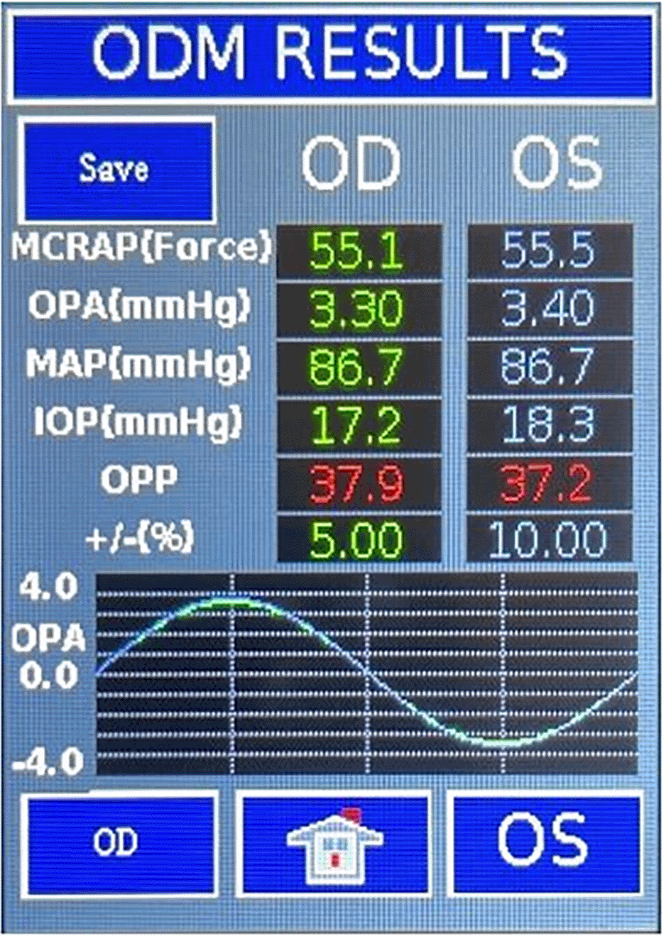
We discussed the effect of obstructive sleep apnea and anti-hypertensive medication on OPP. Added a Figure and explanation showing the screen of the ODM device with all the measurements.
See the authors' detailed response to the review by Andrew Tirsi
See the authors' detailed response to the review by Efe Kanter
Ophthalmodynamometry (ODM) is a non-invasive method to evaluate ophthalmic vascular pressure dynamics in the central retinal artery (CRA). This technique involves increasing the intraocular pressure (IOP) by applying a standardized pressure to the globe. CRA pressure is measured at the point where the lowest standardized pressure induces pulsations.1,2 ODM assessment of CRA perfusion provides insight into other arteries due to their direct anatomical communication. When ODM measures mean central retinal artery pressure (MCRAP), the ocular perfusion pressure (OPP) can be derived by calculating the difference between MCRAP and IOP.3 Because of the anatomical relationship between the CRA, the ophthalmic artery (OA) and internal carotid artery (ICA), narrowing in the OA or the ICA will decrease MCRAP and/or OPP. Therefore, identifying compromised MCRAP or reduced OPP through ODM may suggest systemic cerebrovascular occlusive disease,4 prompting further vascular assessments.
ODM has traditionally been measured using various techniques of raising the IOP, including compression or negative suction pressure. Many of these methods often require a second operator to visualize central artery changes. Recently, the Falck Multifunctional Device (FMD, Falck Medical, CT, USA) has modernized the classic ODM approach by incorporating a digitalized pressure sensor into the holding grip of a slit lamp. This improvement allows a calibrated pressure application to induce fine vascular pulsations in the CRA, eliminating the need for visualizing vessel changes during traditional ODM techniques (Figure 1). In this report, we present one of the first cases of FMD utility in an asymptomatic individual with minimal vascular risk factors. The patient presented with diminished MCRAP and was identified as having focal carotid vascular occlusive disease on further carotid doppler testing. Written informed consent was obtained from the patient to publish this report.

Both the systolic central retinal arterial pressure (SCRAP) and the diastolic central retinal arterial pressure (DCRAP) are detected by the digital infrared optical sensor. The FMD ODM weighted mean central retinal arterial pressure (MCRAP) is calculated by (SCRAP + 2 (DCRAP)) / 3, whereas Mean arterial pressure (MAP=2/3 diastolic blood pressure +1/3 Systolic blood pressure) is measured by blood pressure cuff. If MCRAP is less than 60% of MAP, displayed MCRAP will turn red to indicate measured CRA pressure is lower than it should be. Intraocular pressure (IOP) amplitude varies with the cardiac cycle and percentage (%) variation is displayed. This variation is called the ocular pulse amplitude (OPA). Ocular perfusion pressure (OPP) is displaced as MCRAP minus IOP.
A 70-year-old female in good general health, except with a history of well-controlled hypertension presented for a routine eye exam. She reported no negative effect of taking amlodipine and losartan. Her family history was significant for coronary artery disease, but she was non-diabetic and non-smoking. Her past medical history and surgical history was otherwise unremarkable. Her best corrected visual acuity was 20/20 OU. Slit lamp examination of the anterior segment was unremarkable. Bilateral dilated indirect fundoscopic examination showed arterio-venous nicking, but the retinal nerve fiber layer could be seen clearly. The optic disc margin was sharp and there was no retinal hemorrhage or exudate.
ODM using the FMD was performed because of the presence of vascular risk factors. FMD ODM directly measured the following: IOP of 15 mmHg OD and 14 mmHg OS; and MCRAP of 51.6 mmHg OD and 55.4 mmHg OS. Brachial mean arterial pressure (MAP) measured with automated blood pressure cuff was 102.7 mmHg on right and 93.3 mmHg on left. The right MCRAP (51.6 mmHg), representing 50% of the right MAP, was significantly lower (variability 3%) than the left MCRAP (55.4 mmHg), representing 59% of the left MAP. Similarly, the OPP revealed significantly lower pressure (variability 2%) in the right eye of 36.7 mmHg than in the left eye of 41.2 mmHg. The reduced MCRAP and OPP OD prompted further evaluation with carotid artery duplex scan.
On doppler imaging study, all vessel velocities measured by carotid duplex were significantly higher on the right side than the left. The measured peak velocity of the right internal carotid artery (ICA) was 122.02 cm/s, the external carotid artery (ECA) 118.45 cm/s, and the common carotid artery (CCA) 98.81 cm/s. The left sided measured peak velocities were the ICA of 47.33 cm/s, the ECA of 75.59 cm/s, and the CCA of 68.88 cm/s. In addition, the vessel velocity difference was greater in the ICA at 74.69 cm/s, than in the ECA (42.86 cm/s) or the CCA (29.93 cm/s). The carotid duplex interpretation concluded 50% stenosis in the right ICA ( Figure 2) with heterogenous calcified plaquing and minimal stenosis on the left. The patient was therefore referred for further vascular evaluation.
In this report, ODM using the FMD demonstrated reduced MCRAP and OPP in an otherwise asymptomatic patient. The patient had carotid artery stenosis as determined by carotid duplex ultrasound. Both the right MCRAP and OPP were not only measured low, but also considerably lower than the left MCRAP and OPP readings, suggesting that these parameters can be significantly and linearly linked with the existence and severity of carotid artery stenosis.5
Compared to classic ODM, FMD averages multiple measurements over the cardiac cycle, making it more reproducible. The repeated measurement for each eye in our patient was consistent, with a low variation of 2 to 3%. Notably, systemic blood pressure may significantly influence OPP. A study reported that a nocturnal dip in blood pressure, characterized by a systolic reduction of 10-12% and a diastolic reduction of 14-17%, has been linked to compromised OPP.6 Patients receiving aggressive antihypertensive medication that leads to excessive blood pressure lowering are also at risk of low OPP. If our patient’s systemic blood pressure was not appropriately controlled with amlodipine, it could paradoxically lead to an increase in OPP, potentially masking cardiovascular disease diagnoses. The relationship between OPP and ocular blood flow is complex, largely due to the eye’s autoregulation mechanisms. However, vascular endothelial dysfunction, such as that seen in obstructive sleep apnea (OSA), can impair these autoregulatory capabilities, leading to unstable OPP and significantly impacting ocular perfusion.7
The FMD can also be performed by a single operator as it is mounted to a slit-lamp thereby reducing variability. FMD has the potential to be used in a variety of applications. For example, it can detect patients at risk of having a drop in OPP after anti-VEGF injections, improving safety.8 The FMD can also measure accurate and repeatable applanation forces that compensate for the effect of corneal thickness and curvature.9 FMD can be a promising diagnostic tool for glaucoma screening and glaucoma severity assessment.10 Our case showed that the FMD may also be superior to relying on historical vascular risk factors alone in determining the need for further vascular testing. Our findings align with a recent Turkish prospective study that investigated 65 stroke patients (42 ischemic, 23 hemorrhagic) and 27 stroke-free controls, utilizing Doppler ultrasound (OAD-US) to measure peak systolic velocity (PSV) and end-diastolic velocity (EDV) in the ophthalmic artery.11 The receiver operating characteristic (ROC) analysis revealed high diagnostic accuracy in PSV ratio of 0.913 and the EDV ratio of 0.724 in differentiating stroke patients from controls. The congruence between their utilization of ophthalmic artery diagnostics and our revised FMD ODM method, underscores the ophthalmic artery’s role and potential as a non-invasive access point to the systemic vascular system. This offers a promising pathway for the early detection of conditions such as carotid artery stenosis.
Collectively, these findings render ODM using the FMD as a relatively fast, inexpensive, and reproducible office-based diagnostic test that can help determine a patient’s risk profile for systemic cerebrovascular disease. The association between reduced MCRAP and OPP and carotid artery stenosis suggests that FMD assisted ODM should be further explored as an indicator of compromised cerebrovascular hemodynamic status. Thus, the FMD provides ophthalmologists with an inexpensive non-invasive screening tool for cardiovascular disease.
Institutional approval was waived as our single case report involves retrospective medical record review of one patient and the only interaction with the patient has been for purposes of treating the patient and does not meet the Common Rule definition of research (45 CFR 164.501).
Written informed consent for publication of her clinical details and/or clinical images was obtained from the patient.
Mendeley Data: Murillo, Brian; Cheng, Anny; Samudre, Sandeep; LiVecchi, John; Gupta, Shailesh (2025), “Early Detection of Carotid Artery Stenosis with Falck Multifunctional Device (FMD), A Revised Ophthalmodynamometry Method”, DOI: 10.17632/893knw22df.112
The project contains the following reporting guidelines:
Data are available under the terms of the Creative Commons Attribution 4.0 International license (CC-BY 4.0).
All data underlying the results are available as part of the article and no additional source data are required.
| Views | Downloads | |
|---|---|---|
| F1000Research | - | - |
|
PubMed Central
Data from PMC are received and updated monthly.
|
- | - |
Competing Interests: No competing interests were disclosed.
Reviewer Expertise: Normal tension glaucoma, OPP, electrophysiology in glaucoma, retinal ganglion cell dysfunction in glaucoma, mitochondrial dysfunction in glaucoma and other retinal diseases
Competing Interests: No competing interests were disclosed.
Reviewer Expertise: Emergency medicine, ultrasonography, novel methodology in stroke, non-invasive ophthalmic monitoring.
Is the background of the case’s history and progression described in sufficient detail?
Yes
Are enough details provided of any physical examination and diagnostic tests, treatment given and outcomes?
Yes
Is sufficient discussion included of the importance of the findings and their relevance to future understanding of disease processes, diagnosis or treatment?
Yes
Is the case presented with sufficient detail to be useful for other practitioners?
Yes
References
1. Kanter E, Payza U, Karakaya Z, Acar H, et al.: A new diagnostic method in ischemic and hemorrhagic stroke: Doppler ultrasound of ophthalmic artery. Clinical Neurology and Neurosurgery. 2025; 254. Publisher Full TextCompeting Interests: No competing interests were disclosed.
Reviewer Expertise: Emergency medicine, ultrasonography, novel methodology in stroke, non-invasive ophthalmic monitoring.
Is the background of the case’s history and progression described in sufficient detail?
Yes
Are enough details provided of any physical examination and diagnostic tests, treatment given and outcomes?
Yes
Is sufficient discussion included of the importance of the findings and their relevance to future understanding of disease processes, diagnosis or treatment?
Yes
Is the case presented with sufficient detail to be useful for other practitioners?
Yes
Competing Interests: No competing interests were disclosed.
Reviewer Expertise: Normal tension glaucoma, OPP, electrophysiology in glaucoma, retinal ganglion cell dysfunction in glaucoma, mitochondrial dysfunction in glaucoma and other retinal diseases
Alongside their report, reviewers assign a status to the article:
| Invited Reviewers | ||
|---|---|---|
| 1 | 2 | |
|
Version 2 (revision) 09 Oct 25 |
read | read |
|
Version 1 21 May 25 |
read | read |
Provide sufficient details of any financial or non-financial competing interests to enable users to assess whether your comments might lead a reasonable person to question your impartiality. Consider the following examples, but note that this is not an exhaustive list:
Sign up for content alerts and receive a weekly or monthly email with all newly published articles
Already registered? Sign in
The email address should be the one you originally registered with F1000.
You registered with F1000 via Google, so we cannot reset your password.
To sign in, please click here.
If you still need help with your Google account password, please click here.
You registered with F1000 via Facebook, so we cannot reset your password.
To sign in, please click here.
If you still need help with your Facebook account password, please click here.
If your email address is registered with us, we will email you instructions to reset your password.
If you think you should have received this email but it has not arrived, please check your spam filters and/or contact for further assistance.
Comments on this article Comments (0)