Keywords
pericardial effusion, neonate, E coli, drainage
pericardial effusion, neonate, E coli, drainage
We have made changes to our first draft of the manuscript for more clarity and accuracy.
Grammar and expression errors have been corrected in all sections.
In the introduction, we added a reference explaining the pathogenesis of pericardial effusion caused by central venous catheters.
In the case report, we provided additional data about:
- The mother's serology
- The patient's and her sister's anthropometrics
- The patient's physical examination during her first hospitalization
- The COVID-19 test result
- The discussion and exclusion of the diagnosis of early onset neonatal infection, justifying the non-prescription of antibiotics
- Details of the cardiac ultrasound
- Data from the physical examination at discharge
- Details of follow-up
In the Discussion section, we have:
- Added a reference describing the incidence of PE in neonates with central venous catheters (recommended by reviewer)
- Detailed the rarity of isolation of the pathogen in pericardial fluid according to the literature
- Added a reference (recommended by reviewer) on the rarity of PE in neonatal E. coli septicaemia
- Added a paragraph (recommended by reviewer) on the value of our case report in the context of rational antibiotic prescription to prevent bacterial resistance
- Added a paragraph on the impact on practice, limitations, and recommendations
Figures: we have:
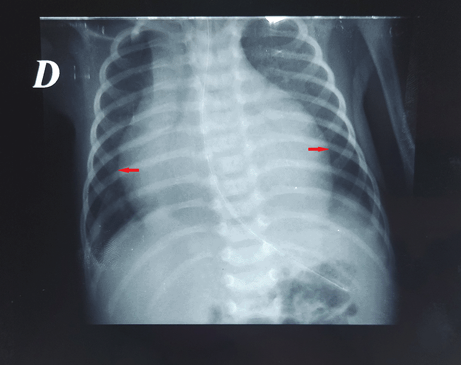
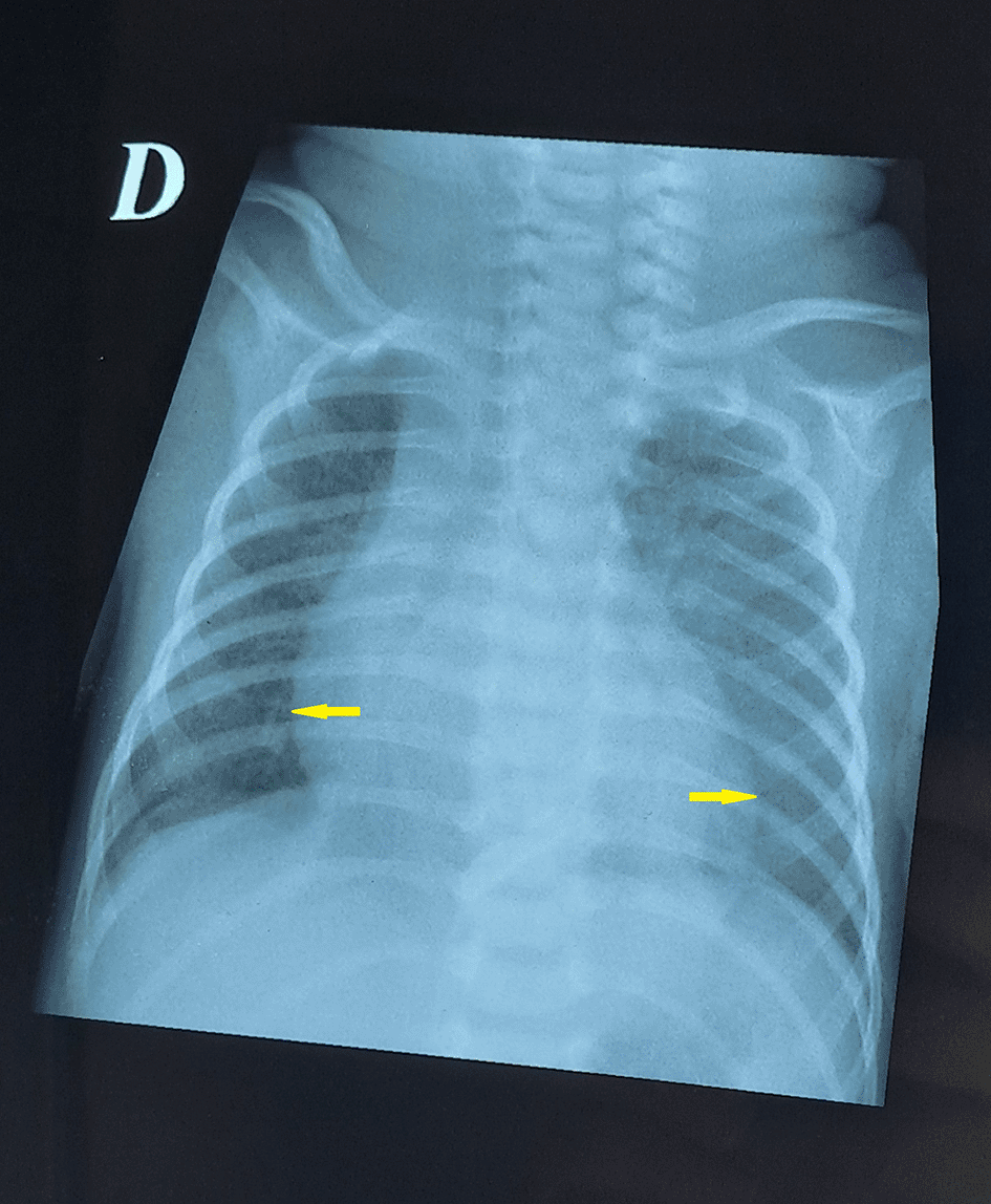
- Modified the titles (recommended by reviewer) to emphasize findings before and after pericardial drainage
- Added arrows on the radiograph showing cardiomegaly
See the authors' detailed response to the review by Khadijat Isezuo and Asma'u Adamu
See the authors' detailed response to the review by Lamin Makalo
Pericardial effusion (PE) is a rare condition in neonates. Symptoms include difficulty breathing and fever. Without appropriate treatment, it can quickly progress to hemodynamic collapse, tamponade, and ultimately, death. The most common cause of pericardial effusion in neonates are central venous catheters. It is due to the mechanical and osmotic injury of the thin and immature myocardium in neonates and especially preterm.1
A preterm female infant and her twin sister were born at 34 weeks of gestation via an emergency caesarean section due to covid 19 infection in the mother. Maternal serologies (toxoplasmosis, rubella, hepatitis B and syphilis) and prenatal ultrasound findings were unremarkable.
The first twin was small for gestational age and had severe respiratory distress. She was referred to the PICU. She was discharged on day 10 of hospitalization. No further symptoms were noted.
Our patient weighted 1600 g (small for gestational age), and Apgar score was 8 at 1 min and 9 at 10 min. She was referred to out neonatal unit in the pediatric ward for respiratory distress. At presentation, respiratory rate was 62 per minute, heart rate was 138 per minute and oxygen saturation was 97%. Chest radiography was normal. Covid 19 polymerase chain reaction test was negative. She was placed on oxygen therapy via high-flow nasal cannula for three days. The patient received fluids through a peripheral venous catheter. The diagnosis of early onset neonatal infection was discussed but excluded based on: rapid improvement of respiratory status, normal CRP levels and negative blood culture. The patient did not receive any antibiotics. Final diagnosis was transient tachypnea of the newborn. As her respiration improved, she was placed on room air on day 3 of life, fluid infusion was discontinued on day 4 of life, and she was gradually fed with milk formula (breast milk was not available as her family lived from our facility). She was fed initially via gastric tube and gradually via bottle. The patient was discharged on day 14 of life.
On day 18 of life, she presented to the emergency room with fever and dyspnea. Respiratory rate was 65/minute with intercostal recession. Oxygen saturation was 97% in room air. The patient was placed on oxygen via a nasal cannula. Chest radiography was normal. Laboratory tests showed a leukocyte count of 26000/mm3 and an elevated reactive protein C level of 220 mg/l. Initially, a nosocomial infection was suspected, and the patient was received imipenem and amikacin. There was no improvement in her respiratory status.
On day 21 of life, two blood cultures obtained on the day of readmission were positive for multidrug-sensitive Escherichia coli (E. coli). The patient was then switched to cefotaxime. On day 23 of life, our patient developed signs of heart failure with tachycardia, hepatomegaly, tachypnea, and a capillary refill of 4 seconds. Chest radiography revealed cardiomegaly (cardiothoracic ratio 0.67) with a globular heart shape (Figure 1). Electrocardiogram was normal. Cardiac ultrasound was not available in our hospital so it was performed by cardiologists in another facility. It revealed echogenic, circumferential pericardial effusion (11 mm) and pre tamponade (protodiastolic right ventricle collapse). It also showed a small patent ductus arteriosus. There were no signs of endocarditis.
A pericardial drain was inserted surgically, and 8 ml of purulent fluid mixed with blood was aspirated. Cytological analysis showed 3 leukocytes per ml. Biochemical analysis was not performed. No bacteria were isolated in pericardial fluid. After pericardial drainage, the respiratory status improved, and the shape of the heart was normal on chest radiography (Figure 2). The pericardial catheter was removed 10 days later. Cefotaxime was continued for a total duration of 14 days. A second cardiac ultrasound was performed at the age of 32 days. It showed no pericardial effusion. The patient was discharged on day 35 of hospitalization. At discharge, her weight was 2550 g and her physical examination showed normal body temperature, respiratory rate 34 per minute and heart rate 124 per minute. She had regular follow-up until the age of 6 months. At that age, her weight was 6700 g and her physical examination was normal. She was lost of follow-up since.
Pericardial effusion (PE) is rare in neonates. Its clinical presentation depends on the speed and amount of fluid accumulation.2 This can lead to tamponade and even death. The most common cause is iatrogenic because of central venous catheters.3 In a meta-analysis of 21 studies and 99 cases, pooled incidence of PE in neonates who had central venous catheters was 3.8‰. Mortality rate was 2.7%.1
In a national American study of all cases of pericardial effusion, the lowest incidence was observed in neonates (0.04%).4 In a literature review, 34 patients were enrolled. The most common causes were central venous catheters (n = 21), Down syndrome (n = 3), and infections (n = 3). The pathogen agent was isolated in pericardial fluid in only one cas (candida albicans). The second case was caused by Escherichia coli isolated in blood culture. In the third case, PE was secondary to parainfluenza virus 3 which was isolated in nasopharyngeal swab.3
Pericardial effusion due to an E. coli infection is rare. To date, only case reports have been published. In 1979, Wynn described purulent pericarditis in a 64 hours aged neonate. Autopsy confirmed the diagnosis of pericardial effusion, and E coli was isolated from the blood culture.5 In 2006, Benjamin described pericarditis in a 10 days old boy. E. coli was isolated from blood samples obtained in the emergency room before referral. He was managed with pericardectomy, pericardial drainage, cefotaxime, and indomethacin. The intraoperative samples were negative.6
Even in E. coli sepsis, pericardial effusion remains rare. Lai described the epidemiology of invasive E. coli infection in 94 Chinese neonates, including early and late onset disease. No case of pericardial effusion was described.7
In our case, initial suspicion of a nosocomial infection was raised given the patient’s recent hospitalization. The isolation of E. coli with multidrug sensitivity underscores the potential for multidrug-resistant strains in neonatal units, especially in settings where broaded-spectrum antibiotics such as carbapenems and aminoglycosides are frequently used empirically. This highlights the importance of strict infection prevention measures, rational antibiotic stewardship and early targeted therapy guided by culture results.
The management of PE is variable, from surveillance in small asymptomatic effusions to pericardiocentesis, pericardectomy, and pericardial drainage.3,8 In an American retrospective cohort, pericardial drainage was performed in 8.5% of neonates.4
Outcomes in case reports of infective pericarditis in neonates were good, except in the case of postmortem diagnosis.5 In an American study, the overall mortality was 6.8%, and mortality was the highest among neonates (12.4%).4
Although neonatal pericardial effusion is rare, it should be considered in neonates with cardiac failure, especially in the context of infection. This diagnosis should be considered in neonates who develop signs of deterioration even if they have no central venous catheters. Infective pericarditis in neonates is a rare condition. Management depends on tolerance of the effusion. Mortality was higher in neonates than in the other age groups.
This case underscores the need for clinicians to maintain high index of suspicion for pericardial effusion in neonates presenting with sepsis and cardiorespiratory deterioration. The absence of central venous catheters cannot reduce the probability. Incorporating point-of-care echocardiography in deteriorating neonates could facilitate earlier detection and intervention. Furthermore, routine review of empirical antibiotic regimens in neonatal sepsis is critical to balance adequate coverage with antimicrobial stewardship.
Neonatal pericardial effusion due to Escherichia coli is an exceedingly rare but potentially life-threatening condition. Early recognition and timely pericardicentesis can be lifesaving.
Recommendations:
• Clinicians should suspect pericardial involvement in neonates with unexplained cardiorespiratory compromise during sepsis, even without central catheters.
• Echocardiography should be integrated in the evaluation of deteriorating neonates with sepsis.
• Infection prevention and control practices should be strengthened in neonatal units to minimize the risk of nosocomial multi-drug resistant infections.
• Empiric antibiotic therapy in neonatal sepsis should be carefully tailored, with early de-escalation based on culture results, to preserve antimicrobial effectiveness.
• Collaborative multicenter registries and pooled case reports are needed to clarify the epidemiology, management and outcomes of neonatal infective pericarditis.
Use of AI tools: The authors did not use AI technology in the writing process.
| Views | Downloads | |
|---|---|---|
| F1000Research | - | - |
|
PubMed Central
Data from PMC are received and updated monthly.
|
- | - |
Provide sufficient details of any financial or non-financial competing interests to enable users to assess whether your comments might lead a reasonable person to question your impartiality. Consider the following examples, but note that this is not an exhaustive list:
Sign up for content alerts and receive a weekly or monthly email with all newly published articles
Already registered? Sign in
The email address should be the one you originally registered with F1000.
You registered with F1000 via Google, so we cannot reset your password.
To sign in, please click here.
If you still need help with your Google account password, please click here.
You registered with F1000 via Facebook, so we cannot reset your password.
To sign in, please click here.
If you still need help with your Facebook account password, please click here.
If your email address is registered with us, we will email you instructions to reset your password.
If you think you should have received this email but it has not arrived, please check your spam filters and/or contact for further assistance.
Comments on this article Comments (0)