Keywords
Alpha-fetoprotein; intrahepatic cholangiocarcinoma; cholangioblastic adenocarcinoma; yolk sac tumor mimic; AFP-producing tumor; non-islet cell tumor hypoglycemia; hypoglycemia; tumor-associated hypoglycemia

This article is included in the Oncology gateway.
Alpha-fetoprotein; intrahepatic cholangiocarcinoma; cholangioblastic adenocarcinoma; yolk sac tumor mimic; AFP-producing tumor; non-islet cell tumor hypoglycemia; hypoglycemia; tumor-associated hypoglycemia
Cholangiocarcinoma is a rare and highly malignant cancer that originates from the biliary tract epithelial cells and is the second most prevalent primary hepatic malignancy after hepatocellular carcinoma.1 Intrahepatic cholangiocarcinoma (ICC) presents with vague symptoms and invades aggressively, leading to diagnosis at advanced stages. Due to this diagnostic complexity, cholangiocarcinoma has a poor prognosis and limited therapeutic interventions. Diagnosis of AFP-producing cholangiocarcinomas is challenging due to high alpha-fetoprotein (AFP) levels that can mimic the more common hepatocellular carcinoma or non-seminomatous germ cell tumors, specifically yolk sac tumors.2–4,8
Uncommon variants of cholangiocarcinoma, such as hepatoid cholangioblastic adenocarcinoma, exhibit immunohistochemical and structural similarities with germ cell tumors due to AFP synthesis and hepatoid differentiation.5 These tumors are known to impersonate metastatic yolk sac tumors clinically, in diagnostic imaging, and histopathologically, commonly in young patients with widespread metastatic disease and significantly elevated AFP levels.2,4 Treatment protocols and success rates vary significantly across the different AFP-producing malignancies.
Severe hypoglycemia can also present in patients with advanced hepatic malignancies due to massive replacement of functional hepatic parenchyma and diminished glucose homeostasis.
A 29-year-old male with a recent diagnosis of hepatitis B on tenofovir initially presented to an outside hospital with progressive shortness of breath, abdominal pain, nausea, and significant unintentional weight loss. Imaging at that time demonstrated a hepatic mass and findings concerning for tuberculosis. No definitive diagnosis was established, and the patient was discharged on amoxicillin–clavulanate, clarithromycin, and systemic corticosteroids.
The patient later developed worsening dyspnea and sought a second opinion at our institution. Imaging performed in the emergency department revealed widely metastatic disease involving the liver and lungs. Laboratory evaluation demonstrated a markedly elevated alpha-fetoprotein (AFP) level of 60,500 ng/mL. The patient was admitted for further evaluation of metastatic malignancy of unknown primary origin.
Computed tomography of the chest, abdomen, and pelvis showed multiple hepatic masses, pulmonary nodules, and a lytic lesion of the left iliac crest, raising concern for disseminated malignancy ( Figure 1A-B). Scrotal ultrasonography showed no evidence of testicular masses or other abnormalities.
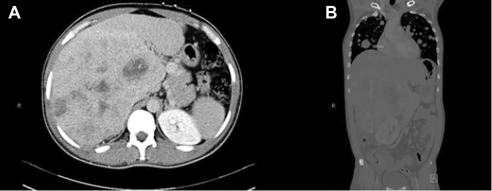
B) Coronal CT with contrast of the abdomen and pelvis demonstrating marked hepatomegaly measuring 24 cm in craniocaudal dimension with diffuse hepatic tumor infiltration.
A liver biopsy initially demonstrated a malignant neoplasm suggestive of a germ cell tumor composed of pure yolk sac tumor elements. Based on this diagnosis and the markedly elevated AFP, the patient was started on BEP chemotherapy (bleomycin, etoposide, and cisplatin). He completed the inpatient portion of cycle one without significant adverse effects, although the final two doses of bleomycin were not administered. Urologic evaluation did not recommend orchiectomy given the absence of testicular lesions, and the patient was considered to have an extragonadal germ cell tumor.
During hospitalization, the patient’s course was complicated by persistent and severe hypoglycemia, resulting in multiple syncopal episodes. Initial management included continuous dextrose 10% (D10W) infusions; however, recurrent episodes of hypoglycemia persisted with glucose levels in the 30 mg/dL range. Endocrine evaluation showed diminished insulin (0.2 μU/mL), C-peptide (0.06 ng/mL), and proinsulin (12.4 pmol/L), findings that excluded endogenous hyperinsulinemia. Serum IGF-1 was diminished (13 ng/mL), while IGF-2 was within the normal range (361 ng/mL), making IGF-2-mediated non-islet cell tumor hypoglycemia unlikely. ACTH and cortisol levels remained normal. Overall, the hypoglycemia was attributed to defective hepatic glucose synthesis and glycogen storage because of the massive tumor burden.
During the same admission, a single sputum culture for acid-fast bacillus (AFB) test returned positive. Mycobacterium tuberculosis PCR testing was negative on two different occasions, and QuantiFERON testing was also negative. A fast-growing nontuberculous mycobacterium, usually seen as an environmental colonizer, was identified on subsequent assessment. The infectious disease team stated that the finding was most likely transient colonization rather than an active infection, as the diagnostic criteria for nontuberculous mycobacterial pulmonary disease were not met. Thus, the infectious disease specialists did not recommend any antimicrobial treatment.
The patient was discharged to outpatient follow-up. Subsequently, the pathology from the liver biopsy was amended. The initial pathology interpretation rendered a preliminary diagnosis of a malignant germ cell tumor composed of pure yolk sac tumor. Given the significantly elevated AFP level and the immunophenotypic profile showing positivity for SALL4, glypican-3, and AFP, the patient received one cycle of BEP chemotherapy. Further pathologic assessment was conducted, including fluorescence in situ hybridization (FISH) for isochromosome 12p [i(12p)], a characteristic cytogenetic abnormality of germ cell tumors. The FISH study was negative, which excluded a yolk sac tumor. Subsequent immunohistochemical analysis showed positivity for CK7, EMA, pankeratin, focal HepPar-1, retained BAP1 expression, and albumin chromogenic in situ hybridization positivity, supporting hepatobiliary differentiation ( Figure 2A-B). These pathologic findings led to revision of the diagnosis to a poorly differentiated adenocarcinoma of cholangioblastic origin.
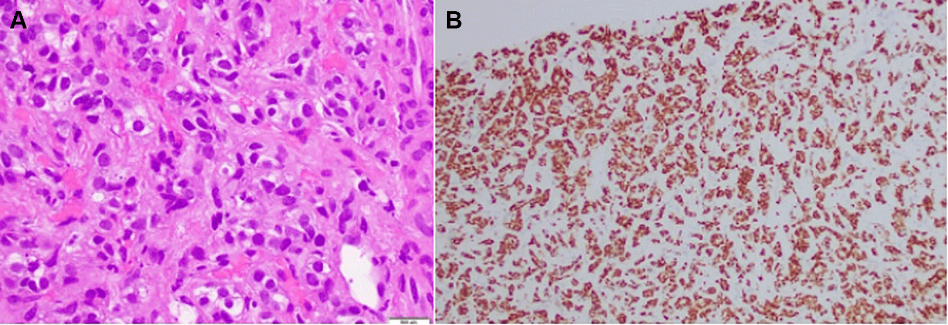
B) Albumin chromogenic in situ hybridization (ISH) showing diffuse tumor cell positivity, supporting hepatobiliary differentiation and cholangioblastic origin.
The patient’s treatment regimen was changed to systemic therapy for cholangiocarcinoma. He was started on gemcitabine, cisplatin, and pembrolizumab, administered on a 21-day cycle (cisplatin on day 1, gemcitabine on days 1 and 8, pembrolizumab every 3 weeks). The patient completed four cycles of this regimen.
Despite treatment, the patient developed progressive abdominal pain and worsening hypoglycemia. Imaging demonstrated progression of metastatic disease within the liver and lungs. Oncology recommended a repeat liver biopsy with next-generation sequencing to identify potential actionable mutations for targeted therapy. Interventional radiology was consulted for a core needle biopsy to be sent for molecular analysis.
Given the patient’s advanced disease, poor functional status, and lack of response to systemic therapy, the prognosis was considered poor. Palliative care was consulted for symptom management and goals-of-care discussions. The patient continued to experience complications related to his malignancy, including cancer-related pain, malignant ascites, severe malnutrition, and persistent hypoglycemia, likely driven by extensive hepatic tumor burden.
Despite supportive care and oncologic management, the patient’s condition continued to deteriorate, and he ultimately succumbed to his illness.
There is significant clinical, serologic, and histopathologic overlap between cholangioblastic adenocarcinoma and yolk sac tumors, creating diagnostic difficulty when determining a definite diagnosis.2,5 This patient was initially diagnosed with pure yolk sac tumor due to the high AFP levels, biopsy results, and widespread metastatic cancer. Although elevated AFP is often associated with nonseminomatous germ cell tumors, it is also elevated in hepatobiliary cancers, particularly cholangioblastic tumors.2–5,9 The potential for misdiagnosis of AFP-producing cholangiocarcinomas, particularly in young patients, is high due to elevated AFP levels that may initially point to hepatocellular carcinoma (HCC) and overlapping radiologic features, compounded by limited literature and low clinical suspicion.3,4
There have been limited reports on AFP-producing cholangioblastic adenocarcinomas. All these cases involve a young patient with significantly elevated AFP levels. However, the severe refractory hypoglycemia and initial treatment for a yolk sac tumor show the unusual and complex characteristics of this presentation. This case makes it evident that maintaining a broad differential diagnosis when assessing AFP-producing tumors is crucial.
This diagnosis was further complicated by the lack of a testicular primary lesion on scrotal ultrasonography. Extragonadal germ cell tumors are extremely uncommon, especially when isolated to the hepatobiliary system, as most occur in the retroperitoneum and mediastinum. The significant morphologic similarity between germ cell tumors and AFP-producing adenocarcinomas is evident on repeat pathologic review, confirming a poorly differentiated metastatic cholangioblastic adenocarcinoma instead.2,5 This case demonstrates the need for repeat pathologic review and multidisciplinary confirmation when there is discordance or atypical presentation.
The complexity of this case is further magnified by the expression of the markers SALL4 and glypican-3. SALL4 is known to be a sensitive marker for germ cell tumors, especially yolk sac tumors; its expression has also been shown in hepatocellular carcinoma and cholangiocarcinoma. Because of this overlap, its specificity in distinguishing these carcinomas is limited.10 In this case, the tumor’s positivity for SALL4, AFP, and glypican-3 ( Figure 3) contributed to the initial misclassification as a germ cell tumor, demonstrating the significance of immunohistochemical panels and molecular testing in establishing the correct diagnosis.
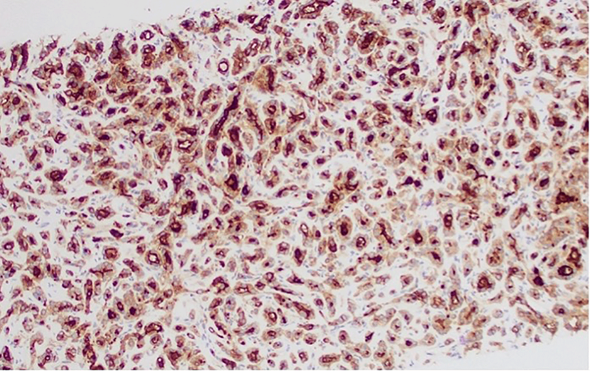
Expression of glypican-3 is a recognized feature shared by both yolk sac tumors and cholangioblastic adenocarcinoma, complicating diagnosis.
Another rare finding in this case was severe recurrent hypoglycemia. Although non-islet cell tumor hypoglycemia (NICTH) was initially considered, endocrine assessment showed normal IGF-2, significantly suppressed insulin, C-peptide, and proinsulin levels, and normal adrenal function. Based on these findings, endogenous hyperinsulinemia and classic IGF-2-mediated NICTH were excluded.6 Instead, the hypoglycemia was most likely related to extensive hepatic replacement by tumor, leading to impaired glycogen storage, decreased gluconeogenesis, and diminished hepatic glucose output. Similar processes have been described in patients with advanced hepatic malignancies and extensive tumor burden.6,7
This case underscores the complexity of treating rare metastatic AFP-producing tumors. Initially, the patient underwent BEP chemotherapy for an expected germ cell tumor before transitioning to cisplatin, gemcitabine, and pembrolizumab upon correct diagnosis. Even with aggressive chemotherapy, the disease rapidly progressed with progressive metastasis and diminished functional status. Due to limited literature on cholangioblastic adenocarcinoma, there are no thoroughly established medical management guidelines.5 To establish therapeutic guidelines and improve diagnostic accuracy, extensive research and case reports must continue.
This case highlights the diagnostic and therapeutic challenges associated with poorly differentiated cholangioblastic adenocarcinoma presenting with markedly elevated AFP and widespread metastatic disease in a young patient. The initial histopathologic interpretation suggested a germ cell tumor, leading to treatment with BEP chemotherapy, before subsequent pathology revision identified cholangioblastic adenocarcinoma. This underscores the importance of careful pathologic evaluation and the potential for diagnostic overlap between AFP-producing malignancies. Additionally, the patient’s persistent and severe hypoglycemia, likely driven by extensive hepatic tumor burden, after exclusion of insulin-mediated and IGF-2 mediated causes, further complicated management and reflects a rare but significant metabolic complication of advanced malignancy. Despite systemic chemotherapy and immunotherapy, the disease demonstrated rapid progression and poor response to treatment. This case emphasizes the need for early multidisciplinary collaboration, consideration of repeat or confirmatory pathology in atypical presentations, and further research into effective therapeutic strategies for aggressive AFP-producing hepatobiliary malignancies.
Written informed consent for publication of the patient’s clinical details and accompanying images was obtained from the patient. A copy of the written consent is available for review by the Editor-in-Chief upon request.
This report describes a single patient and does not constitute human subjects research requiring Institutional Review Board (IRB) approval according to institutional policy. Therefore, formal IRB review and approval were not required. Written informed consent for publication of the patient’s clinical information and images was obtained from the patient.
| Views | Downloads | |
|---|---|---|
| F1000Research | - | - |
|
PubMed Central
Data from PMC are received and updated monthly.
|
- | - |
Provide sufficient details of any financial or non-financial competing interests to enable users to assess whether your comments might lead a reasonable person to question your impartiality. Consider the following examples, but note that this is not an exhaustive list:
Sign up for content alerts and receive a weekly or monthly email with all newly published articles
Already registered? Sign in
The email address should be the one you originally registered with F1000.
You registered with F1000 via Google, so we cannot reset your password.
To sign in, please click here.
If you still need help with your Google account password, please click here.
You registered with F1000 via Facebook, so we cannot reset your password.
To sign in, please click here.
If you still need help with your Facebook account password, please click here.
If your email address is registered with us, we will email you instructions to reset your password.
If you think you should have received this email but it has not arrived, please check your spam filters and/or contact for further assistance.

Comments on this article Comments (0)