Keywords
breast cancer, axillary lymph node dissection, lymphedema prevention, dermofat graft, lymphangiogenesis, reconstructive surgery
This article is included in the Oncology gateway.
breast cancer, axillary lymph node dissection, lymphedema prevention, dermofat graft, lymphangiogenesis, reconstructive surgery
Breast cancer remains the most prevalent malignancy among women worldwide, with approximately 2.1 million new cases reported in 2018, making it the fifth leading cause of cancer-related mortality, with around 620,000 deaths annually.1 Treatment modalities, including surgery, radiotherapy, chemotherapy, endocrine therapy, targeted therapy and immunotherapy have significantly improved survival rates, with a standardized 5-year survival rate approaching 90% globally.1 However, these treatments are often associated with complications, notably lymphedema, a chronic condition characterized by limb swelling, reduced mobility, and diminished quality of life2 and some end up with amputation. Lymphedema arises due to impaired lymphatic drainage, primarily following axillary lymph node dissection (ALND), with incidence rates ranging from 19.3% to 24.6% in ALND patients compared to 2.2%–8.3% in those undergoing sentinel lymph node biopsy (SLNB).3 In addition, Breast Conserving Surgery (BCS) often results in defects such as a sunken skin area, a smaller size of the operated breast, leading to less aesthetic appearance, or the presence of non-physiological structures due to the use of implants, such as silicone, for reconstruction.4
ALND, despite its declining use following trials like ACOSOG Z0011 and AMAROS, remains necessary for patients with axillary lymph node metastases.4 The procedure disrupts lymphatic channels, increasing lymphedema risk, which is further exacerbated by radiotherapy, chemotherapy, obesity, and infections.3 Current lymphedema management, such as compression stockings and manual lymphatic drainage, is primarily symptomatic, time-intensive, and fails to address the underlying pathology, leading to recurrence upon cessation.5 Sentinel Lymph Node Biopsy (SLNB) can reduce the incidence of lymphedema (the risk of lymphedema with axillary dissection is three times higher compared to sentinel node biopsy, with an Adjusted Hazard Ratio [AHR] of 2.71, p < 0.0001) (McDuff et al., 2021). However, SLNB is only effective when performed in early-stage breast cancer and requires specific facilities (such as blue dye and scintigraphy), which are still difficult to obtain in developing countries, including Indonesia. Furthermore, 53% of non-sentinel lymph nodes were found to be positive for metastasis after sentinel lymph node biopsy, necessitating further axillary dissection. Therefore, the use of this procedure remains limited (Sung et al., 2022). Advanced surgical techniques like lymphatic microsurgical preventive healing approach (LYMPHA) and lymphaticovenous anastomosis (LVA) have shown promise but are technically challenging, time-consuming, and resource-intensive, limiting their widespread adoption.6
Dermofat graft (DFG) implantation, involving the transfer of autologous dermis and subcutaneous fat, has emerged as a potential reconstructive technique for post-axillary dissection defects.7 The implanted dermofat tissue will grow and promote lymphangiogenesis through the process of reconnecting and forming new lymphatic channels. The adipose tissue in the dermofat graft can remain viable (non-necrotic) due to the presence of blood vessels that are still connected to the fat tissue. When the dermofat graft is implanted, these blood vessels can provide the oxygen and nutrients needed for the fat cells to survive and function properly8,9,10 Recent studies suggest DFG may promote lymphatic regeneration through adipose-derived stem cells (ADSCs), which differentiate into lymphatic endothelial cells and secrete lymphangiogenic factors like vascular endothelial growth factor-C (VEGF-C).9 VEGF-C is a critical regulator of lymphangiogenesis, facilitating lymphatic vessel formation and potentially mitigating lymphedema risk.10 ADSCs also secrete VEGF-A, which plays a crucial role in the process of angiogenesis.11
Initially popularized for aesthetic corrections, DFG enhances volume restoration and tissue spacing, with minimal volume loss (1–20%) compared to fat grafts (up to 45% loss annually) due to improved vascularization from the dermal layer.7,8 This case series aims to evaluate the feasibility and efficacy of DFG implantation in post-axillary dissection defects for promoting lymphatic regeneration, preventing lymphedema and reconstruction in BCS operation for breast cancer patients.
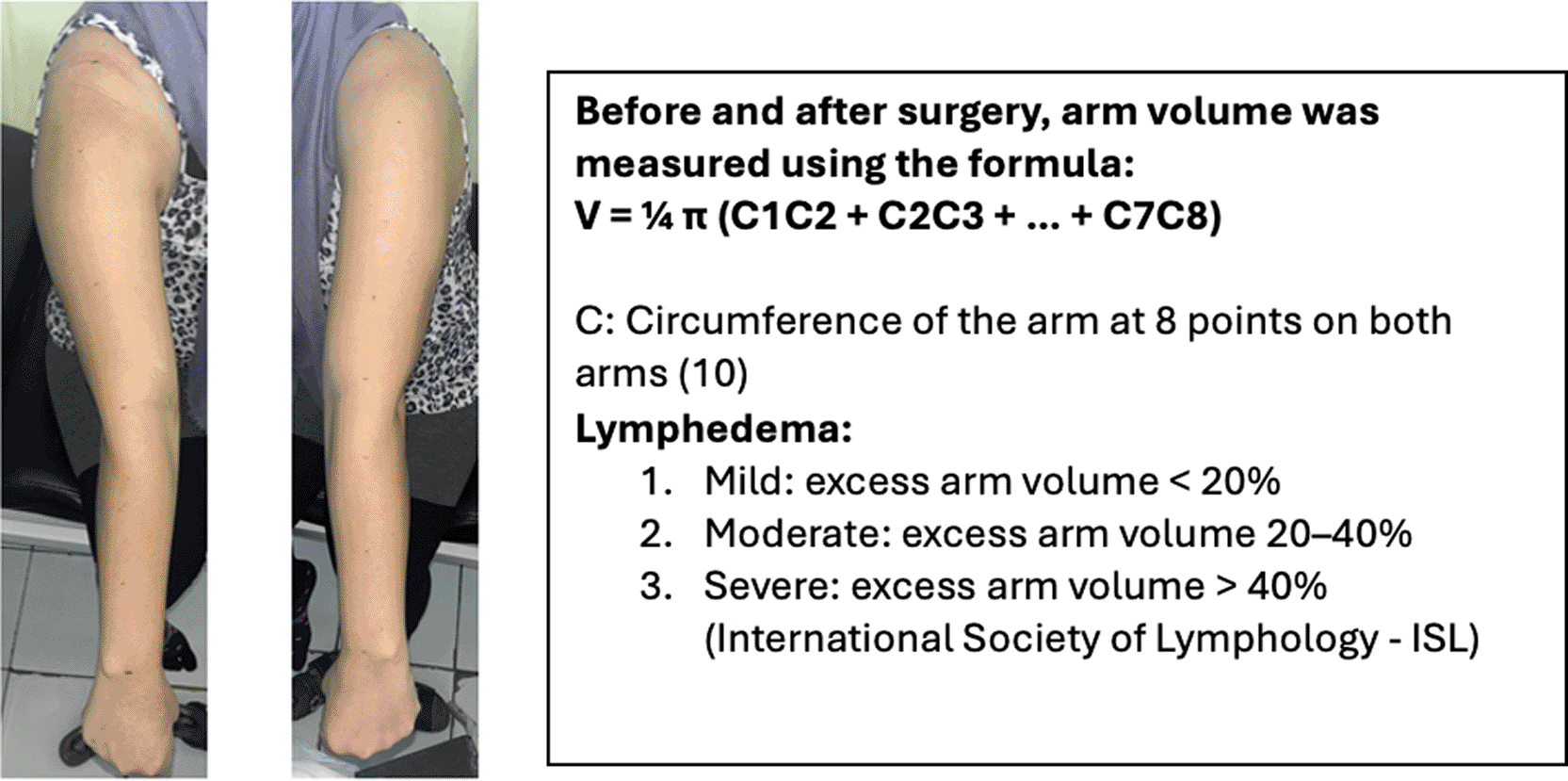
Before surgery, the volume of the right and left arms was measured using circumferential measurements at eight standardized points along the arm ( Figure 1). Intraoperative procedure of lymphedema prevention following Modified Radical Mastectomy (MRM) as follows:
1. After completing the axillary dissection, a drainage tube is inserted. An elliptical deepithelialization is performed in the suprapubic area (lower abdomen) so that the scar will later be hidden by underwear.
2. Harvesting of the DFG (dermis and adipose tissue down to just above the fascia) is performed in the deepithelialized area.
3. The DFG is implanted into the post-dissection defect and fixed to the surrounding tissue, with the dermis positioned inward. The donor graft size is 20% larger than the axillary dissection specimen.
4. The surgical wound is closed with subcutaneous and cutaneous sutures.
5. The donor site defect is closed with primary suturing.
6. A vacuum drainage system is applied.
For lymphedema prevention and breast reconstruction after Breast Conserving Surgery (BCS), the procedure as follows:
1. After completing the lumpectomy and axillary dissection, a drainage tube is inserted.
2. An elliptical deepithelialization is performed in the suprapubic area (lower abdomen).
3. Harvesting of the DFG (dermis and adipose tissue above the fascia) is carried out in the deepithelialized area, with a larger graft size since part of it will be used for reconstruction.
4. The graft is divided into two parts: one portion of the DFG is implanted into the post-dissection defect and fixed to the surrounding tissue with the dermis facing inward. The other portion is implanted into the BCS defect within the breast and also fixed to the surrounding tissue.
5. Surgical wound are closed with subcutaneous and cutaneous sutures.
6. The donor site defect is closed with primary suturing.
7. A Vacuum drainage is applied to both the breast and axillary defects.
One, Three and Six Months Post-Operation:
1. Lymphoscintigraphy is performed to evaluate the formation of lymphatic vessels and the quality of lymphatic flow in the axillary fossa.
2. The volume of the right and left arms is measured and compared to each other, as well as to the preoperative arm volume.
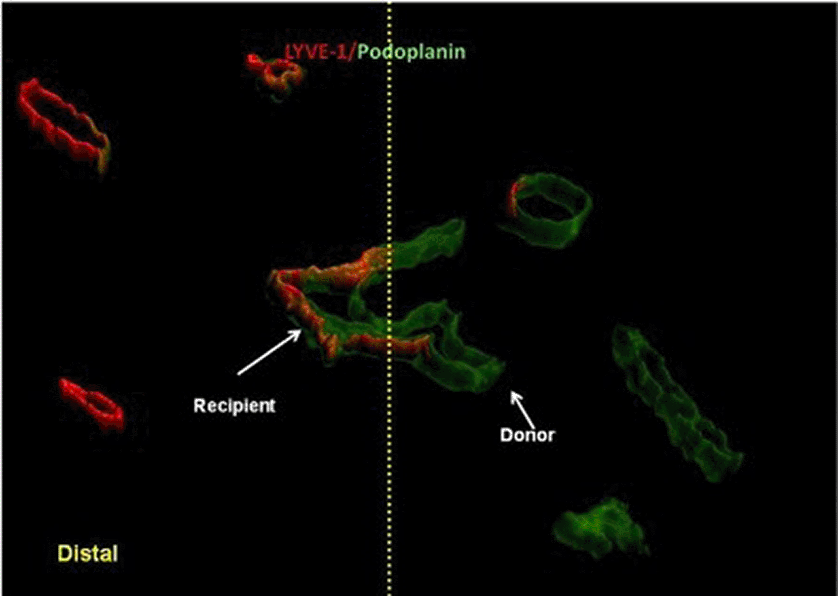
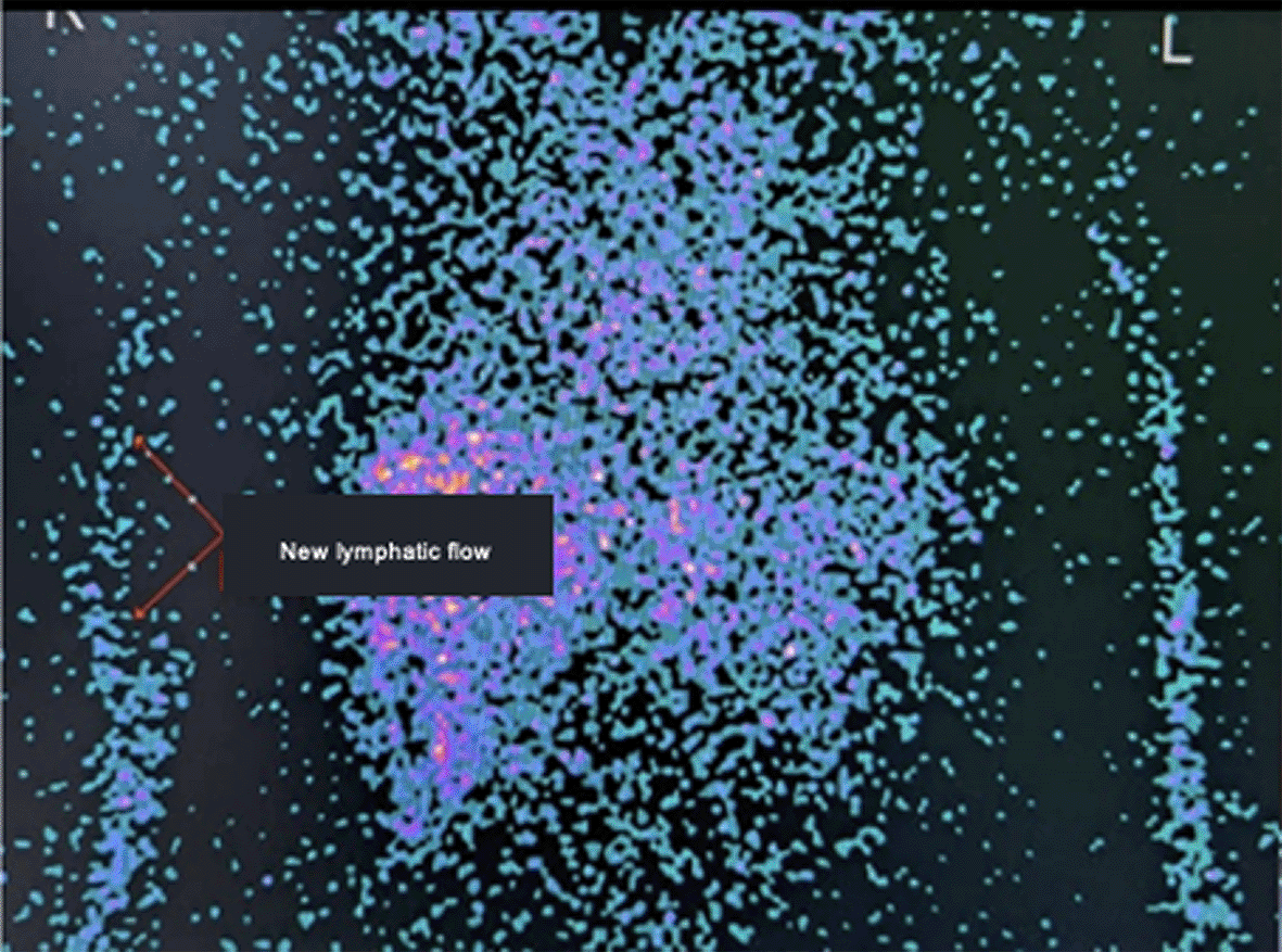
The surgical steps of dermofat graft harvesting from the suprapubic donor site and implantation into the axillary defect following axillary lymph node dissection are illustrated in Figure 2. The surgical steps of dermofat graft implantation for axillary defect filling and breast reconstruction following breast-conserving surgery are illustrated in Figure 3. Lymphoscintigraphy or lymphatic regeneration after tissue transfer occurs through spontaneous reconnection between the donor and recipient lymphatic vessels are illustrated in Figure 4. Figure 5 showed the formation of new lymphatic flow (lymphangiogenesis) was observed following dermofat graft implantation in the axilla.
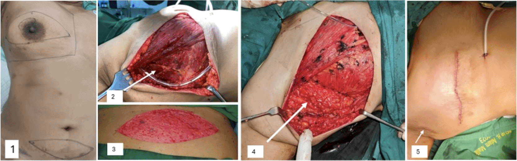
1. Surgical design. 2. Axillary defect. 3. Donor area in the suprapubic region, which has been deepithelialized. 4. DFG filling the axillary defect and fixed to the surrounding tissue. 5. Surgical wound after closure, showing no visible axillary depression.
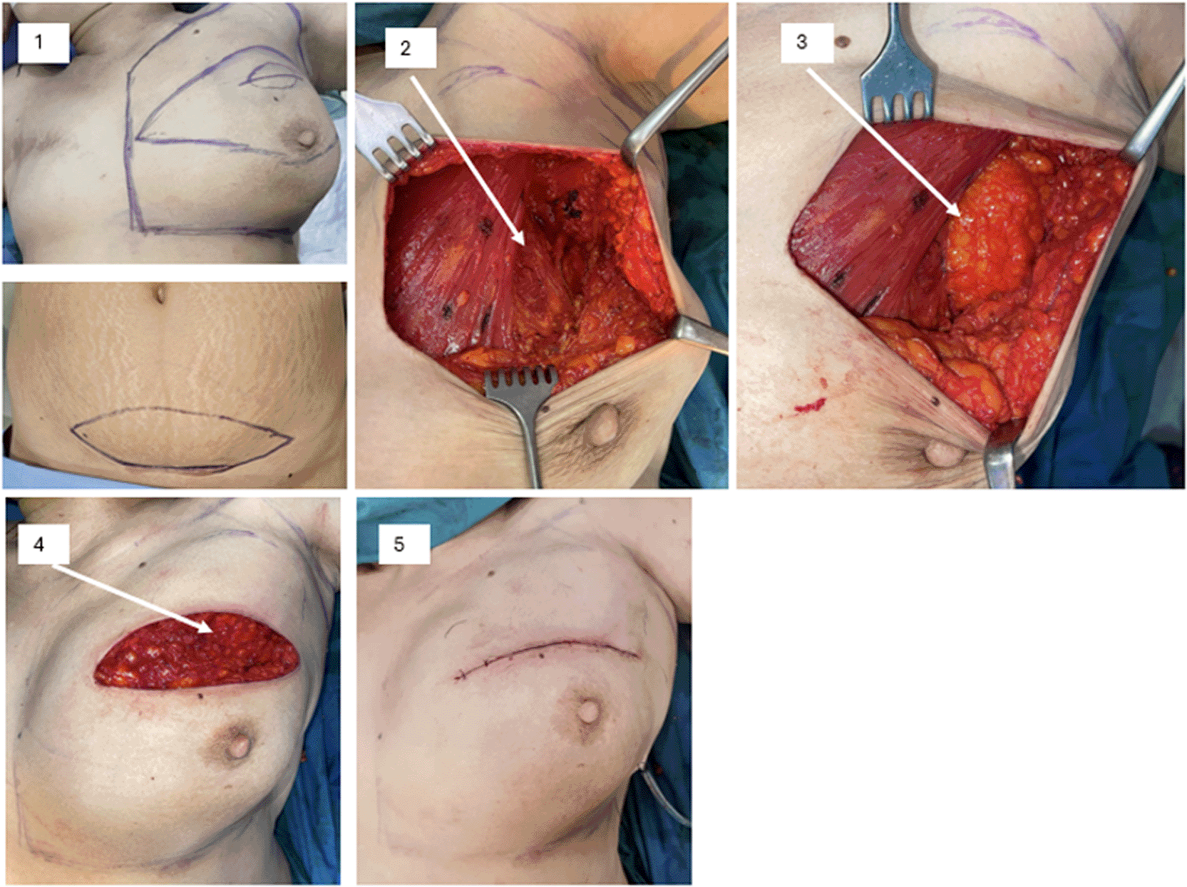
2. Defect following quadrantectomy and axillary dissection. 3. DFG filling the axillary defect. 4. DFG filling the breast defect. 5. Surgical wound after closure.
Patient: FP, 47 years old, treated with MRM with IDFG, 1 month post-operative. November 2023.
This case series includes seven female breast cancer patients, aged 44–66 years, who underwent DFG implantation following ALND at H. Adam Malik General Hospital and Malahayati General Hospital in Medan, Indonesia, between November 2023 and January 2024. Patients were diagnosed with breast cancer at various stages, including early-stage (T2N0M0, T2N1M0, T3N0M0) and locally advanced breast cancer (LABC; T4BN1M0). All underwent modified radical mastectomy (MRM) or breast-conserving surgery (BCS) with ALND, followed by intraoperative DFG implantation harvested from the lower abdomen. Arm circumference measurements were taken at eight points preoperatively and at 1, 3, and 6 months postoperatively to assess lymphedema. Lymphedema was defined as a > 2 cm increase in arm circumference at one or more points compared to the contralateral arm or a volume excess >200 mL or an increase in arm volume > 10%.12 Complications, including seroma, infection, and fat lysis were recorded. Lymphoscintigraphy examination is performed to evaluate the presence of lymphatic regeneration.
A 58-year-old Indonesian woman presented with a progressively enlarging mass in the right breast that had developed over several months. She had no significant family history of breast cancer and had not undergone any prior breast surgery. Clinical examination revealed a large breast mass with ipsilateral axillary lymph node enlargement. The patient worked in a non-specified occupation and had no relevant occupational exposures.
Diagnostic assessment included clinical examination, imaging, and pathological confirmation, which established the diagnosis of locally advanced right breast cancer (T4bN1M0). Differential diagnoses initially considered included benign breast tumors and inflammatory breast disease; however, histopathological findings confirmed invasive breast carcinoma.
The patient underwent modified radical mastectomy (MRM) with axillary lymph node dissection, followed by immediate dermofat graft implantation harvested from the lower abdomen. The rationale for using DFG was to fill the axillary dead space, prevent tissue adhesion, and potentially stimulate lymphangiogenesis through adipose-derived stem cells.
Preoperative arm circumferences ranged from 36 cm to 17 cm on the right arm and 37 cm to 16 cm on the left arm, with estimated arm volumes of 3482.2 cm3 and 3331.6 cm3, respectively.
Timeline of events
• Preoperative: clinical evaluation and baseline arm measurements
• Surgery: MRM with ALND and dermofat graft implantation
• 1-month follow-up: arm circumferences remained symmetrical with no swelling
• 3-month follow-up: stable measurements without evidence of lymphedema
• 6-month follow-up: arm volumes were 3464.8 cm3 (right) and 3314.9 cm3 (left)
No complications such as seroma, infection, or fat necrosis were observed. The patient did not develop lymphedema during the six-month follow-up period.
A 66-year-old Indonesian woman presented with a palpable mass in the right breast that had been present for several months. She had no family history of breast cancer and no previous breast procedures. Physical examination revealed a localized breast tumor with axillary lymph node involvement.
Diagnostic evaluation confirmed right breast cancer stage T2N1M0 (early stage). Differential diagnoses included fibroadenoma and other benign breast lesions prior to histopathological confirmation.
The patient underwent modified radical mastectomy with ALND, followed by dermofat graft implantation in the axillary defect to restore tissue volume and potentially support lymphatic regeneration.
Preoperative arm circumference measurements ranged from 35 cm to 16 cm on the right arm and 36 cm to 17 cm on the left arm. Initial estimated arm volumes were 4466.2 cm3 (right) and 3821.8 cm3 (left).
Timeline of events
• Preoperative: baseline arm measurement and cancer staging
• Surgery: MRM with dermofat graft placement
• Early postoperative period: mild seroma and transient fever on postoperative days 7–8
• 1-month follow-up: no swelling of the affected arm
• 3-month follow-up: stable arm measurements
• 6-month follow-up: arm volumes 4443.9 cm3 (right) and 3802.7 cm3 (left)
The postoperative fever resolved with conservative treatment, and no persistent complications or lymphedema developed.
A 57-year-old Indonesian woman presented with a right breast mass that had progressively enlarged over several months. She had no significant medical or family history of breast cancer. Clinical examination revealed a large breast lesion with axillary lymph node involvement.
Diagnostic assessment confirmed right breast cancer stage T4bN1M0 (locally advanced breast cancer).
The patient underwent modified radical mastectomy with axillary lymph node dissection followed by dermofat graft implantation to reconstruct the axillary defect and reduce the risk of postoperative lymphatic obstruction.
Preoperative arm volumes were estimated at 4143.5 cm3 on the right arm and 4070.5 cm3 on the left arm.
Timeline of events
• Preoperative evaluation and arm measurement
• Surgical intervention with MRM and DFG implantation
• 1-month follow-up: no clinical swelling
• 3-month follow-up: stable arm circumference
• 6-month follow-up: arm volumes 4122.8 cm3 (right) and 4050.1 cm3 (left)
No postoperative complications occurred, and no evidence of lymphedema was observed.
A 57-year-old Indonesian woman presented with a palpable right breast mass. She reported gradual enlargement of the mass over several months and had no family history of breast cancer. Diagnostic evaluation confirmed right breast cancer stage T2N0M0 (early stage).
The patient underwent modified radical mastectomy and dermofat graft implantation following axillary lymph node dissection.
Preoperative arm volume measurements were 3570.3 cm3 (right) and 3397.5 cm3 (left).
Timeline of events
• Preoperative evaluation and baseline measurements
• Surgical treatment with MRM and DFG placement
• 1-month follow-up: no signs of swelling
• 3-month follow-up: stable measurements
• 6-month follow-up: arm volumes 3552.5 cm3 (right) and 3380.5 cm3 (left)
• 8-month follow-up: lymphoscintigraphy demonstrated formation of new lymphatic drainage pathways
No complications or lymphedema were observed.
A 46-year-old Indonesian woman presented with a left breast mass that had progressively enlarged. There was no relevant family history of breast cancer. Diagnostic evaluation confirmed left breast cancer stage T3N0M0. The patient underwent breast-conserving surgery with axillary lymph node dissection, followed by dermofat graft implantation in both the axillary and breast defects to restore breast contour and support lymphatic regeneration. Preoperative arm volumes were 3027.6 cm3 (right) and 3063.8 cm3 (left).
Timeline of events
• Preoperative evaluation and measurement
• Surgery: breast-conserving surgery with DFG reconstruction
• Early postoperative period: surgical site infection and partial fat lysis
• Management: local wound care and debridement
• 6-month follow-up: arm volumes 3012.5 cm3 (right) and 3048.5 cm3 (left)
Although mild skin depression occurred at the incision site, breast symmetry was preserved and no lymphedema developed. Lymphoscintigraphy at nine months did not clearly demonstrate lymphatic regeneration.
A 47-year-old Indonesian woman presented with a right breast mass and was diagnosed with right breast cancer stage T3N0M0. The patient underwent breast-conserving surgery with axillary lymph node dissection, followed by dermofat graft implantation. Preoperative arm volumes were 2233.6 cm3 (right) and 2133.3 cm3 (left).
Timeline of events
• Preoperative diagnosis and baseline measurement
• Surgery with dermofat graft implantation
• Early postoperative period: mild seroma
• Conservative management without intervention
• 6-month follow-up: arm volume 2222.5 cm3 (right) and 2122.7 cm3 (left)
• 9-month follow-up: lymphoscintigraphy showed lymphatic regeneration
No lymphedema developed, as can be seen in Figure 5 and 6.
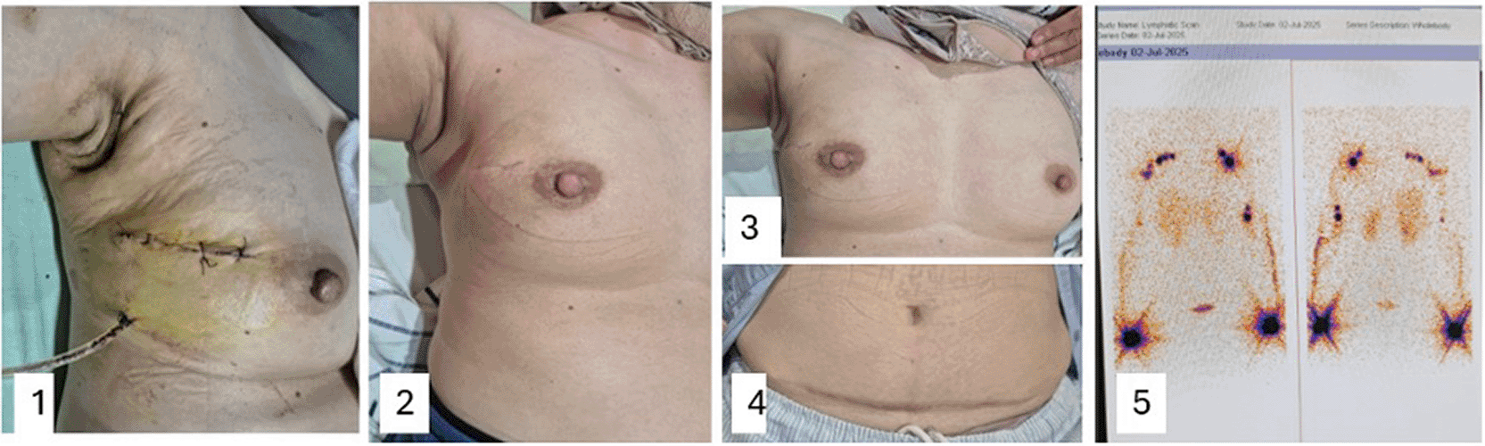
1. Three days post-operation: minimal drainage and no signs of infection. 2, 3, 4. Six months post-operation: no visible breast shrinkage, breast size remains relatively symmetrical, and the donor site scar is covered by underwear. 5.Lymphoscintigraphy result at nine months post-operation: formation of new lymphatic drainage pathways is observed.
A 44-year-old Indonesian woman presented with a right breast tumor. Diagnostic assessment confirmed right breast cancer stage T2N0M0. She underwent breast-conserving surgery with axillary lymph node dissection, followed by dermofat graft implantation to reconstruct both the breast and axillary defects. Preoperative arm volumes were 2055.4 cm3 (right) and 2144.2 cm3 (left).
Timeline of events
• Preoperative evaluation and measurements
• Surgical treatment with dermofat graft implantation
• Early postoperative period: minimal fat lysis mixed with seroma
• Conservative management
• 6-month follow-up: arm volumes 2045.2 cm3 (right) and 2133.5 cm3 (left)
• 9-month follow-up: lymphoscintigraphy demonstrated lymphatic regeneration
The breasts remained symmetrical with no visible skin retraction, and no lymphedema occurred.
Across all cases, no patients developed early lymphedema (stadium 0–II) by 6 months, as defined by the International Society of Lymphology criteria. Two patients experienced seroma with partial fat lysis, and one had a transient fever, but no severe complications (e.g., infection requiring hospitalization) occurred. Lymphoscintigraphy was performed on 4 patients (57.14%), all of whom showed the formation of new lymphatic vessels (lymphangiogenesis).
Lymphedema is a debilitating complication of breast cancer treatment, particularly following ALND, which disrupts lymphatic drainage and predisposes patients to chronic swelling, functional impairment, and recurrent infections.12,13 The pathophysiology involves an overwhelmed lymphatic system unable to clear protein-rich interstitial fluid, leading to tissue fibrosis and progressive morbidity.14,15 Preventive strategies are crucial, as established lymphedema is challenging to reverse, with current treatments like compression therapy and manual lymphatic drainage offering only symptomatic relief without addressing the underlying lymphatic dysfunction.15 The advent of microsurgical techniques such as LYMPHA and LVA has marked progress in lymphedema prevention, but their technical complexity, prolonged operative times (up to 10 hours), and need for specialized facilities limit their applicability, especially in resource-constrained settings.16,17
Dermofat graft (DFG) implantation presents a promising alternative, combining reconstructive and functional benefits. DFG, comprising autologous dermis and subcutaneous fat, has been used historically for aesthetic and reconstructive purposes, offering advantages over fat grafts due to its enhanced vascularization and stability.17,18 The dermal layer improves graft survival by facilitating neovascularization, with reported volume loss ranging from 1% to 20% compared to 45% for aspirated fat grafts within a year.18 In the context of post-axillary dissection defects, DFG serves as a tissue spacer, preventing adhesion of underlying structures and potentially restoring axillary contour, which may reduce mechanical obstruction of residual lymphatic channels.17
The potential of DFG to promote lymphatic regeneration is particularly compelling. Adipose-derived stem cells (ADSCs) within the graft are key mediators, capable of differentiating into lymphatic endothelial cells (LECs) and secreting lymphangiogenic factors, notably VEGF-C.19 in vitro studies have shown that ADSCs co-cultured with LECs upregulate lymphatic-specific markers (e.g., Prox-1, LYVE-1) and enhance lymphatic vessel formation, with effects surpassing those of exogenous VEGF-C.20 Animal models further corroborate these findings, demonstrating spontaneous lymphatic rerouting and increased VEGF-C expression following tissue transfer. These mechanisms suggest that DFG implantation could facilitate lymphatic reconnection or neolymphangiogenesis in the axillary region, mitigating lymphedema risk.21,22
VEGF-C, a critical regulator of lymphangiogenesis, is a promising biomarker for assessing lymphatic regeneration. Its expression is upregulated during wound healing and tissue regeneration, driven by macrophages and ADSCs, which also contribute to lymphangiogenesis through trans-differentiation into LECs.23,24 Elevated serum VEGF-C levels may indicate active lymphatic repair, offering a non-invasive method to monitor DFG efficacy.25 However, the diagnostic threshold for VEGF-C (e.g., ≥10,200 pg/ml) requires validation, as does its specificity for lymphangiogenesis versus angiogenesis, given its interaction with VEGFR-2.26,27
The ADSC (Adipose-Derived Stem Cell) component within the adipose tissue of a Dermofat Graft (DFG) secretes lymphangiogenic growth factors, particularly VEGF-C, and has the potential to differentiate into lymphatic endothelial cells (LECs). The interaction mechanism between ADSCs and VEGF-C in lymphangiogenesis involves several complex stages.28,29,30
Key steps in this mechanism include:
1. Stimulation of VEGF-C Secretion by ADSCs
2. ADSCs can produce and secrete VEGF-C. In response to injury or conditions requiring lymphatic vessel regeneration, ADSCs in the affected area increase their production of VEGF-C.
3. Stimulation of Lymphatic Endothelial Cell Proliferation and Migration
4. VEGF-C secreted by ADSCs stimulates lymphatic endothelial cells to proliferate and migrate to areas in need of regeneration. This process supports the formation of new lymphatic vessels.
5. Increased Permeability of Lymphatic Capillaries
6. VEGF-C also enhances the permeability of existing lymphatic capillaries, facilitating improved lymphatic fluid flow and accelerating the healing process.
7. Differentiation of ADSCs into Lymphatic Vessel Cells
8. In addition to stimulating endothelial cells, VEGF-C can influence the differentiation of ADSCs into lymphatic vessel cells, such as lymphatic endothelial cells or lymphatic progenitor cells.
This helps in the regeneration of damaged or lost lymphatic tissue. Thus, the mechanism of action of ADSCs and VEGF-C in lymphangiogenesis involves a complex collaboration between ADSCs as a source of VEGF-C and VEGF-C as a growth factor that stimulates the proliferation, migration, and differentiation of lymphatic vessel cells. This interaction is crucial in the development of regenerative therapies for lymphedema and other lymphatic disorders.30,31
Lymphatic endothelial cells (LECs) play a key role in lymphangiogenesis. They are specialized endothelial cells found in lymphatic vessels and lymphoid tissues. The following are several key roles of LECs in lymphangiogenesis32:
1. Initiation: LECs are involved in the initiation of lymphatic vessel formation during embryonic development and in response to specific stimuli such as inflammation or tissue injury in adults.
2. Proliferation and Migration: LECs proliferate and migrate to form new lymphatic vessels, expanding the lymphatic network in response to growth factors such as VEGF-C and VEGF-D.
3. Differentiation: LECs differentiate into mature lymphatic endothelial cells, which are essential components of lymphatic vessels.
4. Maintenance: LECs help maintain the integrity and function of lymphatic vessels through various mechanisms, including the secretion of factors that regulate vessel permeability and immune cell trafficking.
5. Interaction with Immune Cells: LECs interact with immune cells such as T cells, B cells, and dendritic cells, playing a role in immune surveillance and immune response.
Overall, LECs are essential for the development, maintenance, and function of the lymphatic system, playing a crucial role in both health and disease. Adipose-derived stem cells (ADSCs) have contributed significantly to advancements in regenerative medicine, as evidenced by the increasing number of publications and clinical trials exploring their use. Recent studies have demonstrated the ability of ADSCs to differentiate into lymphatic endothelial cells (LECs) and to secrete lymphangiogenic growth factors. in vitro studies have been conducted to determine whether ADSCs can be applied in lymphatic tissue engineering.31,32
Compared to LYMPHA and LVA, DFG implantation is less resource-intensive, with shorter operative times and no requirement for microsurgical expertise.33 The procedure leverages autologous tissue, reducing immunogenicity, and the donor site (e.g., lower abdomen or groin) typically yields sufficient graft volume with minimal morbidity.17 Complications, such as fat resorption, donor site scarring, or infection, are less frequent than with flap-based reconstructions, and the absence of complex postoperative monitoring (e.g., flap viability) facilitates early discharge.17,18 However, challenges include the potential for graft instability in highly mobile areas like the axilla and the need for precise donor site selection to optimize dermal thickness and fibroblast content.17 Compared to Sentinel Lymph Node Biopsy (SLNB), Dermofat Graft (DFG) implantation is considered more effective, efficient, and cost-effective. This procedure can be applied in both early and advanced stages, does not require special materials or facilities, and has a relatively simple technique that can potentially be performed by most surgeons.
The clinical implications of DFG implantation extend beyond lymphedema prevention. By addressing axillary defects, DFG may improve cosmetic outcomes and patient satisfaction, critical components of quality of life in breast cancer survivors.2 In Breast-Conserving Surgery (BCS), Dermofat Graft (DFG) implantation not only helps prevent lymphedema but also serves to fill surgical defects in the breast and axilla, resulting in symmetrical breast size and the absence of concave areas in both the breast and axillary regions. Furthermore, its accessibility makes it a viable option in low-resource settings, where advanced microsurgical techniques are often unavailable. Preliminary evidence from case reports supports DFG’s efficacy, with one study reporting a 292 mL reduction in limb volume four months post-procedure in a lymphedema patient, alongside improved clinical symptoms.24
Limitations of DFG implantation warrant consideration. The long-term stability of lymphatic regeneration remains unclear, as does the optimal timing for graft implantation (intraoperative versus delayed). Variability in graft resorption rates and the potential for cyst formation or hair growth from residual follicles require further investigation.27 Additionally, the role of adjuvant therapies (e.g., radiotherapy) in modulating DFG outcomes is poorly understood, as radiation may impair ADSC function and lymphangiogenesis.28 Standardized assessment tools, such as lymphoscintigraphy, are essential for evaluating lymphatic regeneration, but their interpretation can be subjective without quantitative metrics.33
Future research should prioritize randomized controlled trials to compare DFG with standard care or microsurgical techniques, focusing on lymphedema incidence, lymphatic regeneration (via lymphoscintigraphy), and quality of life. Longitudinal monitoring of VEGF-C levels could elucidate its predictive value, potentially guiding patient selection and treatment timing. Multicenter studies are needed to address variability in surgical techniques and patient populations, particularly in high-risk groups (e.g., obese patients or those receiving radiotherapy). If validated, DFG could redefine axillary reconstruction protocols, offering a scalable solution for lymphedema prevention in breast cancer care.
Dermofat graft implantation represents a promising, accessible approach for preventing early lymphedema and promoting lymphatic regeneration in breast cancer patients undergoing axillary lymph node dissection. In addition, DFG is also a good and simple surgical option for reconstructing defects in the breast and axilla following BCS. By leveraging the lymphangiogenic potential of adipose-derived stem cells and VEGF-C, DFG addresses both functional and reconstructive needs, with advantages over complex microsurgical techniques in terms of feasibility and resource requirements. While preliminary evidence is encouraging, further research is essential to establish long-term efficacy, optimize surgical protocols. This case series underscores the potential of DFG to enhance patient outcomes, paving the way for broader adoption in breast cancer management.
| Views | Downloads | |
|---|---|---|
| F1000Research | - | - |
|
PubMed Central
Data from PMC are received and updated monthly.
|
- | - |
Provide sufficient details of any financial or non-financial competing interests to enable users to assess whether your comments might lead a reasonable person to question your impartiality. Consider the following examples, but note that this is not an exhaustive list:
Sign up for content alerts and receive a weekly or monthly email with all newly published articles
Already registered? Sign in
The email address should be the one you originally registered with F1000.
You registered with F1000 via Google, so we cannot reset your password.
To sign in, please click here.
If you still need help with your Google account password, please click here.
You registered with F1000 via Facebook, so we cannot reset your password.
To sign in, please click here.
If you still need help with your Facebook account password, please click here.
If your email address is registered with us, we will email you instructions to reset your password.
If you think you should have received this email but it has not arrived, please check your spam filters and/or contact for further assistance.
Comments on this article Comments (0)