Keywords
clinical, demographics, inflammatory, kidney stones, polymorphism
This article is included in the Genomics and Genetics gateway.
clinical, demographics, inflammatory, kidney stones, polymorphism
Kidney stones are a global health issue, with an annually increasing incidence.1 Kidney stones are one of the most common urological diseases in Asia, and Indonesia is one of the stone-belt areas in Asia.2 In Indonesia, kidney stones remain among the most frequent urological cases, second only to benign prostatic enlargement.3 Kidney stones also have a relatively high recurrence rate globally, which is around 40–50% within 5 years and 50–60% within 10 years.4 Therefore, kidney stones are one of the urological diseases with high prevalence and recurrence.
Although many treatment options for kidney stones are available, prevention is still needed to reduce the physical burden and cost of kidney stones.5 Early identification of high-risk kidney stone patients and the implementation of effective intervention methods can reduce complications and improve patients’ quality of life.6,7 In the past five years, numerous separate studies have been conducted to identify risk factors contributing to kidney stone formation, ranging from genetic to lifestyle factors.8–15 However, the results of these studies have been varied.
These risk factors include genetic, clinical, and demographic factors.16–21 Genetic factors account for 50% of the predisposition to kidney stones.22 The presence of genetic variations, such as single nucleotide polymorphisms (SNPs), plays a significant role in the incidence of kidney stones. Studies on SNPs in several genes have been conducted to help identify individuals at risk of developing kidney stones. Based on several genome-wide association studies (GWAS) and meta-analyses, several gene polymorphisms suspected to be associated with kidney stones include the alkaline phosphatase (ALPL) gene,16 calcium-sensing receptor (CaSR),17 Claudin14 (CLDN14),18 and vitamin D receptor (VDR).19 Some of these gene polymorphisms may increase the risk of kidney stones, while others may provide protection against their occurrence.
In addition to being influenced by gene polymorphisms, the occurrence of kidney stones is also affected by clinical and demographic factors.20,21,23,24 These factors can influence kidney stone formation by modifying gene expression at different biological levels, which ultimately affects the phenotypic effects of the gene products.22 According to the European Association of Urology (EAU) guideline on kidney stones, confirmed by a meta-analysis study, clinical factors influencing kidney stone incidence include fluid intake, body mass index (BMI), waist circumference (WC), history of diabetes mellitus, history of gout, and history of urinary tract infection (UTI).20 Individuals with sedentary occupations, such as office workers, are also associated with a higher risk of kidney stones, although the exact mechanism is still unknown.25,26
Another factor that has a significant impact on kidney stone cases is fluid intake. Increasing fluid intake is universally recommended as a simple and low-cost strategy for kidney stone prevention.27–29 Moreover, from 24-hour urine laboratory parameters, several common metabolic abnormalities associated with kidney stones are hypercalciuria (30–60%), hyperoxaluria (26–67%), hyperuricosuria (15–46%), hypomagnesiuria (7–23%), and hypocitraturia (5–29%).30 Urine pH that is excessively acidic or alkaline is also a risk factor for kidney stone formation.31–33 Based on meta-analysis studies, demographic factors that influence kidney stone incidence include age and education level. The incidence of kidney stones increases with age. Similarly, low education levels have also been reported as a risk factor for kidney stones.34
Identifying patients who are at risk for developing kidney stones can help determine necessary prevention efforts, interventions, and modifications tailored to each patient. To date, no studies have investigated the relationship between gene polymorphisms and kidney stone incidence in Indonesia. Furthermore, there have been no studies analysing the risk factors for kidney stone incidence from genetic polymorphisms, clinical, and demographic perspectives in a comprehensive manner. In this matter, path analysis offers advantages in quantifying direct and indirect relationships between factors, allowing for assessment of how strongly each variable affects the others being analysed. Therefore, this study aims to explore the relationship between kidney stones and genetic polymorphisms, clinical, and demographic factors comprehensively.
The study was conducted at the Cipto Mangunkusumo Hospital, a tertiary referral hospital in Jakarta, Indonesia, between April 2021 and September 2022. The study protocol conformed to the Declaration of Helsinki and was approved by the Ethics Committee of the Faculty of Medicine, Universitas Indonesia (No: KET-544/UN2.F1/ETIK/PPM.00.02/2020). The inclusion criteria for the participants were aged ≥18 years and meeting the diagnostic criteria for KSD based on ultrasonography, computed tomography (CT) scan, kidney, ureter, and bladder X-ray imaging. Patients who met the inclusion criteria were consecutively recruited after providing informed consent. Patients were excluded if they refused to provide informed consent or had a horseshoe kidney, solitary kidney, medullary sponge kidney, polycystic kidney, congenital vesicoureteral reflux, neurogenic bladder, kidney cancer, pyonephrosis, a history of struvite stones, used urinary catheter or nephrostomy. This study included 306 participants (153 cases and 153 controls) who satisfied the criteria and provided written informed consent.
Baseline data, including body mass index (BMI), waist circumference (WC), and blood pressure, were retrieved during the screening process. All eligible patients were Indonesian residents, and BMI was classified based on the Asia-Pacific cut-off criteria.35 High WC was defined as ≥80 cm for females and ≥90 cm for males.35 Patients with at least two symptomatic episodes or new stones after treatment were classified in the recurrence group, while the rest were classified into the first-time stone group. Education levels were classified as follows: high education level for those who completed senior high school or higher, intermediate level for those who completed junior high school, and low education level for those who completed only elementary school or had no formal education.
From each participant, an 8 ml venous blood sample was collected for biochemical and genotyping analyses. Hypercalcemia, high serum creatinine levels, and hyperglycemia were defined as >10.2 mg/dl, >1.3 mg/dl, and > 200 mg/dl, respectively. A cut-off of >6 mg/dl or >7.2 mg/dl was used for hyperuricemia in women and men, respectively. First-voided morning urine samples were collected to measure inflammatory cytokine gene expression. Patients were also required to collect their 24-hour urine samples using the S-Y Easy Fold Urine Collector (Shih-Yung Medical Instrument Co., Taiwan) without additional preservatives and store them in the refrigerator. The cut-off values for interpretation were <2,000 ml/day for low urine volume, hypercalciuria >250 mg/day for females and > 300 mg/day for males, hyperoxaluria >40 mg/day, hyperuricosuria >750 mg/day, hypomagnesuria <60 mg/day, and hypocitraturia <300 mg/day.36,37
Finally, all participants were asked to complete a validated self-administered 7-day fluid record (Liq.In7) to evaluate total daily water and beverage intake.38 All data were collected and managed using the Research Electronic Data Capture (REDCap®, USA) database hosted at Universitas Indonesia.
Blood samples were analyzed to determine random blood glucose (RBG), serum uric acid, and serum calcium levels using an Abbott Architect Chemistry Analyzer (Abbott Diagnostics, USA). Creatinine levels were assessed using isotope dilution gas chromatography-mass spectrometry. Urine pH was determined using a pH paper strip, with acidic urine defined as a pH <5.5. The 24-hour urine excretion of calcium, oxalate, uric acid, magnesium, and citrate was estimated using a colorimetric assay kit (BioVision, USA).
Genomic DNA was extracted from whole blood samples using the QIAamp DNA Blood Mini Kit (QIAGEN, Netherlands). The quantity and quality of the extracted DNA were measured at 260 and 280 nm using a NanoDrop spectrophotometer (Thermo Fisher Scientific, USA). Genotyping of SNPs was performed using the 7500 Fast and 7500 Real-Time PCR Systems (Thermo Fisher Scientific). TaqMan GTXpress Master Mix and SNP genotyping assays (Thermo Fisher Scientific) were used to detect alleles present in the samples.
Data were presented as means and standard deviations, or medians and interquartile ranges for continuous variables, whereas categorical variables were presented as proportions. Statistical comparisons were performed using an independent t-test or the Mann-Whitney U test for continuous variables. All data analyses were completed using SPSS software version 25.0 (IBM Corp., USA), with p < 0.05 indicating statistical significance. Genotypic, dominant, and recessive genotyping models were used to analyze the association of genes with KSD recurrence.
Structural equation modeling (SEM) was used to assess the relationships among variables by integrating principles of regression and factor analysis, allowing simultaneous estimation of multiple pathways and accounting for potential multicollinearity. Model adequacy was judged using standard fit indices, including the comparative fit index (CFI), Tucker–Lewis index (TLI), and the root mean square error of approximation (RMSEA) with the Satorra–Bentler correction. Values of CFI and TLI above 0.90 and RMSEA below 0.08 were taken to indicate acceptable fit. Direct, indirect, and total effects were summarized using unstandardized coefficients, and statistical significance was defined as a two-sided p-value <0.05. All analyses were performed in R version 4.4.3 (R Foundation for Statistical Computing, Vienna, Austria) using the ‘lavaan’ package.
A total of 306 subjects, consisting of 153 case group and 153 control group participants, met the eligibility criteria and agreed to participate in the study. The subjects had a median age of 50 years (range 18–82), with 56.9% being male, 55.2% having higher education (diploma or bachelor’s degree), and 33% working as office employees. A total of 75.8% of the subjects had a body mass index (BMI) classified as overweight or obese, and 61.1% had a high waist circumference. Hyperglycemia and urinary tract infection were present in 6.2% and 11.4% of the subjects, respectively. The average daily fluid intake among the subjects was 1,943 mL (SD 512.98). The demographic and clinical characteristics of the study subjects are presented in Table 1.
Biochemical laboratory examinations of blood and 24-hour urine were conducted on all study subjects. The mean/median values of blood biochemical parameters (calcium, uric acid, and creatinine) for all subjects were within the normal laboratory reference ranges. The 24-hour urine results also showed urinary calcium and uric acid levels within the normal range. On the other hand, urinary magnesium and oxalate levels were high based on the laboratory’s normal values. Meanwhile, low urinary citrate was shown among the subjects. The proportion of subjects with acidic urine pH (<5.5) was 18.3%. The characteristics of urinary and blood laboratory parameters for all subjects are presented in Table 2.
A total of 7 gene SNPs were examined in this study. The G allele was found to have a higher frequency in CaSR rs1801725 and VDR rs2228570, while the A allele was more frequent in VDR rs731236. The C allele was more commonly found in CLDN14 rs219780 and ALPL rs1256328 compared to the T allele. Conversely, the T allele was more frequently observed in VDR rs1544410 compared to the C allele. The distribution of alleles and genotype frequencies for each gene polymorphism is presented in Table 3.
Age was significantly higher in the case group, with a median of 55 years (range 18–82), compared to 43 years (range 20–72) in the control group (p < 0.001). The proportion of office workers was significantly lower in the case group than in the control group (p < 0.001), primarily because most subjects in the case group were retirees, whereas the control group consisted mostly of working-age office employees. Additionally, the proportion of subjects with urinary tract infection (UTI) and high waist circumference were significantly higher in the case group (p < 0.001 and p = 0.010, respectively). Although the case group also had higher proportions of individuals with low-to-moderate education levels, overweight or obesity, hyperglycemia, and acidic urine pH, these differences were not statistically significant. The comparison of proportions for demographic, clinical, and biochemical factors between the case and control groups is shown in Table 4.
Fluid intake was also significantly lower in the case group, with a mean of 1884.77 mL (SD 502.39), compared to 2002.01 mL (SD 518.39) in the control group (p = 0.045). Serum uric acid and serum creatinine levels were significantly higher in the case group (both p < 0.001). Meanwhile, serum calcium was comparable in both groups. Urinary calcium, urinary uric acid, and urinary citrate levels were significantly lower in the case group (p = 0.002, p = 0.004, and p < 0.001, respectively). Urinary magnesium was lower in the case group as well, but this was not statistically significant (p = 0.055). On the other hand, urinary oxalate was significantly higher in the case group (p < 0.001). The comparison of mean values for demographic, clinical, and biochemical factors between the case and control groups is presented in Table 5.
| Parameter | Cases median (min–max) | Control median (min–max) | p-value |
|---|---|---|---|
| Age, years | 55 (18–82) | 43 (20–72) | < 0,001b |
| Fluid intake, mL, mean ± SD | 1884,77 ± 502,39 | 2002,01 ± 518,39 | 0,045a |
| Serum calcium, mg/dL | 9,10 (7,60–12,20) | 9,10 (7,40–10,50) | 0,24b |
| Serum uric acid, mg/dL | 6,60 (1,60–12,50) | 5,90 (2,70–10,90) | < 0,001b |
| Serum creatinine, mg/dL | 1,10 (0,50–6,70) | 0,80 (0,50–2,10) | < 0,001b |
| Urinary calcium, mg/24 h | 128,26 (1,21–1106,95) | 199,36 (1,94–790) | 0,002b |
| Urinary uric acid, mg/24 h | 50,56 (4,49–175,76) | 64,27 (17,40–231,47) | 0,004b |
| Urinary magnesium, mg/24 h | 63,46 (3,37–428,96) | 68,78 (3,75–228,50) | 0,055b |
| Urinary citrate, mg/24 h | 11,88 (0,58–223,18) | 77,63 (0,30–382,53) | < 0,001b |
| Urinary oxalate, mg/24 h | 43,34 (2,63–400,06) | 20,25 (1,07–449,29) | <0,001b |
The distribution of allele proportions and genotype frequencies for each gene polymorphism is shown in Table 6. Bivariate analysis revealed significant differences between the case and control groups.
The CT genotype and T allele of CLDN14 rs219780 were associated with an increased risk of kidney stones, with odds ratios of 2.44 (95% CI: 1.53–3.99 p ≤ 0.001) and 1.55 (95% CI: 1.10–2.19, p = 0.012), respectively, compared to the CC genotype and C allele. Additionally, the CT genotype of ALPL rs1256328 was also associated with an increased risk of kidney stones (OR = 1.75, 95% CI: 1.03–2.96, p = 0.04), although no significant difference in kidney stone occurrence was found between the T and C alleles of this SNP.
The role of VDR polymorphisms in this study showed varying results. The CT genotype of VDR rs1544410 was associated with an increased risk of kidney stones (OR = 2.15, 95% CI: 1.18–3.90, p = 0.01). Conversely, the GG genotype of VDR rs731236 was found to be protective, significantly reducing the risk of kidney stone formation (OR = 0.22, 95% CI: 0.06–0.83, p = 0.02), compared to the wild-type AA genotype. No significant associations were found between kidney stone occurrence and the polymorphisms CaSR rs1042636, CaSR rs1801725, and VDR rs2228570.
Further haplotype analysis was performed for the CaSR and VDR genes to evaluate the combined effects of the studied SNPs within each gene ( Table 7). The SNPs CaSR rs1801725 and rs1042636 showed weak linkage disequilibrium (LD) with D′ = 0.094. Similarly, VDR gene SNPs exhibited weak LD between rs2228570-rs731236 (D′ = 0.184) and rs731236-rs1544410 (D′ = 0.333). A stronger LD was found between rs2228570-rs1544410 with D′ = 0.585.
Notably, the combined presence of the A variant of rs731236 and T variant of rs1544410 was significantly associated with an increased risk of kidney stones (OR = 2.65, 95% CI: 1.38–5.09, p = 0.003). On the other hand, the combination of the G variant of rs731236 and T variant of rs1544410 was associated with a reduced risk of kidney stones (OR = 0.17, 95% CI: 0.03–0.78, p = 0.02). Other haplotypes were not associated with a significant change in kidney stone risk.
Based on the multivariate logistic regression analysis ( Table 8) using the backward LR method, older age was associated with an increased risk of kidney stones. Higher serum creatinine, increased urinary calcium, and increased urinary oxalate levels also significantly elevated the likelihood of developing kidney stones, by 36.775 times, 1.004 times, and 1.011 times, respectively. Patients with urinary tract infections (UTIs) were also more likely to develop kidney stones compared to those without UTIs (aOR 4.062, 1.033–15.970 95% CI, p = 0.045). Conversely, higher urinary citrate levels was associated with a decreased risk of kidney stones.
| Parameter | p-value* | Adjusted OR | 95% CI |
|---|---|---|---|
| CaSR rs1801725 | |||
| GT | 0,002 | 5,829 | 1,867 – 18,197 |
| TT | 0,819 | 0,638 | 0,013 – 30,163 |
| CaSR rs1042636 | |||
| AG | 0,002 | 0,178 | 0,061 – 0,526 |
| GG | 0,153 | 0,452 | 0,152 – 1,344 |
| CLDN14 rs219780 | |||
| CT | 0,005 | 2,840 | 1,379 – 5,848 |
| VDR rs2228570 | |||
| AG | 0,034 | 3,047 | 1,088 – 8,529 |
| GG | 0,014 | 3,515 | 1,283 – 9,625 |
| VDR rs1544410 | |||
| CT | 0,005 | 4,034 | 1,538 – 10,582 |
| ALPL rs1256328 | |||
| CT | 0,026 | 2,749 | 1,127 – 6,705 |
| TT | 0,246 | 0,340 | 0,055 – 2,105 |
| Age | 0,002 | 1,044 | 1,016 – 1,073 |
| UTI | 0,045 | 4,062 | 1,033 – 15,970 |
| Urinary Calcium | 0,001 | 1,004 | 1,002 – 1,007 |
| Urinary Oxalate | <0,001 | 1,011 | 1,006 – 1,016 |
| Urinary Citrate | <0,001 | 0,976 | 0,968 – 0,985 |
| Serum Creatinine | <0,001 | 36,775 | 8,534 – 158,474 |
In terms of gene polymorphisms, the GT genotype of CaSR rs1801725, CT genotype of CLDN14 rs219780, AG and GG genotypes of VDR rs2228570, CT genotype of VDR rs1544410, and CT genotype of ALPL rs1256328 were all associated with an increased risk of kidney stone formation. Meanwhile, the AG genotype of CaSR rs1042636 was associated with a decreased risk of kidney stones.
Other parameters were not found to be independent predictors of kidney stone occurrence. The Nagelkerke R2 value for this model was 62%, indicating a good explanatory power of the model.
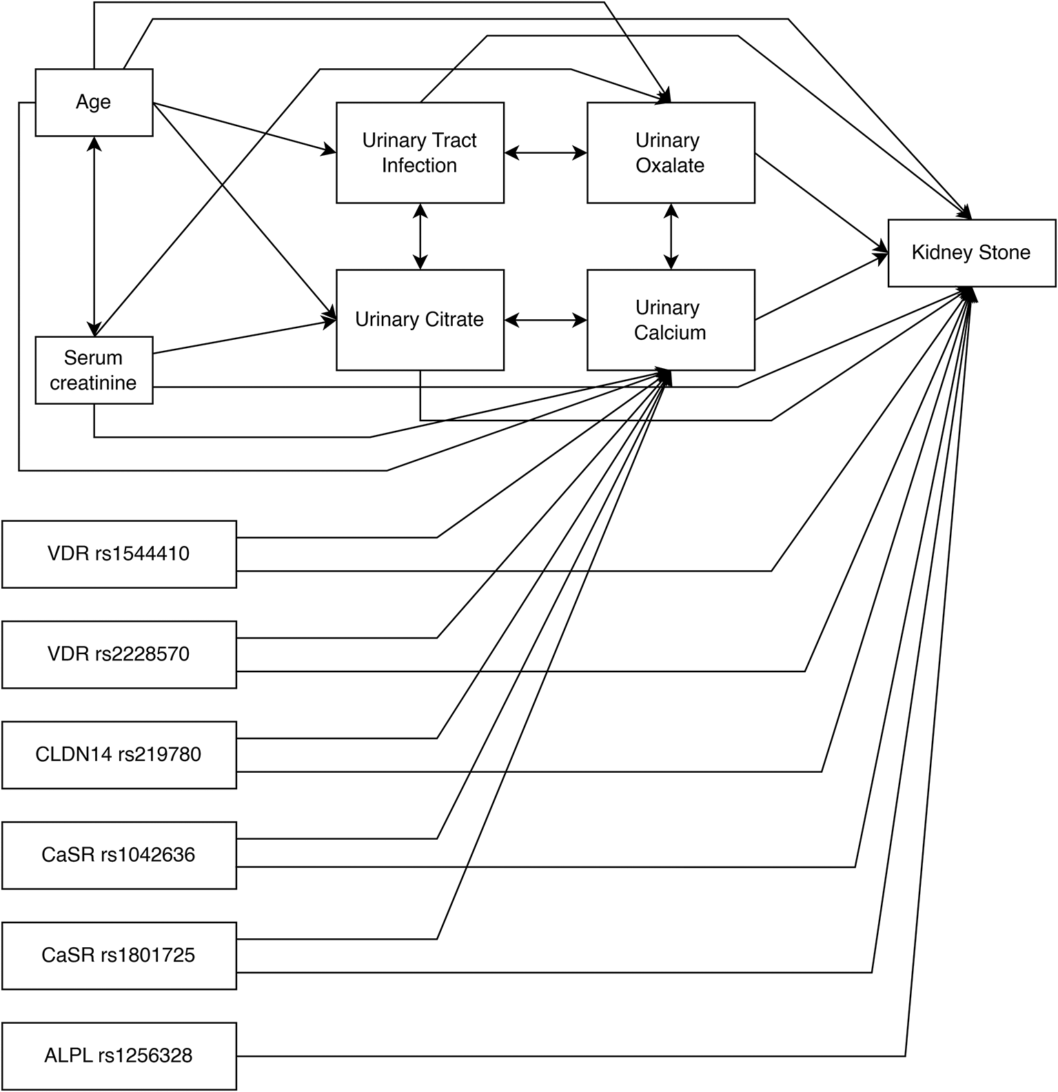
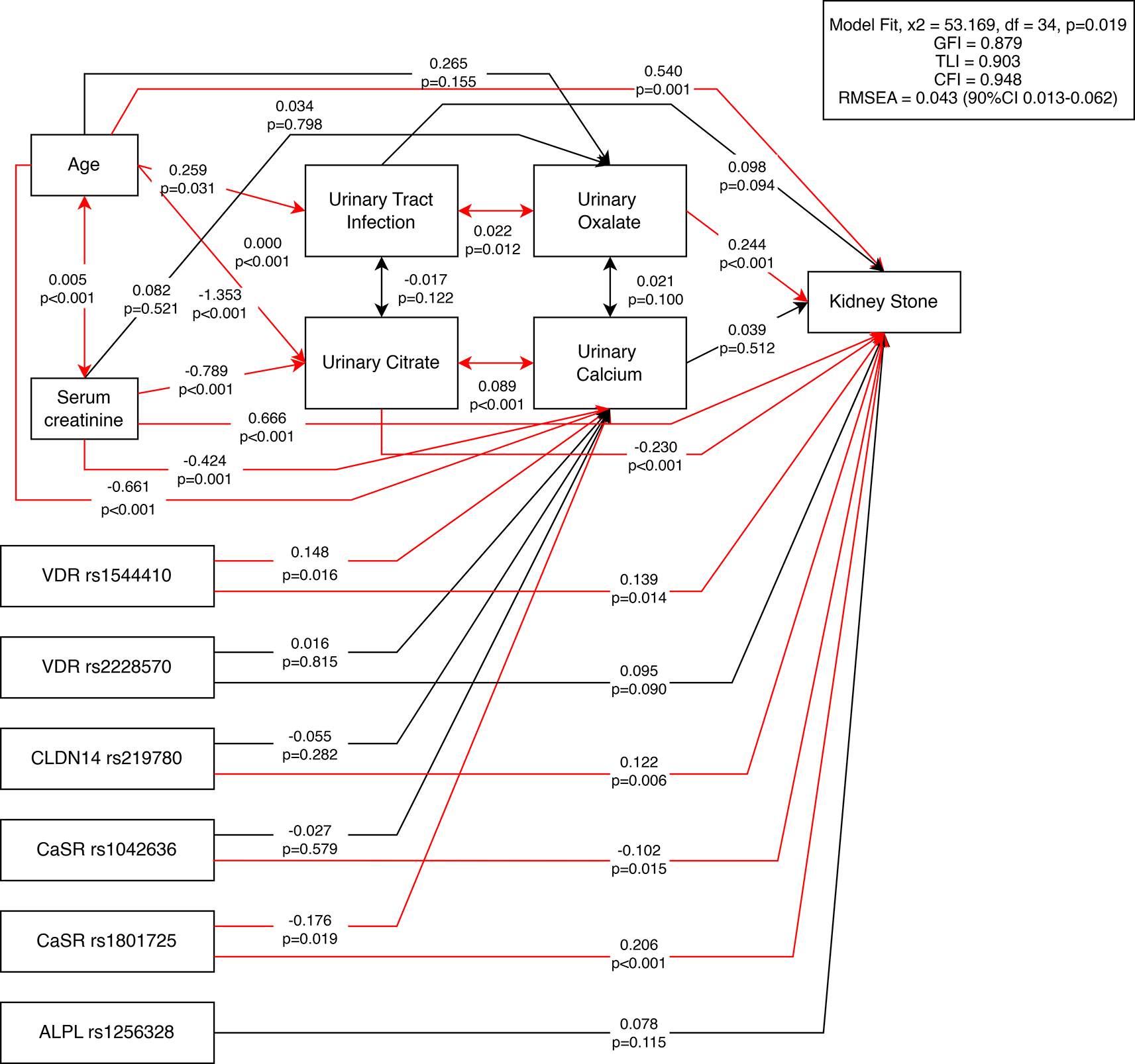
A further analysis was conducted using path analysis to evaluate both the direct and indirect effects of various variables ( Table 9), which were found to be significantly associated with kidney stone occurrence in the previous multivariate analysis ( Table 8). The initial path analysis model was constructed based on theoretical considerations ( Figure 1).
The path analysis revealed significant positive direct effects of age, serum creatinine, urinary oxalate, CaSR rs1801725, CLDN14 rs219780, and VDR rs1544410 on the occurrence of kidney stones. In contrast, urinary citrate and CaSR rs1042636 showed direct inverse associations, suggesting a protective relationship.
Age also exhibited indirect effects on kidney stone formation through UTI, 24-hour urinary calcium, and citrate levels, although these effects were smaller compared to its direct effect. Serum creatinine had a stronger direct positive effect on kidney stone formation than its indirect effects via urinary citrate and calcium. In contrast, the SNPs CaSR rs1801725, CaSR rs1042636, CLDN14 rs219780, and VDR rs1544410 exerted its effects primarily through direct pathways on kidney stone formation, while their indirect pathways through urinary calcium were non-significant.
Among the variables influencing kidney stone formation through urinary calcium, creatinine serum had the most substantial impact, measured by its standardized estimate. A significant correlation was observed between urinary calcium and citrate levels, as well as between age and serum creatinine. Based on the total effect of each variable, serum creatinine emerged as the most influential factor in kidney stone occurrence. Urinary oxalate are shown to be significantly associated with urinary tract infection.
The structural equation model demonstrated an overall acceptable fit to the data. Although the chi-square test was statistically significant (χ2 = 53.169, df = 34, p = 0.019), this is expected in moderately sized samples and does not necessarily indicate poor fit. The comparative fit indices were within acceptable ranges, with a Goodness of Fit Index (GFI) of 0.879, Tucker-Lewis Index (TLI) of 0.903, Comparative Fit Index (CFI) of 0.948, and Root Mean Square Error of Approximation (RMSEA) of 0.043, indicating a good level of approximate fit ( Figure 2).
In this study, the proportion of male subjects was higher among those with kidney stones (56.9% vs 43.1%). A similar finding was reported by Alshehri et al.,39 who found that 65.1% of kidney stone patients were male, with a 1.794-fold increased risk of kidney stones (p = 0.000). This is thought to be related to male androgens promoting the accumulation of uric acid, calcium, and oxalate in the urine, thereby increasing the risk of kidney stone formation.39
The median age of kidney stone patients in this study was 50 years, similar to a previous study by Atmoko et al.,40 which reported a median age of 52 years.40 However, there was a significant age difference between groups, with the control group having a younger median age (43 years), suggesting a possible future risk of kidney stone formation. Kidney stones can occur between ages 20 and 79, with higher incidence in the 30–59 age range. Calcium phosphate and calcium oxalate stones tend to occur in younger individuals (average ages 29.8 and 33.6 years, respectively), whereas uric acid stones are more common in those over 50.41 The analysis showed that age increased a significant 1,044-fold higher risk on kidney stone.
This study showed that office workers had a lower proportion in both groups. Office work was associated with a 0.32-fold lower risk of kidney stone occurrence, even though it is not showing any significance in the multivariate analysis. This may be influenced by a high proportion of “other” occupations in the case group, which includes housewives, entrepreneurs, drivers, retirees, and laborers. These occupations may involve sedentary behavior. In Turkey, Güller et al.41 found that housewives tend to lead a sedentary lifestyle despite performing household chores, and retirees generally show reduced physical activity.42 Sedentary behavior and physical inactivity can lead to weight gain and metabolic syndrome. Furthermore, job types related to limited access to toilets or exposure to high-temperature environments may influence kidney stone risk. Workers like drivers may intentionally limit fluid intake due to restricted toilet access, and laborers in hot environments may lose fluids through sweating, increasing urine concentration and promoting salt supersaturation.43
Mean fluid intake was significantly lower in the case group. Alshehri et al. reported that low daily fluid intake (<1 liter) was strongly associated with kidney stones (OR 28.398, 95% CI: 18.158–44.403, p = 0.000).39 Every 0.5 L increase in fluid intake reduced the risk by 0.93 times. Intake above 2 L reduced the risk by 8% compared to 1.5 L.39 Reduced fluid intake decreases urine output, leading to more concentrated urine, which increases supersaturation and stone formation risk.44 However, fluid intake did not appear to be significant in the multivariate analysis, this may due to the close volume gap between both groups.
This study also found that having a UTI increased the risk of kidney stones by 4.06 times, a statistically significant result. Kidney stones and UTIs often coexist and can influence each other. While kidney stones often lead to recurrent UTIs, UTIs themselves can promote stone formation. UTIs have traditionally been linked to infection-related stones (struvite and uric acid stones) via urease-producing bacteria like Proteus, Staphylococcus, Pseudomonas, Providencia, Ureaplasma, and Klebsiella. However, non-urease-producing bacteria like E. coli can also contribute to metabolic stone formation. Some bacteria can promote crystal aggregation by lowering urinary citrate and increasing calcium oxalate levels. E. coli and Klebsiella pneumoniae have been found to enhance calcium oxalate crystal aggregation, adhere to tubular epithelium, and stimulate expression of stone matrix proteins in renal tubular or inflammatory cells.45
In this study, we have found that only urine calcium appeared to significantly increase 1,004 times risk of kidney stone. An elevated 24-hour urinary calcium was defined as >250 mg/day in females and > 275–300 mg/day in males.46 The median urinary calcium levels in both the kidney stone and control groups did not exceed these reference ranges. While there was no significant difference in serum calcium between groups, the median urinary calcium level was significantly lower in the case group than in the controls. This is contrary to the expected physiological mechanism, where high urinary calcium excretion increases the risk of calcium-based kidney stone formation.47 The lower urinary calcium in the stone-forming group may be attributed to comorbidities, dietary factors, elevated serum creatinine, and timing of sample collection (pre-operative). A low-calcium diet can result in urinary calcium levels between 50–150 mg/day, and even 5–40 mg/day with calcium-free diets. Additionally, low sodium intake is associated with reduced urinary calcium excretion. Medications such as thiazide diuretics, benzothiazides, and estrogen may also influence urinary calcium levels. Higher serum creatinine in the stone group may reflect impaired renal function and reduced calcium excretion. Conditions such as hypoparathyroidism, pseudohypoparathyroidism, rickets, hypothyroidism, steatorrhea, and nephrosis can also cause hypocalciuria, yet were not assessed in this study.48 Moreover, calcium values may vary before and after kidney stone surgery. A study by Zhu et al.49 reported a 9.4% difference in 24-hour urinary calcium before and after surgical stone removal, possibly due to stones absorbing urinary solutes. Thus, most subjects in this study being preoperative may have contributed to the observed lower calcium levels.
Even though uric acid, both in urine and serum, did not show a significant result when compared with other variables, higher uric acid in serum were found in kidney stone patients without any hyperuricosuria. Hyperuricemia is defined as serum uric acid ≥7 mg/dL in men and ≥6 mg/dL in women, while hyperuricosuria is defined as >800 mg/day in men and >750 mg/day in women.50 Serum uric acid levels were significantly higher in the stone group (6.60 mg/dL vs. 5.90 mg/dL), whereas urinary uric acid was significantly higher in the control group (51.07 mg/24 h vs. 64.10 mg/24 h). Uric acid stone formation is closely linked to low urinary pH, and can occur even with normouricosuria.51 Pak et al.52 reported high serum uric acid, low urinary pH, and low urinary uric acid in patients with uric acid stones. Primary gout does not always present with hyperuricosuria, which may also apply to the subjects in this study. Decreased renal function and genetic variations in urate transporters may cause reduced urinary excretion despite elevated serum uric acid. Elevated serum uric acid alone can contribute to stone formation. Uric acid stone prevalence correlates more with serum than urinary uric acid levels.53
Serum creatinine was associated with a markedly elevated risk of kidney stone disease, with a 36-fold increase in odds for each 1 mg/dL rise. A similar result was found by Shen et al.54 who reported higher creatinine in cases (≈0.94 mg/dL) compared to controls (≈0.89 mg/dL, p < 0.001). Elevated creatinine reflects declining renal function, which is a common complication of kidney stones due to delayed or inadequate treatment.54 Stones increase intratubular pressure, inducing renal vasoconstriction, reducing GFR and renal blood flow, and ultimately leading to glomerulosclerosis. This process inhibits creatinine excretion, raising serum levels. Coexisting pyelonephritis can further damage renal parenchyma.55 Sui et al.56 linked worsening kidney function with decreased urinary excretion of calcium, citrate, and calcium supersaturation. The decline in calcium excretion likely reflects reduced GFR and may also explain decreased citrate.57 However, the exact mechanism for reduced citrate in renal failure remains unclear.58 Nonetheless, Sui et al.56 emphasized that citrate supplementation may be more beneficial than calcium reduction in preventing stones.
Urinary citrate is considered low when <320 mg/24 h.59 Its excretion is influenced by thiazide diuretics, UTIs, and systemic diseases (e.g., chronic diarrhea, distal renal tubular acidosis). Low citrate is found in 5–11% of stone formers and is a primary cause in 10%.60 In this study, urinary citrate was significantly lower in cases (12.15 vs. 77.61 mg/24 h). Ferraro et al.61 also found higher citrate in controls (777 vs. 668 mg/24 h). However, both groups in this study had hypocitraturia, possibly due to population-specific normal ranges. He et al.53 demonstrated significant differences in urinary citrate between Chinese and Swedish men (382 vs. 586 mg/24 h, p < 0.01). Diet and metabolic differences may also contribute, high animal protein, low fruit/vegetable, and high sodium intake can reduce urinary citrate.62 This study found urinary citrate showed a protective role in kidney stone (aOR 0.976, 0.968–0.985 95% CI, p value < 0,001).
The CaSR gene is located on chromosome 3q13.3–21 and encodes a 1078-amino-acid protein belonging to the G-protein-coupled receptor family. It consists of two promoters, containing vitamin D-responsive elements, and eight exons. CaSR regulates calcium homeostasis, particularly in the parathyroid glands and distal renal tubular cells. Its activation inhibits parathyroid hormone secretion and calcium reabsorption in the thick ascending limb of Henle (TALH), promoting urinary calcium excretion. Notably, two SNPs, rs1801725 (G > T, Ala986Ser) and rs1042636 (A > G, Arg990Gly) located in exon 7, have been associated with kidney stone risk.1,63 1,25-dihydroxyvitamin D3-induced upregulation of CaSR mRNA in renal tissues is associated with increased VDR expression, contributing to hypercalciuric stone formation in mice.63 Additionally, CaSR activation promotes CLDN14 expression, reducing paracellular calcium permeability and increasing urinary calcium.64
The T allele (Ser986) is associated with reduced inhibition of renal tubular calcium reabsorption and impaired PTH suppression, resulting in higher serum and urinary calcium. This study found that GT genotype conferred a 5.829-fold increased risk of kidney stone. On the other hand, TT genotype showed no difference on both groups. Ethnic differences may account for the variability, as studies in European, Iranian, and Chinese populations identified GG as high-risk, while East Indian populations found GT to be more predictive, and Egyptian populations highlighted TT.65
In vitro studies suggest the G allele (990Gly variant) increases CaSR sensitivity, inhibiting tubular calcium reabsorption and promoting urinary calcium excretion, potentially increasing stone risk.66 However, in this study, AG and GG genotypes were associated with a lower risk than AA, although only AG results were statistically significant. A study by Patel et al.67 found that AA genotype increased risk 2-fold compared to AG, suggesting AA may lead to higher PTH secretion and indirectly elevate calcium levels.
This synonymous SNP is located in exon 3 of the CLDN14 gene (c.687C > T, p.Thr229), which encodes claudin-14, a tight junction protein interacting with claudin-16 and claudin-19. Although the nucleotide change does not alter the amino acid, it may alter mRNA splicing and gene expression.10 This study found that the CT genotype significantly increased the risk of kidney stones (aOR 2.840), consistent with data from Pakistani and Egyptian cohorts, where the T allele also conferred elevated risk.68
The ALPL gene (chromosome 1p36.1–34) encodes tissue-nonspecific alkaline phosphatase (TNSALP), produced in renal proximal tubules. TNSALP hydrolyzes pyrophosphate, a potent inhibitor of hydroxyapatite crystallization. The rs1256328 polymorphism, particularly the CT genotype, was associated with a 2.749-fold increased risk in this study. This aligns with findings by Elshamaa et al., where CT increased risk over sixfold.68 TT genotype was not showing any significance compared to other risk factors.
This SNP alters the start codon, creating two isoforms: a longer form (A allele) and a shorter, more transcriptionally active variant (G allele). The increased transcriptional activity is potentially enhancing serum and urinary calcium levels. This study found that AG and GG genotypes were significantly associated with stone risk, trends were consistent with previous studies showing increased risk.69
A synonymous variant in exon 9 (p.Ile352=), where the GG genotype conferred a significant protective effect (OR 0.22) although it did not show any significant result when compared to other factors. AG also trended protective. This is supported by Asian population studies and in silico predictions that AA genotype may reduce VDR mRNA stability via nonsense-mediated decay (NMD).70–72
Located in intron 8 near the 3′ end, potentially affecting gene expression. This study found that the CT genotype significantly increased kidney stone risk (aOR 4.034, 1.538–10.582, p value 0.005), while the T allele showed a non-significant trend toward higher risk. Findings differ from Gonzales et al., who reported a protective effect of the CC genotype.73,74 Ethnic and environmental factors (e.g., diet, latitude) may explain the variation.
Meta-analyses have yielded mixed results. Imani et al.75 found associations of all three VDR SNPs with nephrolithiasis in East Asian and Caucasian populations. In contrast, Seyedzadeh et al.76 found only rs2228570 (FokI) significant in Asians. Although some SNPs do not alter protein structure directly, they may affect mRNA stability and translation efficiency, possibly explaining their protective or pathogenic roles.
The path analysis did not reveal any significant indirect associations via urinary calcium between the gene variants VDR rs2228570, CLDN14 rs219780, and CaSR rs1801725 and the occurrence of kidney stones. However, each of these variants showed a significant direct positive effect on kidney stone formation, consistent with the findings of both the bivariate and multivariate analyses. Although this model did not support a calcium-mediated mechanism, these variants still demonstrated an increased risk for kidney stones, suggesting that other unexplored pathways may be involved.
In contrast, the ALPL rs1256328 variant was not directly associated with kidney stones in this path model, differing from the significant association observed in the bivariate and multivariate analyses.
The VDR rs1544410 variant exerted a significant indirect effect via urinary calcium as a mediator, indicating that this polymorphism may contribute to elevated urinary calcium levels. Furthermore, it showed a direct effect on kidney stone occurrence in this model, similar to the multivariate analysis result.
Interestingly, CaSR rs1801725 showed a statistically significant negative effect on urinary calcium levels (i.e., reduced urinary calcium), yet still exerted a dominant direct positive effect on kidney stone risk. Similarly, CLDN14 rs219780 also showed a statistically significant positive effect to kidney stone occurrence without significant effect on the urinary calcium levels. This suggests that these variances may promote stone formation through mechanisms independent of urinary calcium, which were not captured in this study. Consistent with the multivariate results, CaSR rs1042636 demonstrated a negative direct association with kidney stone formation and showed no meaningful influence on urinary calcium.
Serum creatinine and age both had significant negative effects on urinary calcium, indicating that increases in either variable were associated with decreased calcium excretion. Additionally, serum creatinine had a significant negative effect on urinary citrate levels and a strong direct positive effect on kidney stone formation. However, it is important to note that elevated serum creatinine is more likely a consequence of kidney stone-induced renal damage rather than a cause, a limitation of the path analysis method, which cannot infer reverse causality.
Although urinary calcium was not a significant mediator in this model, the multivariate analysis did show a positive association with kidney stone risk. The R2 value of 62% from the multivariate model suggests that 38% of the variability remains unexplained, pointing to other unmeasured factors that may influence calcium’s role in stone formation. Notably, urinary calcium showed a strong positive correlation with urinary citrate.
Age had significant indirect negative effects on both urinary citrate and calcium, suggesting that increasing age leads to reductions in these protective urinary components. Age also had a significant positive indirect effect via UTI, meaning older individuals had a higher likelihood of UTI, which in turn increased stone risk. The direct effect of age on kidney stone formation was stronger than its indirect pathways, mirroring the multivariate and bivariate results. Moreover, age and serum creatinine were positively correlated with one another.
Urinary oxalate demonstrated a significant positive association with kidney stone formation, consistent with the multivariable analysis. Urinary citrate, on the other hand, showed a significant negative effect, reinforcing its well-established protective role. In this model, UTI did not exhibit a direct significant effect on kidney stone risk; however, it was significantly associated with urinary oxalate, suggesting that its influence may operate indirectly through oxalate levels.
Among all variables in the model, the strongest positive effect on kidney stone formation was caused by increase of serum creatinine, age, and urinary oxalate. Conversely, urinary citrate is the most protective of kidney stone development, followed by CaSR rs1042636.
One of the key strengths of this study is its novelty in exploring the association between genetic polymorphisms and kidney stone formation, which has not previously been conducted in the Indonesian population. Furthermore, this study employed a comprehensive risk factor analysis, integrating genetic variants, clinical characteristics, and demographic variables to understand their collective impact on nephrolithiasis. The study also utilized path analysis, aiming to delineate the direct and indirect effects among measurable and quantifiable variables. This analytical approach allowed for the evaluation of the strength of interrelationships among multiple factors.
This study, while offering important insights into the genetic and clinical risk factors for kidney stone formation, is not without limitations. Several key constraints must be acknowledged to interpret the findings appropriately and guide future research. Phosphate level, which may relate to genetic polymorphisms and calcium metabolism, were not assessed. Phosphate plays a role in systemic calcium homeostasis and its absence limits interpretation of calcium-phosphate balance. Only a single 24-hour urine sample was collected, although current recommendations suggest repeating this test for improved accuracy. This limitation may have introduced variability in urinary biochemical parameters. The history of UTI and prior antibiotic treatment was not evaluated, particularly in kidney stone patients who had previously received care. Thus, the study could not distinguish whether UTIs were a cause or consequence of kidney stones at the time of evaluation. Calcium measurements in serum and urine did not account for potential confounders such as high-calcium diets, calcium supplements, or vitamin D intake. Likewise, lower levels of calcium, uric acid, and magnesium observed in this study population could not be fully explained, as dietary patterns and comorbid conditions were not explored. The younger age of the control group may mean that some individuals could still develop kidney stones later in life. Age-related changes in diet, lifestyle, and fluid intake might also influence future stone risk.
This study found that individuals with kidney stones were more likely to have higher mean age, elevated serum uric acid and serum creatinine levels, and a greater incidence of urinary tract infections (UTIs). They also had lower fluid intake and reduced levels of urinary calcium, uric acid, and citrate compared to those without kidney stones. Interestingly, office-based occupations were associated with a lower risk of kidney stone formation. However, there were no significant associations between kidney stone occurrence and factors such as educational attainment, high waist circumference, hyperglycemia, urinary pH, body mass index (BMI), serum calcium, or urinary magnesium levels.
In terms of genetic predisposition, several polymorphisms were found to be significantly associated with kidney stone risk. Variants in CaSR rs1801725, CLDN14 rs219780, VDR rs2228570, VDR rs1544410, and ALPL rs1256328 were linked to an increased risk of stone formation, while CaSR rs1042636 and VDR rs731236 appeared to confer a protective effect. Path analysis provided further insights, showing that serum creatinine, age, and genetic variants in VDR rs1544410, CLDN14 rs219780, and CaSR rs1801725 exerted significant direct positive effects on the likelihood of developing kidney stones. In contrast, CaSR rs1042636 showed direct negative effects, while ALPL rs1256328 showed no direct association. The variant VDR rs1544410 was found to influence stone risk indirectly through increased urinary calcium, Additionally, urinary citrate had direct associations with kidney stone formation acting as a protective factor.
These findings underscore the multifactorial nature of kidney stone formation, where genetic predispositions interact with clinical and lifestyle factors, and highlight the potential utility of genetic screening and integrative risk modeling in enhancing preventive strategies.
| Views | Downloads | |
|---|---|---|
| F1000Research | - | - |
|
PubMed Central
Data from PMC are received and updated monthly.
|
- | - |
Provide sufficient details of any financial or non-financial competing interests to enable users to assess whether your comments might lead a reasonable person to question your impartiality. Consider the following examples, but note that this is not an exhaustive list:
Sign up for content alerts and receive a weekly or monthly email with all newly published articles
Already registered? Sign in
The email address should be the one you originally registered with F1000.
You registered with F1000 via Google, so we cannot reset your password.
To sign in, please click here.
If you still need help with your Google account password, please click here.
You registered with F1000 via Facebook, so we cannot reset your password.
To sign in, please click here.
If you still need help with your Facebook account password, please click here.
If your email address is registered with us, we will email you instructions to reset your password.
If you think you should have received this email but it has not arrived, please check your spam filters and/or contact for further assistance.
Comments on this article Comments (0)